Membrane Transport Part 1
 Call Kai
 Learn
 Practice Test
 Spaced Repetition
 Match
 Flashcards
 Knowt Play
1/45

There's no tags or description
Looks like no tags are added yet.
46 Terms
Selective Permeability
cell determines when they do and don’t want to let molecules in
impermeable to charged molecules (ions) no matter how small: the charge and high degree of hydration of such molecules prevent them from entering the hydrocarbon phase of the bilayer

What factors determine the rate of diffusion
polarity and charge
What molecules are more/less permeable
Most
non polar
polar
Larger molecules
charged molecules
Least
What are the two classes of transport proteisn
Transporters and carriers
Transporters
Also known as carriers or permeases
Like substrate enzyme binding, the solute binds to the binding site and then causes a conformational change in the transporter

Carrier
Weaker interaction with the solute
Let the channels cross the membrane passively “downhill”
Forms pores instead of using solute binding sites
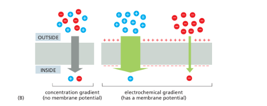
Concentration gradient
Concentration Gradient - the difference in concentration on the two sides of the membrane
Drives passive transport and determines its direction
Electrochemical Gradient
difference in membrane potential across the membrane; concentration + change = electrochemical gradient
Uniporter
type of transporter where only one solute is moved
Symporters
Simultaneous movement of more than one solute in the same direction
Antiports
simultaneous movement of more than 1 solute in opposite directions
Multispanning transmembrane
function by undergoing reversible conformational changes that alternately expose the solute binding site to one side of the membrane and then the other
Simple Diffusion
passive
Solute crosses the membrane without a transport protein (no energy required)
movement of solute down concentration gradient
Examples of simple diffusion
Rate determined by hydrophobicity and size:
Fastest:
Nonpolar gases (Oxygen and CO2)
Small steroid hormones
Small uncharged polar (small amounts of water), ammonia
Larger, uncharged polar: Glucose (very slow, needs facilitated transport)
Ions: relatively impermeable
Facilitated Diffusion (PASSIVE)
Transport protein moves solutes down their concentration gradient (Energy not required)
DOWN concentration gradient
Examples of facilitated diffsuion
Aquaporins
Glucose transporter
Voltage gated na+ channel
VOltage gated K+ channels
K+ leaky channelschannelropsins
Aquaporins
type of facilitated diffusion
water channel
Blocks passage of ions
Why can’t aquaporin pass protons through
Two asparagine in the center of the pore prevent protons from passing through the channel
Na+ Glucose transporter
type of facilitated difussion
Alternates between conformations
Solute binding site is sequentially accessible to one side of bilayer, then the other
All channels (passive)
Na+ drives the movement of glucose against it’s concentration gradient
binding of na+ and glucose is cooperative
Voltage gated Na+ channel
type of membrane diffusion
(3 conformations ), open in response to depolarization , Na+ rushes into cell
Voltage gated K+ channels
type of facilitated diffsuion
efflux of K+ to restore resting membrane potential
K+ leaky channels
type of faciliatetd diffusion
Do not need stimulus to move K+ down concentration gradient
(opens in resting cell): Maintains resting membrane potential
Primary Active Transport
Requires ATP
against the concentration gradient
ex. Na+ K+ pump, calcium pump , ABC transporter, CTFR protein, V type pump
Na+ K+ pump (P-type):
primary active transport
antiport direction of solutes, both move against concentration gradient. ATP hydrolysis required.
self phosphorylate
3 Na+ out, 2 K+ in.
Na+ concentration is high outside cell, K+ high inside cell
(pumping cycle)
The concentration of K+ ions are higher inside than outside , and vice versa for Na+ ions
Na - gradient drives transport of nutrients into animal cells
Pump is electrogenic (produces a change in the elcyrical potential of the cell)
Calcium pump
primary active transport
(P-type) Detailed description of pumping cycle
maintain Ca 2+ cell gradient

Ca 2+ in the Sarcoplasmic Reticulum
Sarcoplasmic Reticulum: specialized endoplasmic reticulum in muscle cell cytoplasm, intracellular storage of calcium ions
When the action potential depolarizes the muscle cell plasma membrane, Ca 2+ is released into cytosol from the sarcoplasmic reticulum through Ca 2+ release channels, which stimulates muscle to contract
The Ca2+ pump moves Ca2+ from cytosol back to sarcoplasmic reticulum, end of muscle contraction

Sarcoplasmic Reticulum
specialized endoplasmic reticulum in muscle cell cytoplasm, intracellular storage of calcium ions
ABC transporter
primary active trasnport
aka ATP-binding cassette transporters
ATP binding and hydrolysis influences conformational changes to expose binding sites.
differ structurally from P-type ATPases and primarily pump small organic molecules across cell membranes
Examples: MDR protein
Clinical Importance of ABC Transporters
Multi-drug resistance (MDR) proteins also called P-glycoprotein: high levels in cancer cells, makes cells resistant to cytotoxic drugs (chemo)
Pasmodium falciparum (causes malaria) transporter pumps
CTFR Protein
Primary active transport
(why unique?) Acts like a channel
balances the cl- ions in the cell
V type pump:
primary actove transport
Generate H+ gradient (acidify lysosome, vesicles)
to aicdifiy the interior of these organelles
acidify vacuole compartments (lysosomes, endosomes golgi)
Uses the Gibbs free energy of ATP hydrolysis to make an H+ gradient
What do uncouplers do?
collapse the proton gradient by equalizing proton concentrations on both sides of the membrane
How is DNP an uncoupler
Equalize proton concentration on both sides of the membrane
caused increased oxygen consumption and an increased metabolic rate, but a decline in ATP production
Secondary active trasnport
Ion gradient (potential energy) required to moves another solute against its concentration gradient.
Indirect use of ATP (pump that maintains ion gradient)
AGAINST concentration gradient
ex. Na+/Glucose transorter
Na+/Glucose transporter
Use Na+ gradient (provided by Na+ K+ pump) to drive glucose against its gradient (symporter)
Na⁺/K⁺ ATPase pumps 3 Na⁺ OUT and 2 K⁺ IN; both move uphill.
Na⁺ is higher outside; K⁺ is higher inside -> electrogenic.
Na⁺ moves downhill to help glucose move uphill into the cell.
Na⁺ and glucose bind cooperatively
Occupied symporter becomes occlude

Transcellular transport of glucose
Asymmetric distribution of transporters in plasma membrane
The Na/+K+pump helps maintain the Na+ gradient
It’s found exclusively in basal and lateral domains
Mechanism of Na+ driven glucose transporter
secondary active transport
movement of Na+ (from Na+ K+ pump) down its concentration gradient is used to move glucose AGAINST its concentration gradient
binding of Na+ and glucose happens through symporter that switches between occluded (closed) and non-occluded states and utilizes the cooperative binding nature of Na+ and glucose
Mechanism fo Na+ Glucose Symporter
Symporter is in an occluded-empty state and opens to the intestinal lumen
Na+ and glucose bind to the symporter
symporter closes and enters an occluded-filled state
symporter opens to the intestinal lumen, allowing the Na+ and the glucose to enter the lumen
lastly, the symporter will return to its original occluded-empty state and the cycle starts again
GLUT
Na+/ Glucose trasnporter
Glc fits into the GLUT binding site,
Binding causes a conformational change
Low [Glc] causes dissociation
GLUT bounces back to it’s original conformation

Ways to use Active transport
Coupled transporters harness the energy stored in concentration gradients to couple the uphill transport of one solute across the membrane to the downhill transport of another.
ATP-driven pumps couple uphill transport to the hydrolysis of ATP.
Light- or redox-driven pumps, which are known in bacteria, archaea, mitochondria, and chloroplasts, couple uphill transport to an input of energy from light, as with bacteriorhodopsin and photosystem II (discussed in Chapters 10 and 14, respectively), or from a redox reaction, as with cyto chrome c oxidase (discussed in Chapter 14).
Classes of ATP driven pumps
p-type
ABC
V-type pumps
MDR protein (ABC transporter)
pump that hydrolyzes two ATP molecules per transport cycle
channel proteins
do not possess a binding site for a specific solute, and instead interact weakly with the solute
can only carry out passive transport
What happens after Ca2+ binds ATP and is phosphorylated?
a conformational change that opens a passageway to the sarcoplasmic reticulum lumen, causing two Ca 2+ ions to exit into the sarcoplasmic reticulum
(not a typo)
Mechanism of Glucose Transporter on basal side of intestinal cell
passive transport through a uniporter
high concentration of glucose through cell → too big to leave w/o a transporter
Glucose transporter open to intestinal cell
allow the glucose to bind to the interior of the transporter → causes conformational change → transporter opens toward extracellular fluid → Since lower conc. i extracellular fluid, glucose will detach and leave → loss of glucose will cause a consequent conformational change and the transporter will open to the intestinal cell
How would the inhibition of the Na+ K+ pump affect the abortion of glucose in the cell
would inhibit absorption of glucose
if the Na+ K+ pump is inhibited , Na+ wouldn’t be removed from cell → collapses the gradient needed for Na+ glucose symporter