Lower GI pathology and Image critique
 Call Kai
 Learn
Learn Practice Test
 Spaced Repetition
 Match
Match Flashcards
 Knowt Play
1/54

There's no tags or description
Looks like no tags are added yet.
Name | Mastery | Learn | Test | Matching | Spaced | Call with Kai | Chat |
|---|
No analytics yet
Send a link to your students to track their progress
55 Terms
what is chron’s disease
chronic inflammatory disease of the intestines
what is the radiogrpahic appearance of chron’s disease
inflammation and fibrosis with narrowing
string sign, skipped pattern
cobblestone appearance

what is this an example of?
chron’s disease

what are these a picture of
chron’s disease
what side of the body is most likely to have chron’s disease
right side
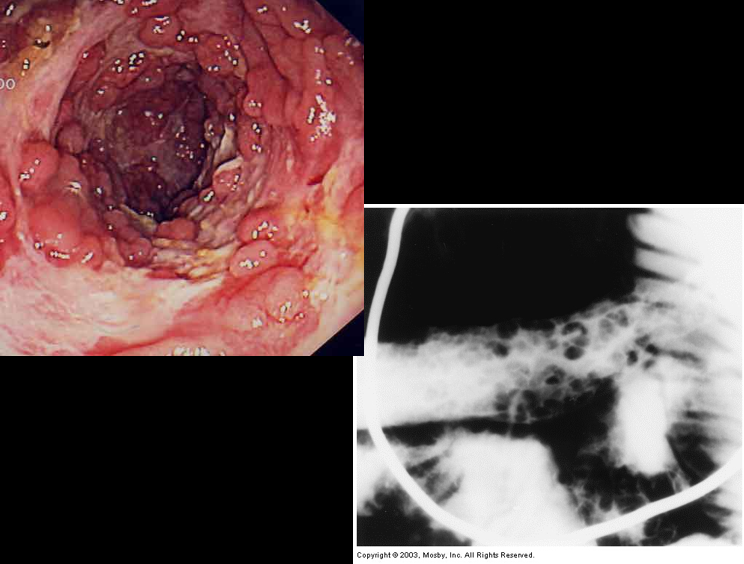
what are these an image of?
the cobblestone effect of chron’s disease
what is colonic diverticula
outpouchings, herniation of mucosa and submucosa through the muscular layers at points of weakness of the bowel wall
condition of: diverticulosis
what is the radiographic appearance of colonic diverticula
round or oval outpouchings, tend to be in clusters
abscesses
extravasation
what is diverticulitis
complication of diverticulosis; inflammation of the diverticula
typically in the sigmoid region
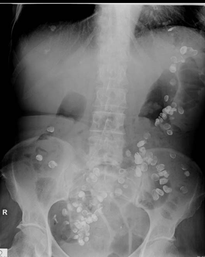
what is this an example of?
retained barium in diverticulum

what is this an image of?
colonic diverticula

what is this an image of?
perforation with intramural absecc (diverticula)
what is ulcerative colitis
inflammatory bowel disease
can lead to toxic mega colon, perforation
what is the radigraphic appearance of ulcerative colitis
earliest detection: fine granulatrity of the mucosa
chronic: haustra pattern absent, tubular looking colon

what is this an image of?
ulcerative colitis (haustra smooth)

what is this an example of?
ulcerative colitis- granularity
what is colon cancer?
cancer found in the colon
what is the radiographic appearance of colon cancer?
polyps
apple core

what is this image of?
colon cancer (apple core)
what is intussusception
telescoping of one part of the intestinal tract into another becuase of the peristalsis which forces the proximal segment of bowel to move distally w/ in the ensheathing outer portion
what is the radiographic appearance of intussusception
coiled- spring

what is this an imge of?
intussusception (barium)
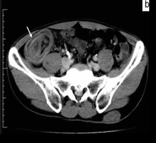
what is this an image of?
intussusception (CT)

what is this an image of?
intussusception (ultrasound)
what is disk herniation
protrusion of an intervertebral disk
L4-L5, L5-S1, C5-C6, C6-C7, T9-T12
what is the radiographic appearance of disk herniation
requires CT, MRI, or myelo to demostrate impression of hte disk on the spinal cord or individual nerve roots

what is this an image of?
disk herniation
for the small bowel PA stomach what do you annotate
0 minute
what is best seen of a PA stomach (0min image)
body of stomach
medial and lateral margins
what is best seen on an RAO stomach
the duodenal bulb filled with barium
what is best seen on a right lateral stomach
9see the retrogastric area and loop in profile; anterior and posterior margins
you do not need to annotate times for which views
RAO stomach
right lateral stomach
what do you need to see on the PA/AP follow through image
small intestines/ small bowel
ok if you cut stomach
what are the views for a single BE
scout
PA
LPO
RPO
PA axial sigmoid
Left Lateral rectum
post evacuation PA
BE scout (single) image requirements
all bowel pattern and need to have symph
for RPO and LPO BE (single) you do not need ____
symph (do not need rectum)
for an RPO BE (single) you will see
right pelvic wing flattened
LEFT splenic flexure open
for an LPO BE (single) you will see
left pelvic wing flattened
RIGHT hepatic flexure open
PA BE (single) you need
entire large intestine
symphysis
RH: can also see the enema tip
PA axial (sigmoid) angle (single)
30 degrees caudad
17×17 collimation
PA axial (sigmoid) you will see (single)
all of the signoid colon and rectum included
can see end of descending colon
Left lateral rectum RH protocol (single)
need to deflate balloon prior to exposure
what is seen on a Left lateral rectum (single)
rectosigmoid area in the center of the image
leave the tip in

what is circled in red
the BE balloon
Post Evac PA (single) what is seen
entire large intestine
symph (need rectum)
POST marker
residual barium will be in the bowel
views for a double BE
scout
PA
AP
RLD
LLD
PA axial sigmoidCross table lateral rectum
Right lateral decub Double BE
air inflated portion of the colon is of primary importance- descedning colon
splenic to symph
mark side up (looking at air side)
DONE AP
Left Lateral Decub double BE you will see
the air inflated portion of hte colon is primary importance
ascending colon
mark side up (right)
RH pt faces the IR
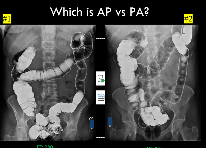
#1
prone/PA

#2
supine/AP
AP supine double BE what is seen
need the entire colon
air seen in transverse colon
PA prone double BE what is seen
the entire colon
barium seen in transverse colon
PA axial sigmoid double BE
rectosigmoid area
30 degree cauda
all sigmoid colon and rectum included
can see end of descending colon
right lateral X-table rectum double BE what is seen
rectosignoid area visualized, air fluid level demonstrated
deflate balloon and remove tip prior to exposure
if the patient cannot lay prone for the PA axial (sigmoid images what direction do you angle)
30 degrees cephalad