PBL 2: HeLa Cells
 Call Kai
 Learn
 Practice Test
 Spaced Repetition
 Match
 Flashcards
 Knowt Play
1/55

There's no tags or description
Looks like no tags are added yet.
56 Terms
What are HeLa Cells?
the first immortalised line of endothelial human cancer cells derived from cervical carcinoma
this culture adequately stimulates a simplified human body under lab conditions
Characteristics of HeLa Cells:
Can be frozen for decades, thawed and grown again in separate batches so they are easy to culture
Have a wide range of cell surface receptors, making them useful for studying the effects of cytokines
The cells proliferate twice as fast in vitro than in normal tissues, while the intracellular growth suppression program was turned off
More stable than other cancer cells and don’t have Haylick limit
How can Cell Lines be Created
the individual cells need to be isolated from tissue
the cells must be grown in culture
Can be transferred to secondary cultures
How are individual cells remove from tissues
Isolation requires disrupting extracellular matrices and cell–cell junctions that hold cells together.
Proteolytic enzymes (trypsin and collagenase): Break down proteins in the extracellular matrix.
Agents such as EDTA binds to Ca²⁺ ions, which are necessary for cell–cell adhesion, helping cells separate.
Gentle agitation: Tissue can then be physically separated from a part into individual cells by gentle agitation
What are Primary and Secondary Cultures
Primary culture: Cells grown directly from tissue obtained from an organism.
Cells may first be separated into different cell types.
They often maintain characteristics of their original tissue.
Secondary culture: Cells taken from a primary culture and transferred to a new culture dish.
Passaging (subculturing): Repeatedly transferring cells to fresh culture dishes to allow continued growth over weeks or months.
Cells can retain specialized functions
How can you make human cells immortal
The application of in vitro culturing of animal and plant cells makes it possible to grow the cells of various human tissues on selective media
Numerous human cell cultures are derived from cancer tumors, and these cells are capable of endless division and are therefore called ‘immortal’
What is Replicative Cell Senescence
Cells stop dividing after a finite number of cell divisions
the limited proliferation capacity reflects a progressive shortening and uncapping of the cell’s telomeres
What are Telomeres and its Sequence
the repetitive DNA sequences and associated proteins that cap the ends of each chromosome (GGGTTA)
What is telomerase and what is it’s role in human cells
an enzyme that maintains the telomeres by maintaining special telomeric DNA sequences that promote the formation of protein cap structures to protect chromosome endings
Human cells have turned off production of the enzyme
What does the deficiency of telomerase do?
The deficiency in telomerase causes protective caps to deteriorate and telomeres to shorten at each division
This creates a DNA damage signal because the unprotected chromosome ends resemble double-strand breaks, and lead to permanent cell-cycle arrest or cause the cell to die
Could all Human Cells be immortalised by this trick
Not all human cells can be immortalised by this trick
Although their telomeres remain long, they still stop dividing after a limited number of divisions because culture conditions cause excessive stimulation of cell proliferation, which activates a poorly understood protective mechanism that stops cell division, this process is called culture shock
What is another mechanism to immortalise human cells
Other than providing cells with the gene that encodes the catalytic subunit of telomerase
one must also inactivate the protective mechanisms, which can be done by introducing certain cancer promoting oncogenes, additional to activation telomerase
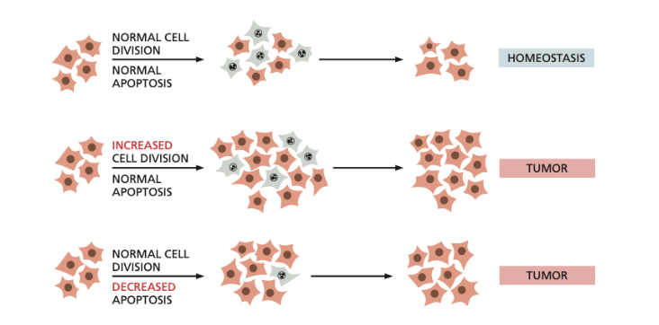
What is the selective advantage possessed by mutant cancer cells
This selective advantage increases the rate at which a clone of cells proliferates or enabling it to continue proliferating when normal cells would stop or die
An important property of cancer cells is that they fail to undergo apoptosis, when a normal cell would do so
What are Transformed Cell Lines?
Transformed cell lines are populations of cells that have undergone a permanent, heritable change in their phenotype—allowing them to divide indefinitely
Cancer cells that can be grown in culture or cultured cells artificially engineered to contain the types of mutations encountered in cancers typically show a transformed phenotype
What are Characteristics of Transformed Phenotypes:
Transformed phenotypes are abnormal in their shape, motility, responses to growth factors in a culture medium and in the way they react to contact with the culture dish and with one another
Differences between normal cells and transformed cells
The manner in which they divide, negative influences and behaviour in natural environments
Normal cells will not divide unless they are attached to the surface, transformed cells will often divide even if held in suspension
transformed cells no longer require all of the positive signals from their surroundings that normal cells require.
Transformed cells also fail to recognise negative influences
For Example: normal cells become inhibited from moving and dividing when the culture reaches confluence (where the cells are touching one another), while transformed cells continue moving and dividing even after confluence, and so pile up in layer upon layer in the culture dish
Cancer cells also misbehave in their natural environment
Normal cells are constantly turning over through rounds of cell division and death, but maintain their barrier function by extruding old or damaged cells, and then these cells go through apoptosis.
However transformed cells override normal death signals, therefore they may be able to survive after their ejection. They are however still sent to the waste canal
However some mutations selected for during cancer progression can change the direction of extrusion thereby surrounding tumor cells to cross the basement membrane and invade surrounding tissue
How do Cancer Cells Bypass these Negative Influences (Bypass Death Signals)?
A large multicellular organism requires powerful safety mechanisms that guard against damaged and deranged cells.
Normally, internal disorder gives rise to danger signals in the faulty cell, activating protective measures or, failing that, leading to cell death by apoptosis.
To survive, cancer cells require mutations to elude or break through these defenses designed to eliminate defective cells.
To survive and divide without limit, a prospective cancer cell must accumulate changes that disable the normal safety mechanisms that would otherwise cause a stressed cell to commit suicide by apoptosis.
How can Cancer Cells Escape Built-in Limit to Cell Proliferation
Cancer cells avoid replicative cell senescence in one of two ways
They reactivate their telomerase gene as they proliferate, so that telomeres do not become capped or do not shorten (already discussed, same as with normal cells)
They can also evoke a mechanism based on a DNA repair process that depends on homologous recombination for elongating their chromosome ends (Called ATL)
What is Necrosis and How do Tumors Grow?
Necrosis is the premature, accidental death of cells and living tissue in the body caused by external or internal damage
this is how cancer cells in the interior of a large solid tumor die
The tumor grows because the cell birthrate outpaces the cell death rate.
How are HeLa Cells connected to HPV
The karyotype of normal human cells contains 46 chromosomes, while HeLa cells have 76 to 80 chromosomes that are significantly mutated
This deviation is associated with human papillomavirus (HPV), which is responsible for almost all cases of cervical cancer
HPV inserts its DNA into the host cell, therefore the host cell begins to synthesise a protein that binds and inactivates p53 protein (which usually prevents mutations and suppresses tumors), therefore leading to consequences
Why are HeLa Cells Important
The cell line allowing to grow cells in vitro gave rise to new biomedical scientific research in molecular biology
They gave scientists an opportunity to cultivate hundreds of other cancer cell lines
Scientists have not yet found conditions that would allow normal cells to grow indefinitely in culture, making cancer cells useful models for research.
HeLa cells became important for the isolation and cultivation of a number of other viruses, as well as production of antibodies and antitumor chemotherapy drugs
What else are HeLa cells research used for?
The cells are commonly used in virology research, to study diseases such as cancer and AIDS, to assess the effects of radiation and toxic substances, to construct genetic maps, to develop cell engineering methods, and to solve a great number of other scientific problems
How were HeLa Cells Important for Polio
The creation of a vaccine from inactivated viruses against polio has been created due to the HeLa cell culture
But before testing the new vaccine out on humans, it had to be tested on living human cells
The population of HeLa cells proved to be an ideal tool
They grew rapidly, which made it possible to assemble a huge number of cells necessary for research sufficiently quickly
Moreover, it also turned out that they were easily infected with the polio virus. In less than a year, the vaccine was ready for use in patients
Why is cDNA useful?
it allows scientists to study, clone, and express genes without the introns found in eukaryotic genomic DNA.
this is because cDNA is made from mRNA transcripts which have already undergone splicing removing introns
What is cDNA
an alternative strategy to cloning that enriches for protein-coding genes
It begins the cloning process by selecting only those DNA sequences that are transcribed into mRNA and therefore correspond to protein-encoding genes
This is done by extracting the mRNA from cells and then making a DNA copy of each mRNA molecule present, this is called complementary DNA
What are the Steps of cDNA creation
First, mRNA is isolated, and hybridised with oligo T primer (poly T primer) the mRNA strand has a poly-A-tail
The copying reaction is catalysed by a reverse transcriptase enzyme of retroviruses, which synthesises a complementary DNA chain on a RNA template
The RNA strand is degraded with Ribonuclease (H), and keeps the DNA intact
Then the single stranded cDNA molecules are converted by DNA polymerase into double stranded cDNA molecules, and these are inserted into a plasmid or virus vector and cloned
DNA polymerase binds to a leftover RNA fragment acting as a primer
Each clone is called a cDNA clone, and the entire collection of clones constitutes a cDNA library

Why was cDNA used in the expirement rather than for ex normal DNA?
The researchers used cDNA instead of genomic DNA because cDNA contains the protein-coding sequence of the CD4 gene without introns, making it easier to introduce into HeLa cells and express the CD4 receptor.
Genomic DNA could potentially have been used, but it would require appropriate regulatory sequences and correct RNA processing, including removal of introns.
cDNA was more convenient because it already contained the processed protein-coding sequence needed to produce CD4.
What is the relationship between HIV and the CD4 gene
HIV can primarily infect cells that have the T4 glycoprotein (now called CD4) on their surface, because HIV uses this receptor to attach to the cell
Researchers wanted to investigate whether CD4 itself was responsible for this susceptibility.
They also wanted to investigate whether introducing CD4 into cells that normally lack it would make them susceptible to HIV infection.
Specifically, whether HIV could infect non-T lymphocytes (non-immune cells), including epithelial cells such as HeLa.
Determine whether additional T-cell-specific proteins were necessary for infection.
Why were HeLa Cells Used?
They are cancer cells not T lymphocytes, which are the main targets of HIV.
Unmodified HeLa cells do not normally express T4 gene (CD4).
They were therefore not susceptible to HIV infection under the experimental conditions.
This made HeLa cells useful for testing whether CD4 alone could make non-immune cells susceptible to HIV infection.
Researchers could introduce the CD4 gene into HeLa cells and compare them with unmodified HeLa cells.
This helped determine whether HIV infection depends on the presence of CD4 rather than on the cell being a T lymphocyte.
HeLa cells are also immortal and can be maintained in culture, making them practical for laboratory experiments.
Researchers isolated the cDNA encoding the T4 (CD4) protein.
Previously, scientists isolated the c-DNA sequence that codes for the T4 (CD4) protein and used it to determine the protein's amino acid sequence.
This led to the ability to clone T4 in new cellular environments and to examine the role of this molecule in the pathogenesis of AIDS
They inserted the CD4 gene into a retroviral expression vector (T4-pMV7).
Full-length cDNA clone encoding the surface protein T4 (CD4 gene) was subcloned into the retroviral expression vector, pMV7
The gene was introduced into HeLa cells, producing HeLa-T4+ cells expressing CD4 on their surface.
What were the three finding of the experiment?
Introducing CD4 made HeLa cells susceptible to HIV infection
HeLa cells expressing CD4 became susceptible to productive HIV infection.
Unmodified HeLa and HeLa-T8+ control cells remained resistant.
CD4 acts as a cell surface receptor for HIV, allowing the virus to attach to human cells
HIV bound to HeLa-T4+ cells but not to unmodified HeLa cells or HeLa-T8+ controls.
HIV infection was not restricted to T lymphocytes
Introducing CD4 into both HeLa epithelial cells and Raji B lymphocytes made them - susceptible to HIV infection.
HIV susceptibility depended strongly on CD4 expression rather than exclusively on the identity of the human cell type.
What is Sanger Sequencing
strategy used for determining nucleotide sequence of any purified DNA sequence
Needs to be used because manipulating DNA, RNA and proteins rely on prior knowledge of the nucleotide sequences of the genome of interest
Also Known as Dideoxy Sequencing
What are Requirements for DNA Sanger Sequencing
In classical Sanger sequencing, four separate DNA synthesis reactions are performed. Each reaction contains the same components
DNA template strand, DNA primer, DNA polymerase and deoxyrionucleosides (dATP, dTTP, dCTP, dGTP)
What is the Difference between the four reaction
The difference between the four reactions is the type of dideoxyribonucleosides (ddNTPs) added in small amounts
What is the function of ddNTPs
ddNTPs are modified nucleotides that lack a 3′ OH group.
When a ddNTP is incorporated into a growing DNA strand, DNA synthesis terminates.
This happens because the missing 3′ OH group prevents the formation of a phosphodiester bond with the next nucleotide.
How does random termination occur and what does it produce
DNA polymerase begins synthesising a new DNA strand complementary to the template strand.
Since normal dNTPs are present in large amounts, DNA polymerase usually incorporates a normal dNTP, allowing DNA synthesis to continue.
However, DNA polymerase will occasionally incorporate a ddNTP instead of a normal dNTP.
When a ddNTP is incorporated, DNA synthesis immediately terminates because no 3′ OH group is available.
As a result, DNA fragments of different lengths are produced.
Each reaction contains DNA fragments with the same starting point but different endpoints.
Together, the four reactions produce fragments terminating at every nucleotide position in the sequence being analysed.
What happens after the creation of all fragments
The DNA fragments from the four reactions are loaded into four separate lanes of a polyacrylamide gel.
Polyacrylamide gel electrophoresis separates the DNA fragments based on their size.
Smaller DNA fragments migrate faster and travel further down the gel.
Larger DNA fragments migrate more slowly and remain closer to the top.
This allows DNA fragments differing by only one nucleotide to be separated
How are the DNA fragments visualised
In the original Sanger sequencing method, radioactive labels were used to visualise the DNA fragments.
The DNA primers were radioactively labelled, meaning the resulting DNA fragments also contained the radioactive label.
An autoradiograph of the gel was obtained, revealing the positions of the DNA fragments as bands.
Each band represents a DNA fragment that terminated at a particular nucleotide position.
The lane containing the band identifies which nucleotide caused termination.
How are the DNA sequences determined
The DNA bands are arranged according to fragment size.
The sequence is read from the bottom to the top of the gel, starting with the shortest fragment and ending with the longest.
Because DNA synthesis occurs in the 5′ → 3′ direction, reading the gel from bottom to top gives the 5′ → 3′ sequence of the newly synthesised DNA strand.
The lane in which each band appears identifies the final nucleotide of that fragment.
The original DNA template sequence is complementary and antiparallel to the newly synthesised DNA strand.

What is the New Method of Sanger Sequencing
This method was replaced, robotic devices mix the reagents, including the four dideoxyribonucleotides, each tagged with a different coloured fluorescent dye
