619 9/29 GLA Integument
 Call Kai
 Learn
 Practice Test
 Spaced Repetition
 Match
 Flashcards
 Knowt Play
1/79

There's no tags or description
Looks like no tags are added yet.
80 Terms
Components of Integument
skin (epidermis and dermis)
hypodermis
hair
nairs
sweat glands
sebaceous glands
Epidermal Appendages
outgrowths of the epidermis into other areas
includes hair, nails, sweat glands, sebaceous glands
Integument Importance
largest organ in the body (15-20% of total body weight)
the interface between our bodies and the external environment
4 Integument Functions
protection
homeostasis
sensory
metabolic
Protection Function of Integument
barrier: physical, chemical, and biological barrier against external environment
immunologic: provides info through antigen processing to effector cells
Homeostasis Function of Integument
waterproof to prevent loss of bodily fluids
temperature regulation: provides insulation and mechanisms for heat diffusion (sweating)
Sensory Function of Integument
monitors external environment via receptors
Metabolic Function of Integument
endocrine: secretes hormones, cytokines, and growth factors/converts precursor molecules into active molecules (vitamin d)
excretion: exocrine secretion of sweat, sebaceous glands, and apocrine glands
Integument Layers Diagram

Integument Layers (Superficial to Deep)
epidermis (epithelium)
dermis (layer of loose ct and dense irregular ct)
hypodermis (superficial fascia)
deep fascia
skeletal muscle
Epidermis
epithelium composed of keratinocytes (epithelial cells)
Epidermal Layers Diagram
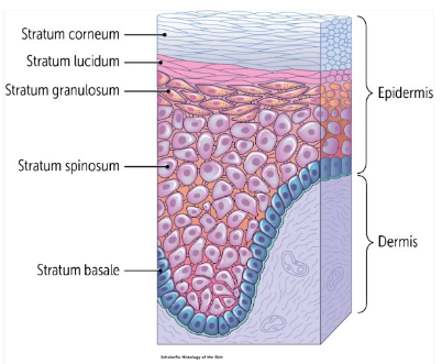
Epidermal Layers (Superficial to Deep)
stratum corneum
stratum lucidium
stratum granulosum
stratum spinosum
stratum basale
(come let’s get some beer)
Stratus Basale Histology
single layer of cuboidal/columnar cells, appears basophilic (purple)

Stratum Basale
contains stem cells that create new keratinocytes (the main epithelial cells located in the epidermis)
keratinocytes replicate and migrate upwards
Stratum Spinosum Histology
have desmosomes (“prickly” keratinocytes)
keratins are actively being synthesized by free ribosomes
slowly changing to eosinophilic cytoplasm in the upper layers of the stratum spinosum as intermediate fillaments assemble and fill more of the cytoplasm

Stratum Spinosum
upper layer of keratinocytes being to synthesize keratohyalin granules (aid in keratinization)
Stratum Granulosum Histology
1-3 layers of flat keratinocytes, granular appearance caused by cytoplasm filled with dark keratohyaline granules

Stratum Granulosum
keratinization beings, granules begin to promote formation of keratin filaments that are not fully keratinized yet
cells close to the stratum corneum start undergoing apoptosis and a decrease in pH from neutral to between 4.5-6
Keratohyaline Granules
contain filaggrin and trichohyalin, in stratum granulosum
Lamellar Bodies
in keratinocytes and stratum granulosum to form epidermal water barrier, secreted by exocytosis between the stratum granulosum and stratum corneum
2 Components of Water Barrier Whose Formation is Initiated by Lamellar Bodies
cell envelope
lipid envelope
Cell Envelope
component of water barrier whose formation is initiated by lamellar bodies
insoluble protein deposited on the inside of the plasma membrane of the bottom row of stratum corneum cells, creates the strong components of the mechanical barrier
Lipid Envelope
component of water barrier whose formation is initiated by lamellar bodies
layer of lipids attached to the cell surface producing “teflon-like” coating
Burn Victims Loss of Epidermal Water Barrier
puts them at risk for dehydration
Stratum Lucidium Histology
translucent layer of dead, flattened keratinocytes
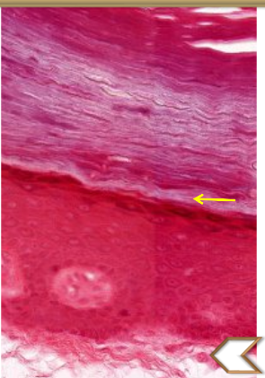
Stratum Lucidium
only present in thick skin, fully keratinized because nuclei and organelles are lost and replaced by densely packed keratin filaments
Stratum Corneum Histology
16-20 layers of dead keratinized cells
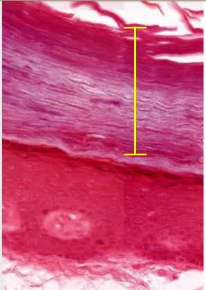
Stratum Corneum
provides protection against abrasion, superficial layers continuously shed at the same rate new cells are being produced in deeper layers
Formation of Integument
occurs from deep to superficial layers
Thick vs Thin Skin
determined by the thickness of the epidermis
Thick Skin
on palms of hands and soles of feet, no hair follicles
5 well-defined layers
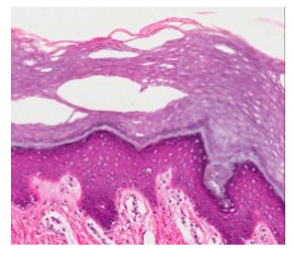
Thin Skin
everwhere else on the body, has hair follicles, lacks stratum lucidium
4 well-defined layers
4 Epidermal Cells
keratinocytes
langerhans cells
melanocytes
merkel cells
Keratinocytes
epidermal cell, epithelial cells, most abundant cells found in the epidermis
produce keratins and form epidermal water barrier
Langerhans Cells
epidermal cell, dendritic cells that migrate into the epidermis and become resident cells, originate from bone marrow and travel to epidermis via bloodstream
Langerhans Cells Function
phagocytize foreign structures in the epidermis and present the antigents to T lymphocytes in regional lymph nodes (adaptive immunity)
Langerhans Cells Appearance
hard to see with H&E because cells are clear, dendritic processes that extend into stratum corneum and “sense” the microenvironment

Melanocytes
epidermal cell, neural crest cells that produce pigment, in stratum basale
produce and then distribute melanin granules (via melanosomes) to neighboring keratinocytes
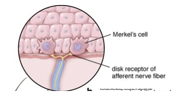
Merkel Cells
epidermal cell, mechanoreceptor cells (touch cells), in the stratum basale in regions with high tactile sensitivity
slowly adapting mechanoreceptors that function in cutaneous sensation
Merkel Cells Location
next to nerve endings
Keratinocyte Formation
stem cells are found in the deepest layer residing on the basement membrane
they differentiate as they migrate from the stratum basale to the corneal surface
the cells change both biochemically and morphologically as they ascend thorugh the layers of the epidermis
Melanocytes Appearance
dark, dense nucleus with clear cytoplasm

Melanin
a dark pigment that protects the skin from non-ionizing uv radiation
keratinocytes absorb melanin, which surrounds the nucleus and protects it
Skin Color Variation
everyone has approximately the same number of melanocytes, what varies is the production of melanin
What affects the melanin production rate?
both intrinsic and extrinsic factors including age, ethnicity, gender, variable hormone levels and well as UV radiation, climate and season changes, and chemical exposures
Tanning
UV light darkens existing melanin and increases the rate of melanin synthesis
Melanin and Skin Tone
most melanin stays in the basal portion of all skin types, however some can be found in upper layers of dark skin
in lighter skin, the melanin that is produced tends to be less concentrated and is more uniformly distributed within the lower layers of the epidermis, as a result these cell have little to no melanin remaining when they reach the stratum corneum

Dermis
layer of ct that mechanically stabilizes and metabolically sustains the overlying epidermis, also assists in themoregulation and sensation
2 Dermal Layers
papillary layer of type iii and iv collagen
reticular layer of type i collagen
Epidermal Ridges ad Dermal Papillae
projections off of the layers of the dermis that interlock with one another to stabilize the layers and prevent separation during abrasion
Why is it important that the papillary layer is loose CT?
all epithelium is avsacular, so there must be a layer of loose ct beneath with lots of space between the cells and blood vessels to allow diffusion of substances to and from the epithelium
Dermis Diagram

Dermal-Epidermal Junction
enhanced adhesion between the dermis and epidermis, involves dermal papillae and epidermal ridges
more prominent and abundant in skin commonly exposed to high friction, causes fingertips
3 Dermal Cells
fibrocytes/fibroblasts
macrophages
immunologic cells
Fibrocytes/Fibroblasts
dermal cell, most common cell in ct, create and repair extracellular matrix
Macrophages
dermal cell, phagocytic cells that digest pathogens, dead cells, and debris
Immunologic Cells
dermal cell, mast cells and b lymphocytes, contribute to skin homeostasis and immunosurveillance
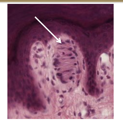
Meissner’s Corpuscles
in dermal papillae, detect light touch (low frequency stimuli)
Meissner’s Corpuscles Appearance
look like swirled cotton candy
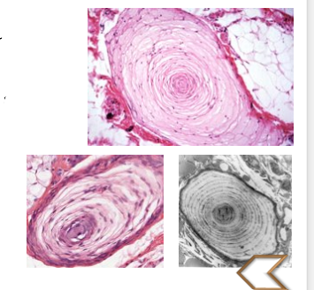
Pacinian Corpuscles
in reticular layers of dermis and hypodermis (deeper than meissner’s corpuscles), transmit vibration and pressure
Pacinian Corpuscles Appearance
lamellar organization, “onion-shaped”
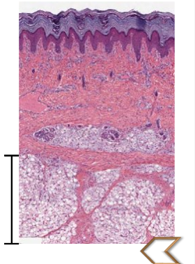
Hypodermis (aka Superficial Fascia)
subcutaneous layer, technically not part of the skin
Hypodermis Appearance and Function
loose ct that reduces friction between the dermis and deep structures, various amounts of adipose tissue for thermoregulation
sometimes contains pacinian corpuscles, highly vascularized
Products of Epidermal Appendages
hair follicles → hair
sebaceous glands → sebum
eccrine sweat glands → sweat
apocrine sweat glands → mixed sweat and carbs, lipids, proteins
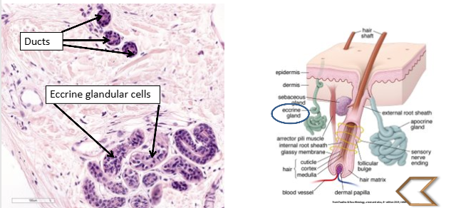
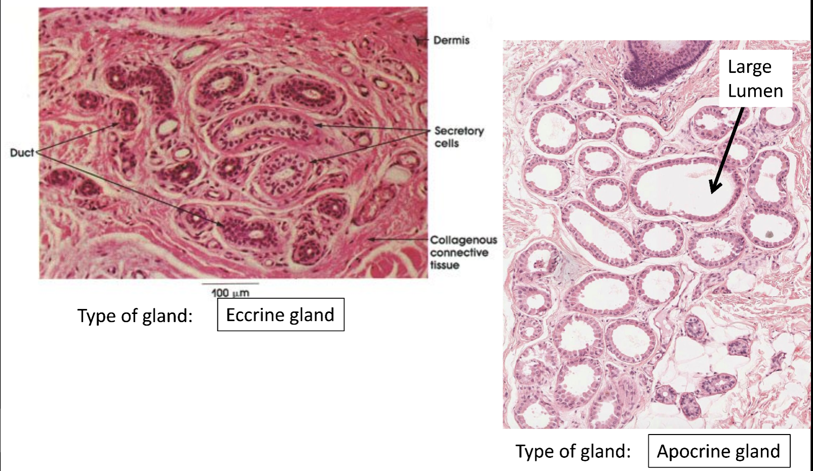
Eccrine/Merocrine Sweat Glands
duct of gland extends to skin surface, distributed throughout the body
secrete watery substance for thermoregulation
Eccrine/Merocrine Sweat Glands Appearance
sqiggly pasta
Apocrine Sweat Gland
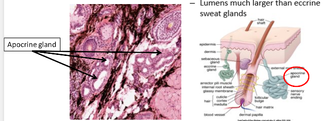
actually merocrine secretion via exocytosis, ducts empty into hair follicles
secrete pheromones (stimulated by sex hormones), secretion is initially odorless but broken down by bacteria (smelly)
Apocrine Sweat Gland Location
mostly in As (axilla, areola of nipple, anal regions) and genitals
Apocrine Sweat Gland Appearance
lumens much larger than eccrine sweat glands
Eccrine vs Apocrine Gland
Components of Pilosebaceous Unit
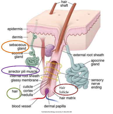
hair shaft, hair follicle, arrector pili muscle, sebaceous gland
Pilosebaceous Unit Diagram
Pilosebaceous Unit Hair
hair is modified, keratinized epithelium
the hair follicle surrounds the root of the hair

Arrector Pili Muscle
smooth muscle of pilosebaceous unit, when contracted the hair stands up on end and “goose bumps” are visible
Secaceous Gland in Pilosebaceous Unit
produce and excrete sebum
Sebum
an oily/lipid substance released via holocrine secretion that lubricates the hair and surrounding skin
Pilosebaceous Unit Histology

Skin Vasculature
capillaries extend into the papilla to interact with the epidermis

Skin Innervation Review
