Health and Illness 3 Exam 2
 Call Kai
 Learn
 Practice Test
 Spaced Repetition
 Match
 Flashcards
 Knowt Play
 Podcast
1/57

Earn XP
Description and Tags
10/19 - Includes review from last exam
Name | Mastery | Learn | Test | Matching | Spaced | Call with Kai | Chat |
|---|
No analytics yet
Send a link to your students to track their progress
58 Terms
Chronic Stable Angina vs Unstable Angina
Chronic Stable Angina:
Decreased blood flow to heart from CAD
Often occurs at predictable times (ie during exercise)
Position pt upright, encourage rest
Apply O2
Nitroglycerin (can pre-treat)
As plaque increases and blood flow decreases over time, unstable angina may occur
Unstable Angina:
Partial rupture of artery - no permanent damage - a type of acute coronary syndrome
Often new onset
Less predictable
Occurs at rest
More prolonged and severe >10 min
Cardiac markers not elevated - distinguishes from NSTEMI and STEMI
ST segment depression, T wave inversion
STEMI vs NSTEMI
STEMI:
Classic heart attack - extensive muscle damage
Occur abruptly from stopped blood flow - usually from disrupted arthroscelertoic plauqe that becomes a thrombus and occludes blood flow
Cardiac markers are elevated
ST segment elevation. - appears as tombstones - T wave inversion
NSTEMI:
Intermediate form of ACS - less extensive heart damage
Cardiac markers are elevated
ST segment depression, T wave inversion (also seen in unstable angina)
Medications for HF
Reduce afterload (lowers heart workload)
Via vasodilation
ACEs/ARBs - Stop RAAS system - Monitor creatinine and BUN for kidney function
Nitrates
Hydralazine
Valsartan
CCBs (not diltiazem)
Reduce preload (lowers heart workload)
Diuretics
Inhibit SNS
BETA blockers
Improve Contractility (Makes heart beat BETTER, not faster) (+ Inotropes)
Dobutamine (increases contraction strength and vasodilates)
Milrinone (increases contraction strength and vasodilates)
Digoxin (watch potassium lab!) (if on furosemide or HCTZ especially) (low K = digoxin toxicity)
Norepinephrine and epinephrine - weaker but used to vasoconstrict during shock
Sepsis 1-Hour Bundle
Measure lactate levels (repeat if >2)
Draw blood cultures (2 sets peripherally from different sites) (drawn before antibiotics)
Administer antibiotics
Fluid resuscitation (isotonic crystalloids LR or NS) (30mL/kg)
Vasopressor administration if fluids fail to restore a MAP >65 (norepinephrine, epinephrine, vasopressin)
Intracranial Pressure
Normal 5-15 mmHg
Monroe-Kellie Doctrine says that if there is an increase in one factor (CSF, intravascular blood, or brain tissue) there must be a corresponding decrease in one or both of the other factors to maintain normal intracranial pressure.
If there is too much increase, displacement occurs which leads to herniation and brain death
Cerebral Perfusion Pressure Calculation
MAP - ICP = CPP
Normal CPP is 60-70 mmHg
If abnormal - interventions to either increase MAP or decrease ICP (or both) are done
Clinical Manifestations of Increased ICP
Loss of consciousness
Dizziness and fainting
Change in memory
Headache
Projectile vomiting
Cushing Triad (late sign) - Impending Brainstem Herniation
Profound bradycardia (<50)
Abnormal respirations (cyanne-stokes)
Increasing pulse pressure (distance between diastolic and systolic increases) (high systolic and low diastolic) (ie 160/70)
Glasgow Coma Scale

3 is worst, 15 is best
13-15 Mild TBI
9-12 Moderate TBI
3-8 Severe TBI / Coma
External Ventricular Device (EVD)
Placed into ventricles of brain where CSF resises
Mesures ICP and drains excess
Brain ICP goes above a set # (for example 20) then the EVD drains off some CSF until ICP is back below desired number
Must stay above pts ear
Only waveform to know: Impending herniation
Waveform giving you the middle finger (see photo)

Nursing Interventions to Reduce + Maintain Low ICP
Head midline - avoid neck flexion
AIRWAY!
HOB 30 degrees (if no spinal)
DO NOT cluster care - ICP goes up when you interact with patient - sustained increased ICP = BAD. Take breaks
Avoid suctioning
Keep lights off, no talking - educate families
Paralytic medications to reduce agitation (propofol, fentanyl)
No bearing down/straining/coughing
Treat pain promptly (avoid opiates w/ pt’s not on the vent)
Mannitol (osmotic diuretic) to decrease brain swelling
Hypertonic saline (3% or higher) (not too much - bad for kidneys)
Phenytoin for seizure prevention
Concussion
Minor TBI - no identified brain damage - may result in post concussion syndrome >3mo
Diffuse axonal injury
Diffuse axonal injury -
widespread axonal damage (trauma changes function of axon resulting in swelling and disconnection)
not seen right away on CT
approx 90% of patients will remain in vegetative state
decortication, decerebration
Contusion
Bruising of brain tissue
Often associated with coup (primary impact) and countercoup (brain hitting back of head/secondary impact) injury
Associated with closed head injuries
Can re-bleed (blossoming) - monitor for seizures
Epidural Hematoma
Bleeding between dura and inner surface of skull
Brief lucid interval followed by decreased LOC - ie pt wakes up fine immediately, then rapidly deteriorates and doesn’t recover
Neurological emergency - needs surgical intervention/evacuation
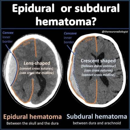
Subdural Hematoma
Bleeding between dura and arachnoid layers
Acute (24-48h) - immediate deterioration - craniotomy, evacuation, decompression
Subacute (within 2-14 days) - s/p severe trauma - AMS as hematoma develops - evacuation, decompression
Chronic (weeks-months s/p injury) - usually older adults - risk for misdiagnosis as initial injury seemed benign - evacuation, decompression

Emergency Management of a TBI
Stabilize cervical spine until spinal injury r/o
Airway support - intubate if GCS less than 8
Control external bleeding
Maintain warmth
Caution with fluid admin
CT scan
PREVENT SECONDARY INJURY - prevent seizures, hypotension
Basic Brain Anatomy
Frontal lobe: cognition, language, motor
Temporal: sensory info processing, understanding language
Parietal: interpreting sensory information
Occipital: interpreting visual information
Brainstem: bodies vital functions
Cerebral Edema vs Increased ICP
Cerebral edema is swelling of the actual brain r/t increased ICP
High ICP and rigidity of the skull interfere with BBB and prevent blood and CSF from traveling around
Causes O2 deprivation and tissue damage
Mannitol notes
May cause hyponatremia r/t diuretic properties
Watch for pulmonary edema (lung sounds, JVD)
Watch for AKI
Use filtered IV line
Seizure Phases
Prodromal - anxiety, mood changes (may be hours-days before)
Early ictal/aura - (seconds to minutes before)
Ictal - 1st manifestation until seizure ends
Postictal - until pt reaches baseline again
Types of Generalized Seizures
Tonic - Sudden onset of increased tone that is maintained
Clonic - Begins with loss of consciousness, sudden loss of muscle tone - rhythmic limb jerking
Myoclonic - sudden excessive jerk or twitch of body/extremities (maintains consciousness and some awareness)
Atonic - Involves tonic episode followed by sudden loss of muscle tone, resulting in a fall.
Tonic clonic:
Loss of consciousness
Body stiffens (tonic) with jerking (clonic)
Fencing position
Subclinical Seizures
Typical absence seizures - staring spell, only a few seconds but can occur many times if untreated - usually only peds
Atypical absence seizures - staring spell with other manifestations (blinking, lip movement, >10 seconds, continues into adulthood)
Status Epilepticus
Seizure activity longer than 5 min within rapid succession w/o return to consciousness between seizures
Neurons can become exhausted leading to permanent brain damage
Hypoxemia, dysrhythmia, increased ICP, rhabdomyolysis, acidosis, SUDEP
Give lorazepam first
Treatment/Interventions for Seizures
Maintain airway
Protect head - soft items under and around
Turn to side (not when actively seizing)
Loosen clothing
Suction after - not during
Time the seizure
Supplemental O2
Medications:
Lorazepam: Benzodiazepine used for acute seizure control, acting quickly to calm the nervous system via GABA enhancement. Watch for respiratory depression, hypotension, tachycardia, dysrhythmias
Phenytoin: Blocks repetitive activation of sodium channels - watch for gingival hyperplasia, hirsutism, and cardiac effects. Regular monitoring of serum levels is necessary.
Acute Kidney Injury
Rapid loss of kidney function - hours to days - impaired fluid, electrolytes, acid base and waste regulation (acute or chronic)
Dx Criteria:
Acute decrease in UOP (<0.5 for >6 hours) (may have normal UOP)
And/OR
Elevation in serum creatinine >0.3 mg/dl within 48 hours or >1.5x baseline within 7 days
S/S of AKI
Hypertension
Dysrhythmias
Edema, crackles, JVD, S3 sounds
Too little or too much UOP
Muscle twitching, seizures
Increased BUN, Creatinine, Potassium and Phosphate
Decreased calcium and HCT
Types of AKI
Pre-renal
Before the kidney - not enough blood flow causing decreased GFR - r/t dehydration, hypovolemia, hemorrhage, shock, sepsis - fluid challenge
Intrarenal
Direct kidney tissue damage - acute tubular necrosis impairs filtration - r/t nephrotoxic, contrast, rhabdomyolysis, glomerulonephritis
Postrenal
Urine cannot get out and backs up - raises kidney pressure - obstruction, kidney stones, tumor, kinked/clotted foley, bladder retention - if obstruction is removed within 48 hours damage can be avoided
1st Phase of AKI
Oliguric - begins with kidney insult
Lasts 10-14 days
UOP <400 mL/day
Fluid volume overload - edema, crackles, JVD, S3 gallop
Hyperkalemia
Hyponatremia
Metabolic acidosis
Azotemia (nitrogenous waste build up leading to LOC changes)
2nd Phase of AKI
Diuretic
Lasts 1-3 weeks
UOP 1-5 L/day - lots of UOP but not good quality waste clearing urine
BUN/Creatinine begin to normalize
Hypovolemia
Hypotension (admin IVF)
Hyponatremia
Hypokalemia
3rd Phase of AKI
Recovery
May take up to 12 months
GFR increases steadily
BUN/Creatinine plateau then fall - some may never go back to baseline
Tubule function restores
Watch for residual CKD - CKD occurs if recovery does not
Assessment Priorities for Patient in AKI
Strict I/O - hourly UOP if unstable w/ foley
Monitor electrolyte labs closely (sodium and potassium especially)
Daily weights - best fluid trend indicator
Avoid foods high in potassium and sodium
High protein diet r/t rate of protein breakdown that occurs with AKI
RED FLAGS
Potassium elevation - ECG changes
New pulmonary edema/hypoxia
Rapidly decreasing UOP
Severe metabolic acidosis
Altered LOC, seizures (r/t sodium changes)
Treatment for AKI
Stabilize ABCs/hemodynamics (O2, IVF, Pressors)
Stop the HIT (hold nephrotoxic meds, tx sepsis, relieve obstruction, renal dose meds)
Manage complications (hyperkalemia, acidosis, pulmonary edema, uremia, infection risk (kidneys play role in immunity)
Dialysis (intermittent, peritoneal, CRRT)
Hyperkalemia management:
Give calcium gluconate to buy time during hyperkalemia to protect the heart from potassium effects
Insulin + dextrose to force potassium to move into cells (also buying time)
Loop diuretics/K-exulate to make potassium leave in urine and feces
Intermittent Hemodialysis
Done for stable patients, CKD
Over a few hours 3x/week
Fastest type of dialysis - via fistula, graft, CVC
Anticoagulants to prevent filter clotting
Watch for:
Hypotension (slow rate if occurs)
Disequilibrium syndrome (r/t solutes moving out of body too quickly) (HA, vision changes, nausea r/t brain swelling)
Bleeding/infection
Peritoneal Dialysis
Uses patient’s perineum as a filter - works via gradient system from solute solution
Stable patients, can be done at home (over 8 hours) slowly!
Sterile procedure
Watch for infection/peritonitis - pain, drainage
Keep overflow bag below abd
Warm dialysate prior to instillation
Color should be light yellow/clear
Continuous Renal Replacement Therapy (CRRT)
For the most unstable patients - ICU patients that can’t handle large fluid shifts
Continuous, gentle filtration 24/7
Not as effective
Slowest form of dialysis
Via CVC (needs big vein, jugular, subclavian, femoral)
Watch for:
Filter clotting (anticoagulation therapy prophylaxis)
Nursing Interventions for Dialysis
Assess fistula (joined artery and vein) for thrill (feel) and bruit (sound) of whooshing
Don’t use fistula arm for BP, venipuncture
Don’t sleep or put pressure on site
Watch for infection at CVC catheter sites (kidneys play a large role in immunity)
Assess baseline vitals and weight before dialysis - reassess after
Hold dialyzable meds
Monitor B/P during
Monitor CBG (dialysate contains glucose)
No NSAIDs for AKI patients
Ischemic Stroke
Inadequate blood flow
Can be a thrombus (formed in brain)or embolus (traveling clot)
May be preceded by TIA
Facial drooping is hallmark
Hemmoragic Stroke
Bleeding into brain tissue
Intracerebral hemorrhage - sudden onset “worst headache of life” - HTN is most common cause
Subarachnoid hemorrhage- bleeding into CSF filled space - trauma, aneurysm burst
Keep MAP >70 for first 14 days s/p subarachnoid hemorrhage
Causes displacement of tissues - causes herniation and death if not treatment
Hemorrhagic type stroke usually has more loss of LOC

TIA
Usually lasts <60 min and resolves completely with no deficits
Hx of TIA is associated with increased risk of stroke later on (highest risk within 48 hours)
Ischemia without infarction
Treatment:
Antiplatelet - ASA, clopidogrel
Antihypertensives if indicated (HTN is common cause)
Afib meds as indicated (a-fib is common cause)
Stroke Localization Deficits
Right sided damage:
Left sided hemiplegia, neglect
Impulsivity, impaired judgment
Deficits begin at the nose level - eyebrows are spared but mouth droop occurs
Left sided damage:
Right sided hemiplegia
Aphasia/language impairment
Receptive aphasia (can’t understand you)
Expressive aphasia
Slower and more cautious
Diagnosis of Stroke
Non contrast CT within 25 min of ED arrival - shows hemorrhagic stroke w/o contrast but not ischemia
CT with contrast if no hemorrhage on initial NoCo CT to look for ischemia
Hx if able - onset, duration, progression - ‘last known well’ is hour 0
NIH stroke scale
Nursing Interventions for Stroke/S/P
Draw labs - coag studies
CBG (hypoglycemia can mimic stroke)
CT scanner ASAP
Last known well assessment
Airway protection
Careful fluid management
NPO with swallow eval before oral feedings - aspiration pneumonia is big risk after stroke
Tx fever quickly - can raise ICP
Have suction ready during mealtimes
Place food on unaffected side
Dress affected side first
Support limp extremities to prevent dislocation
Swallow with neck flexed forward
Teach scanning side-side if patient has homonymous hemianopsia
Acute Care for Ischemic Stroke
Goal: preserve life - prevent further brain damage and reduce disability
ABCs
Baseline neuro assessment - patients may worsen in 24-48 hours
Thrombolytic therapy - tPA - must be given within 3-4.5 hours of symptom onset
Monitor for conversion (switch between ischemic to hemorrhagic stroke r/t tPA)
After stabilization:
Antiplatelets
Anticoagulants (for life if patient has stent placed)
Stains
Nimodipine to prevent vasospasm
Keep BP a little higher to increase perfusion
Acute Care for Hemorrhagic Stroke
Tx based on cause of bleed - evacuation of hematomas, AVM resection, catheter embolization, clipping/coiling of aneurysm
No anticoags or antiplatelets - admin antidote if patient has taken any
Nicardipine for hypertension management (keep BP in lower range to reduce bleeding)
Seizure prophylaxis - suction setup - keppra admin ?
tPA Notes
Do not administer with antiplatelets or anticoagulants within 24 hours
Monitor for hypotension
Monitor for intracranial hemorrhage and bleeding
Arterial vs Venous Thrombosis
Arterial thrombosis:
Blood can’t go to extremities
Absent pulses
More uncommon r/t fast arterial blood flow
atherosclerosis, tissue ischemia, polycythemia and A-fib are causes
Venous thrombosis:
Blood can’t get OUT of extremities
Superficial and deep
Virchow’s Triad
Virchow’s Triad:
Virchow’s Triad:
Endothelial damage (smoking, HTN)
Hypercoagulability (hereditary, cancer, chemo, pregnancy, obesity)
Stasis (immobility, polycythemia)
A risk factor for increased clotting/venous thrombosis
Clotting Lab Tests
Prothrombin time (PT)
How long blood takes to clot via the extrinsic and common pathways (clotting factors I,II,V,VII and X)
Timed study - how long to clot (in seconds)
International normalized ratio (INR)
Clotting time compared to normal average
Warfarin needs INR between 2-3
Partial thromboplastin time (pTT)/(aPTT)
How long blood takes to clot via the intrinsic and common pathways (clotting factors VIII, IX, XI, XII)
Timed study - how long to clot (in seconds)
Prolonged time indicates problem in secondary hemostasis
Venous Thrombosis
Superficial: not as concerning
May extend deeper if untreated
Cord-like to the touch
rendess, pain warmth (usually no edema)
Confirmed via duplex ultrasound
Tx with compression stockings/bandages, elevation, mild exercise/walking, NSAIDs
Deep VTE:
Tenderness, redness, edema, fast cap refill
Confirmed with duplex ultrasound and increased D-dimer
Can cause PE, post thrombotic syndrome (chronic inflammation)
Heparin
Pulmonary Embolism
Blockage of one or more pulmonary arteries by thrombus, fat or air embolus that obstructs alveolar perfusion
Dyspnea
Chest pain
Tachycardia/tachypnea
Crackles, wheezing, pleural friction rub
Change in LOC
Cough with hemoptysis (r/t blood in alveolar space)
Refractory hypoxia (O2 application not working)
Can cause
Pulmonary infarction
Abscess secondary to infection
Pleural effusion
Pulmonary HTN r/t hypoxemia
Diagnosis of PE
D-dimer
CT scan WITH contrast
Ventilation perfusion (V/Q) study if contrast is contraindicated
Nursing Management of PE
Oxygen support - mechanical ventilaltion if indicated
Fluids and vasopressors
Diuretics
Analgesics
Surgery for massive PE
Apixaban, alteplase
DIC
Widespread, uncontrolled clotting that consumes all clotting factors and platelets - leads to uncontrolled bleeding
Never a primary dx, always secondary to underlying problem
Triggering event/cause - sepsis, trauma, OB emergency
Thrombin overproduced - coagulation cascade activated systemically
Microvascular clots - organ ischemia leading to MODS
Factors and platelets depleted - consumed by widespread clotting - UNCONTROLLED HEMMORAGE :(
DIC Clinical Manifestations
Thrombotic Manifestations:
Cyanosis, mottling
Gangrene of fingers and toes
Oliguria/anuria (AKI)
Decreased LOC
Stroke
Bleeding Manifestations:
Petechiae, purpura
Oozing from IV sites, wounds, gums
Hematemesis, melena, hematuria
Intracranial hemorrhage
Hemoptysis
Diagnostic Studies for DIC
D dimer elevated
Fibrinogen decreased
Platelets decreased
PT/PTT prolonged
RBC, Hgb, Hct decreased
Treatment of DIC
Treat underlying cause - DIC will not resolve until trigger is eliminated
Thrombosis dominant DIC:
anticoagulation with heparin
antithrombin 3 concentrate
monitor PTT
Bleeding dominant DIC:
Cryoprecipitate (replaces fibrinogen and factor VIII)
FFP (replaces all clotting factors)
Plt transfusion if <50,000 or active bleed
PRBCs for significant anemia/hemorrhage
Usually FFP, fluids and heparin all done at the same time
Nursing Interventions for DIC
O2 therapy/mechanical ventilation
Large bore IV access
Fluids/pressors for shock
Cardiac, o2 UOP monitoring
Inspect all skin for petechiae, purpura and ecchymosis
Check all IV sites for oozing
Assess neuro status
Avoid unnecessary injections
Pressure >5min to all sites
Soft mouth care, electric razor only
No rectal temps, smallest gauge needles
Heparin Induced Thrombocytopenia (HIT)
Similar presentation to DIC- autoimmune reaction to heparin
Usually occurs 5-10 days after heparin admin
Platelet drop >50% from baseline
Thrombosis despite low platelets
DVT, PE and arterial clots
STOP heparin and never give again
switch to argatroban or bivalirudin for anticoagulation
monitor platelets until >150,000
Protamine sulfate is heparin antidote