seidels chap 15 (heart) - skills unit 3
 Call Kai
 Learn
 Practice Test
 Spaced Repetition
 Match
 Flashcards
 Knowt Play
1/205

There's no tags or description
Looks like no tags are added yet.
206 Terms
what is the name of the area that the heart is located
mediastinum
dextrocardia
heart positioned to the right as a mirrored image

sinus inversus
when the heart and stomach are placed to the right and the liver to the left

pericardium
tough, double-walled fibrous sac encasing and protecting the heart - fluid is between outer and inner layers of pericardium for movement

epicardium
thin outermost muscle layer that covers surface of heart and extends onto great vessels

myocardium
thick muscular middle layer responsible for pumping action

endocardium
innermost layer that lines chambers of heart and covers valves + small muscles associated w/ opening/closing of valves

interventricular septum
blood-tight partition that divides the left and right heart

coronary sulcus
seperates atria from the ventricles on the anterior external surface of the heart

order of cardiac valves
"try pulling my arm"
tricuspid, pulmonic, mitral, aortic
what happens during systole
ventricles contract -- ejects blood from R ventricle into pulmonary artery
what happens during diastole
ventricles dilate -- draws blood into ventrocles as atria contract and move blood from atria to ventricles
what causes the S1 (lub) sound
mitral and tricuspid valve closure from raised ventricular pressure during contraction to prevent backflow

what causes the S2 (dub) sound
aortic and pulmonic valve closure when empty ventricles have low pressure
S2 sound components
A2 = produced by aortic valve closure
P2 = pulmonic valve closure
what can cause an S3 sound
filling of ventricle during diastole
what can cause an S4 sound
atria contracting to ensure ejection of any remaining blood
what causes an S2 split
aortic valve closing slightly before pulmonic (pressures on the right side are lower and events can occur slightly later on right side vs left)
process of electrical activity for contraction and the order of structures
1. impulse originates and paced by sinoatrial node (SA) located on wall of R atrium
2. impulse travels through both atria to atrioventricular node (AV) in atrial septum
3. AV node delays impulse then passes it down bundle of His to purkinje finbers in ventricular myocardium
4. ventricular contraction initiates @ apex and goes up to base of heart
what is an electrocardiogram (ECG)
graphic recording of electrical activity during cardiac cycle from movement of ions in/out of myocardial cell membranes
what is depolarization
spread of stimulus through heart muscle
what is repolarization
return of stimulated heart muscle to resting state
what does the P wave on ECG represent
spread of a stimulus through atria AKA atrial depolarization
what does the PR interval on ECG represent
time from initial stimulation of atria to initial stimulation of ventricles
what does the QRS complex on ECG represent
spread of stimulus through ventricles AKA ventrical depolarization
what does the ST segment and T wave on ECG represent
return of stimulated ventricular muscle to a resting state AKA ventricular depolarization
what does a U wave on ECG represent
small deflection rarely seen just after T wave thought to be related to repolarization of purkinje fibers
what conditions would present with a U wave
bradycardia mostly
sometimes seen with electrolyte abnormalities, hypothermia, hypothyroidism
what does the QT interval on ECG represent
time elapsed from onset of ventricular depolarization until completion of ventricular repolarization
fetal circulation
blood flows from right atrium - L atrium via foramen ovale -- R ventricle --- patent ductus arteriosus (instead of to lungs)
possible cardiac causes of chest pain
angina
acute myocardial infarction
coronary insufficiency
myocardial infarction
nonobstructive/nonspastic angina
mitral valve prolapse
possible aortic causes of chest pain
aortic dissection
possible pleuropericardial causes of chest pain
pericarditis
pleurisy
pneumothorax
mediastinal emphysema
possible GI disease causes of chest pain
hiatus hernia
reflux esophagitis
esophageal rupture
esophageal spasm
cholecystitis
peptic ulcer disease
pancreatitis
possible pulm disease causes of chest pain
pulm HTN
pneumonia
PE
bronchial hyperreactivity
tension pneumothorax
possible MSK causes of chest pain
cervical radiculopathy
shoulder disorder/dysfunction (arthritis, bursitis, rotator cuff injury, biceps tendonitis)
costochondral disorder
xiphodynia (pain @ breastbone)
possible psychoneurotic causes of chest pain
drug use (cocaine)
herpes zoster (when lesions occur in thoracic region)
characteristics of cardiac chest pain
substernal
provoked by effort, emotion, eating
relieved by rest, nitroglycerin
can be accompanied by diaphoresis and nausea
characteristics of pleural chest pain
precipiated by breathing or coughing
sharp, present during respiration, absent wwhen breath held
characteristics of esophageal chest pain
burning, substernal, occasional radiation to shoulder
nocturnal occurance
daytime attacks relieved by food
unrelated to activity
characteristics of peptic ulcer chest pain
infradiaphragmatic and epigasteric
nocturnal occurance and daytime attacks relieved by food and unrelated to activity
characteristics of biliary chest pain
usually under right scapula
prolonged in duration
often occuring after eating
will trigger angina more often than mimic it
characteristics of arthritis/bursitis chest pain
lasts for hours
local tenderness/pain with movement
characteristics of cervical chest pain
associated with injury
provoked by activity and persists after activity
painful on palpation/movement
characteristics of musculoskeletal chest pain
intensified/provoked by movement like twisting/bending
long lasting
often w/ focal tenderness
characteristics of psychoneurotic chest pain
associated w/ anxiety
poorly described
located in intramammary region
risk factors of cardiac disease
gender (men > women)
hyperlipidemia
smoking
elevated homocysteine level (metabolizes proteins)
family history
DM
obesity
sedentary lifestyle
examples of light exercise intensity
walking 10-15 steps
preparing simple meal for 1
retrieving newspaper
pulling down bedspread
brushing teeth
examples of moderate exercise intensity
making the bed
dusting/sweeping
walking a level or short block
office filing
examples of moderately heavy exercise intensity
climbing one/two flights of steps
lifting full cartons
long walks
sexual intercourse
examples of heavy exercise intensity
jogging
vigorous activity
cleaning entire house
raking leaves
mowing lawn w/ push mower
shoveling deep snow
hypovolemia vs autonomic failure (orthostatic hypotension)
hypovolemia increases HR as BP falls
orthostatic hypotension has no reflex-induced increased HR as BP falls = autonomic failure
what is a thrill
grade 4 or higher heart murmur that is felt on palpation
what conditions can increase intensity of S2 sound
systemic hypertension
syphillis of aortic valve
exercise
excitement
what conditions can decrease intensity of S2 sound
shock like state w/ arterial hypotension
immobile/thickened valves
aortic stenosis (affects A2)
pulm stenosis (affects P2)
overlying fat/tissue/fluid
where is a split S2 best heard
pulm area during inspiration
how are heart sounds characterized
pitch
intensity
duration
timing
S1 sound is ____ in pitch and heard ____ than S2
S1 is lower in pitch and heard longer than S2, occuring right after diastole (bc mitral valve shuts more vigorously)
S2 is _____ in pitch and heard ____ than S1
S2 is higher in pitchc and shorter duration than S1
what is wide splitting
split of S2 becomes wider when delayed activation of contraction or R ventricle emptying slows pulmonic valve closure
ex: during RBBB
what is fixed splitting
S2 splitting unaffected by respiration
ex: septal defects
what is paradoxical (reversed) splitting
closure of aortic valve is delayed so P2 occurs 1st followed by A2
where is mitral stenosis best heard
with bell at apex, pt in left lateral decubitus
findings on exam for mitral stenosis
low diastolic rumble w/o radiation
palpable thrill @ apex in late diastole
mitral stenosis description/causes
narrowed valve restricts foward flow and makes forceful ejection into ventricles
often occurs w/ mitral regurg
caused by: rheumatic fever or cardiac infection

where is aortic stenosis best heard
aortic area w/ ejection sound at second right intercostal border
findings on examination for aortic stenosis
midsystolic ejection murmur w/ medium pitch crescendo-decrescendo
radiates along L sternal border and carotid w/ thrill
description/causes of aortic stenosis
calcified valves restrict forward flow = forceful ejection from ventricle to circulatory system
causes: congenital bicuspid (instead of tricuspid), rheumatic heart disease, atherosclerosis

where is subaortic stenosis best heard
at apex and along left sternal border
findings on examination for subaortic stenosis
crescendo-decrescendo during systole w/ medium pitch and thrill at apex
S2 usually split and S3/S4 often heard
jugular venous pulse prominent
description of subaortic stenosis
fibrous ring below aortic valve becomes more severe

where is pulmonic stenosis best heard
pulmonic area radiating to left and neck
thrill in second/third left intercostal spaces
findings on examination for pulmonic stenosis
systolic ejection murmur, crescendo-decresendo, medium pitch usually w/ thrill
dimished S2 with wide split
description/cause of pulmonic stenosis
valve restricts forward flow = forecful ejection from ventricle into pulmonary circulation
causes: almost always congenital

where is tricupsid stenosis best heard
with bell over tricuspid area
findings on physical exam for tricuspid stenosis
diastolic rumble louder on inspiration
diastolic thrill palpable over R ventricle
jugular venous pulse prominent
description/causes of tricuspid stenosis
calcification of valve restricts forward flow = forceful ejection into ventricles
usually seen w/ mitral stenosis and rarely occurs alone
causes: rheumatic heart disease, congenital defect, R atrial myxoma

where is mitral regurgitation best heard
best heard/loudest at apex and can transmit to left axilla
findings on physical exam for mitral regurgitation
holosystolic high pitch blowing murmur that can obliterate S2
radiates from apex to base or to left axilla
thrill may be present at apex
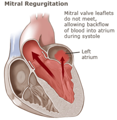
mitral regurgitation description/causes
valve incompetence allows backflow from ventricle to atrium
causes: rheumatic fever, MI, myxoma (benign tumor), rupture of chordae
where is mitral valve prolapse best heard
apex and left sternal border
easily missed in supine
findings on PE for mitral valve prolapse
late systolic murmur preceded by midsystolic clicks
description of mitral valve prolapse
valve is competent early in systole but prolapses into atrium later in systole, often concurrent w/ pectus excavatum

where is aortic regurgitation best heard
with diaphragm, pt sitting and leaning forward
ejection click heard @ second intercostal space
findings on PE for aortic regurgitation
early diastolic high pitch blowing w/ crescendo-descrecendo midsystolic murmur
can have austin-flint murmur (low pitch rumbling @ apex)
description/causes of aortic regurgitation
valve incompetence allows backflow from aorta to ventricle
caused by: rheumatic heart disease, endocarditis, aortic diseases (Marfan), syphilis, ankylosing spondylitis, dissection, cardiac trauma

description/causes of pulmonic regurgitation
valve incompetence allows backflow from pulm artery to ventricle (hard to distinguish from aortic regurg)
causes: secondary to pulm hypertension or bacterial endocarditis

where is tricuspid regurgitation best heard
left lower sternal border sometimes slightly radiating
findings on PE for tricuspid regurgitation
holosystolic murmur over R ventricle, blowing & increased on respiration
S3 and thrill over tricuspid area
description/causes of tricuspid regurgitation
valve incompetence allows backflow from ventricle to atrium
causes: congenital defects, bacterial endocarditis (especially IV drug users), pulm HTN, cardiac trauma
structural heart disease causing a murmur in a child sounds like...
holosystolic or diastolic
grade 3 or higher
systolic click associated
increased in intensity when standing
harsh quality
SAFER approach for history taking for high risk children w/ underlying structural heart defect
S- Syndromic features (kids w/ syndromes/chromosomal abnormalities)
A- Age (murmurs in infants = pathological)
F- Family history (3-5% risk passed onto offspring)
E- Evaluation of feeding/growth
R-Rheumatic fever/significant pervious med history (Kawasaki disease)
cyanosis at birth or shortly after indicates...
transposition of great vessels
tetrology of fallot
tricuspid atresia (defect/missing valve)
severe septal defect
severe pulmonic stenosis
cyanosis after neonatal period indicates...
pulmonic stenosis
Eisenmenger complex (defect that leads to right-to-left shunt)
tetrology of fallot
large septal defects
a pneumothorax in a child shifts the heart...
shifts apical impulse away from area of pneumothorax
a diaphragmatic hernia in a child shifts the heart...
to the right (hernias commonly found on the left)
dextrocardia in a child shifts the heart...
shifts apical impulse to the right
are S3 and S4 commonly heard in peds?
yes
are murmurs commonly heard in peds?
yes, within 48 hours of age (grade 1-2)
most are not pathological and caused by transition from fetal to pulmonic circulation
what accounts for most acquired murmurs in children
Kawasaki disease - rare/ acute illness affecting kids under 5 that causes inflammation of vessels