Skeletal Muscle Physiology - Kin 2204 Lecture Content
 Call Kai
 Learn
Learn Practice Test
 Spaced Repetition
 Match
Match Flashcards
 Knowt Play
1/45

There's no tags or description
Looks like no tags are added yet.
Name | Mastery | Learn | Test | Matching | Spaced | Call with Kai | Chat |
|---|
No analytics yet
Send a link to your students to track their progress
46 Terms
What is the functional unit and smallest aspect of a Muscle?
the sarcomere
What is the are strands of sarcomeres called in a Muscle?
myofibrils
What are the cells which house the myofibrils called in a Muscle? what connective tissue layer bundles/covers them? how many myofibrils are typically housed in 1 of these cells?
muscle fibres/cells, the endomysium (fascia) covers each fibre
1 muscle fibre can be hundreds of myofibrils

what are the key characteristic features of a myofibril?
cylindrical, runs alon longitudinal axis of fibre
contains sarcomeres with thick and thin filaments
has light and dark bands creating visually striping / striations in the muscle
what is a bundle of muscle fibres called and what connective tissue layer bundles/covers them?
bundle of muscle fibres = fascicle, covered by perimysium

what is a bundle of fascicles called and what connective tissue layer bundles/covers them?
a whole muscle, covered by epimysium

in a myofibril which of the following are thick versus thin filaments?
actin
myosin
What is the A-band of a sarcomere?
what is the I-band of a sarcomere?
What is the H zone?
What is the Z line of the sarcomere?
what are the M lines of the sarcomere?
H-zone = only thicc filament
M-line = supports thicc filament at centre of sarcomere

what are thick filaments (myosin) a collection of? what are the structural aspects of a myosin dimer? what binds to the myosin head?

What are 3 main components of the thin filaments? What are their roles?

what is titin? what does it do in the sarcomere (2)?
titin is are elastic proteins which extend from the M-line along the the length of the thicc filaments to the Z line.
serves to stabilize the position of thicc filaments in relation to thin gilaments
to act as a spring to provide elasticity to the muscle to be able to passively recoil or spring back to resting length after stretching
how does contraction influence overlap of thicc and thin filaments? what happens to sarcomere?
guiding questions:
what happens to the I band?
what happens to the A band?
What happens to the H zone?

what is the sliding filament theory and how does it describe skeletal muscle contractions?
what is a cross-bridge? what are the 4 steps within the cross bridging cycle?

what is ATP? how does it function within the body?

what is the role of ATP in the cross-bridge cycle? how does calcium support this cycle?

what is a rigor complex and what is it a result of?

What are the t-tubules of muscle fibres? what function do they serve in the excitation - contraction coupling?
t - tubules (transverse tubules) are tubes which are continuous with the cell membrane, running perpendicular to the surface, wrapping around the myofibrils
(Transverse tubules are extensions of the plasma membrane that travel into the muscle fibre to the myofibrils and transfer the action potential into the fibre)
bc they are extensions of the membrane, they help to propograte action potentials (change in permeability of ions) into the tubules across the network of myofibrils to assist in signaling
part of the linkage / path of the excitation - contraction coupling by carrying the action potential (electrical signal) through to the sarcoplasmic reticulum

what is the sarcoplasmic reticulum and how does it contribute to the excitation - contraction coupling?
SR = modified smooth endoplasmic reticulum that consists of a fine network of inter connected compartments surrounding each myofibril like a mesh sleeve
This membranous network encircles the myofibril throughout its length but is not continuous. Separate segments of SR are wrapped around each A band and each I band.
act as web-like sacs (lateral sacs / terminal cisternae) which store calcium ions (Ca 2+)
stored calcium will be released into the cytosol of a muscle fibre in the prescence of an action potential for muscle contraction
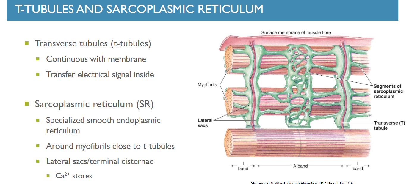
what purpose do the “foot proteins” / Ryanodine receptor have and what do they link with? how does this assist the Excitation - contraction couplng?
Ryanodine Receptors / foot proteins / Calcium release channels are imbedded in the Sarcoplasmic reticulum to bridge the gap between it and the T - tubules
the dihydropyridine receptors ar voltage gated sensors which have a similar foot-like composition to the SR foot proteins which they link to.
when an action potential is propagated through the t-tubules, the local depolarization activates the dihydropyridine receptors which then trigger the Ryanodine receptors to release calcium from the lateral sacs into the muscle fibres in the excitation - contraction coupling
what are the sequence of events for excitation - contraction coupling beginning from signaling at the NMJ to the end of a power stroke (combines events at NMJ, t-tubule + SR and power stroke)? (7 stages)

What is the latent period with regards to muscle contraction?
the latent period is the 0.5msec interval between onset of the excitation at the neuromuscular joint to the onset of muscle contraction (excitation - contraction coupling responsible)

define contraction time of a muscle. how does it differ in slow vs fast twitch muscle fibres?
the time it takes for a muscle via sequential power strokes to reach maximum tension. fast twitch fibres relative to the slow twitch fibres have a much shorter contraction time due to the difference in ATPase activity between the fibre types
when does the contractile response end? what is the time from peak contraction to reuptake of calcium called?
only once
ACh stops releasing and is degraded by Acetylcholinesterase
Calcium seperates from receptors on actin
the calcium ions released in response to the AP are removed from the cytosol via the calcium ATPase pumps in the Sarcoplasmic reticulum and.
Peak tension to reuptake of calcium is called Relaxation time
what defines a motor unit?
an alpha motor neuron (only 1) and all the muscle fibres it innervates
what is the relationship between the number of muscle fibres per motor unit and movement precision? what about the strength of contractions?
the greater the amount of fibres per motor unit, the less precise a movement is but the greater contractile strength is
thus less fibres = more precision, but less contractile strength
how do motor units compliment the all or nothing principle of action potentials?
if a motor neuron does not fire, then non of the fibres in the motor unit will, but if it does, then all of them will.
Other than the # of fibres in a given motor unit how else can we produce stronger contractions in a muscle?
we can acheive stronger contractions also by recruiting more motor units
what is the size principle and how does it describe motor unit recruitment?

why is asynchronous contraction of motor units important for muscle contraction? (2)
by varying motor units recruited in contractions we can prevent fatiguing immediately
allows for smoother muscle contractions which may otherwise become jerky if motor units were recruited simultaneously

what 5 factors influence the development of tension (force output)
1.) frequency of stimulation
As the firing frequency of the nerve increases, the force developed by the muscle increases, and at about 60 Hz no further increase in firing frequency will elicit a greater increase in force.
2.) # of motor units recruited
more = more tension
3.) fibre length at the start of a contraction (lengthened vs shortened vs optimal)
Length impacts # of cross bridges that can form possibly reducing ability to develop tension
4.) Extent of muscular fatigue
5.) thickness of the muscle fibre
thicker fibres have greater cross-sectional area due to large content of myofibrils which increase contractile strength
what is a twitch in a muscle?
a single twitch is equivalent to a singular action potential
what is twitch summation? what frequencies are required to sum and what frequency is required for moderate force output?
occurs when a muscle fibre is restimulated before it relaxes wherein subsequent twitches add onto the magnitude of the first twitch (or summate)
the minimum range for twitches to be able to summate are between 8 - 12 Hz (stimulations per second)
for a moderate amount of force to be produced it requires ~30Hz
What is tetanus? can you stimulate a muscle greater than 60Hz to increase force output beyond tetanus
Tetanus = when a muscle is stimulated so rapidly and repeatedly before it has any time to relax between stimuli
is maximal sustained contraction of a muscle at 60Hz
no greater increase in force output beyond 60Hz
what is the relationship between muscle length and tension? what about optimal length? what about when a muscle is stretched beyond its maximum stretch capability?
length-tension relationship states that there is an optimal length with the most available cross bridges, changes from optimal length decreases the available cross bridges, therefore decreasing tension capacity

What are the differences between all the types of muscle contractions:
isometric?
concentric?
eccentric?
isokinetic?
Isotonic?

what is the force velocity relationship?
describes that the amount of force a muscle can develop depends on its speed of the contraction.
In eccentric contractions (muscle lengthening), force generating capacity increases with velocity;
in concentric contractions (muscle shortening), force generating capacity decreases with velocity

what are the key differences between Slow twitch type 1, fast twitch type 2a and fast twitch type 2x muscle fibres from a structural, functional (speed/fatigue), and metabolic standpoints generally speaking?
Slow twitch (Type I) muscle fibres are smaller with ample mitochondria, blood supply and aerobic enzymes, making them contract slower/weaker but they are very resistant to fatigue.
Fast twitch X (Type IIx) fibres are larger with less mitochondria and blood supply but lots of anaerobic enzymes, making them contract faster/stronger but are very quick to fatigue.
Fast twitch A (Type IIA) fibres are the largest with lots of mitochondria and blood supply but a medium relaxation time and force production making them an intermediate in force production and fatigability
what are the more specific structural, functional and metabolic properties of a type 1 muscle fibre?
4 structural
5 functional
just need to know general metabolic details so dont memorize allat.

what are the more specific structural, functional and metabolic properties of a type 2x muscle fibre?
4 structural
5 functional
just need to know general metabolic details so dont memorize allat.

what are the more specific structural, functional and metabolic properties of a type 2a muscle fibre?
4 structural
5 functional
just need to know general metabolic details so dont memorize allat.

what are the ways in which muscles can become fatigued from the PNS? (4)
what is central fatigue?
What ways from the CNS / Psychological factors cause muscle fatigue? (2)
Heres what textbook says:
Peripheral (glycogen depletion or metabolic byproducts):
The local increase in ADP and inorganic phosphate from ATP breakdown may directly interfere with cross-bridge cycling and/or block Ca21 release and uptake by the SR.
Accumulation of lactic acid may inhibit key enzymes in the energy-producing pathways and/or excitation–contraction coupling process.
The accumulation of extracellular potassium (K+) that occurs in the muscle when the sodium-potassium pump cannot actively transport K+ back into the muscle cells as rapidly as this ion leaves during the falling phase of repeated action potentials (p. 63), causes a local reduction in membrane potential.
This altered potential may decrease the release of Ca 2+ intracellularly by impairing coupling of the voltage-gated dihydropyridine receptors in the T tubules and the Ca21-release channels in the SR.
Depletion of glycogen energy reserves may lead to muscle fatigue in exhausting exercise.
CNS / Central Fatigue:
occurs when the CNS no longer adequately activates the motor neurons supplying the working muscles. The person slows down or stops exercising even though the muscles are still able to perform. Central fatigue often is psychologically based.
Psychological central fatigue factors:
boredom
discomfort / pain

Describe ways in which muscle plasticity and interconversion are influenced by aerobic training adaptation and resistence training adaptatations.
Can fast twitch fibres convert from 1 to the other? Ex 2a to 2x?
can slow fibres become fast twitch fibres through exercise adaptations?
Aerobic training improves the metabolic properties of all muscle fibres but switching
between fibre type only occurs between fast twitch X and fast twitch A

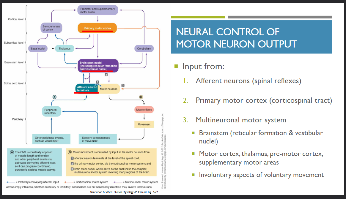
what sources of input feed the neural control of a motor neuron’s output? (3 categories)
what components of the brain are involved in the Multineuronal motor system? (4)
What components of the brainstem are involved in the Multineuronal motor system? (2)
consult the CNS cue cards for further details
alternatively heres the textbook:
“Three levels of input control motor-neuron output:
1. Input from afferent neurons, usually through intervening interneurons, at the level of the spinal cord—that is, spinal reflexes (p. 107).
2. Input from the primary motor cortex. Fibres originating from neuronal cell bodies, known as pyramidal cells, within the primary motor cortex (p. 115) descend directly without synaptic interruption to terminate on motor neurons. These fibres make up the corticospinal (pyramidal) motor system.
3. Input from the brain stem as part of the multineuronal motor system. The pathways composing the multineuronal (extrapyramidal) motor system include a number of synapses that involve many regions of the brain (extra means “outside of”; pyramidal refers to the pyra midal system). The final link in multineuronal pathways is the brain stem, especially the reticular formation (p. 110), which in turn is influenced by motor regions of the cortex, the cerebellum, and the basal nuclei. In addition, the motor cortex itself is interconnected with the thalamus as well as with premotor and supplementary motor areas, all part of the multineuronal system.
What are muscle spindles and what role do they serve?
Muscle spindles are modified muscle fibres with contractile ends, innervated by (efferent) gamma motor neurons, and a non-contractile centre that detects length and speed of stretch in the muscle
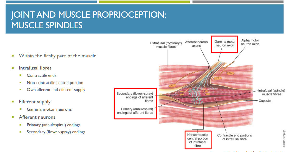
what are the 2 afferent Neuronal endings of the muscle spindle? where do they attach to the spindles and what do they do?
both are activated by stretch.
The primary (annulospiral) endings are wrapped around the central portion of the intrafusal fibres; they detect changes in the length of the fibres during stretching as well as the speed with which it occurs.
The secondary (flower-spray) endings, which are clustered at the end segments of many of the intrafusal fibres, are sensitive only to changes in length.

gamma motor neuron vs alpha motor neuron?
The efferent neuron that innervates a muscle spindle’s intrafusal fibres is known as a gamma motor neuron,
whereas the motor neurons that supply the extrafusal fibres are called alpha motor neurons
What are Golgi tendon organs and what do they do?
Goldi-tendon tendon organs detect and relay the tension in a tendon to the brain to modify tension and create a smooth movement
Because a number of factors determine the tension developed in the whole muscle during contraction (e.g., frequency of stimulation or length of the muscle at the onset of contraction), it is essential that motor control systems be apprised of the tension actually achieved so that adjustments can be made as necessary.
