Anatomy 6 (Cardiovascular)
 Call Kai
 Learn
 Practice Test
 Spaced Repetition
 Match
 Flashcards
 Knowt Play
1/73

There's no tags or description
Looks like no tags are added yet.
74 Terms
Mediastinum
Superior thoracic aperture to the diaphragm
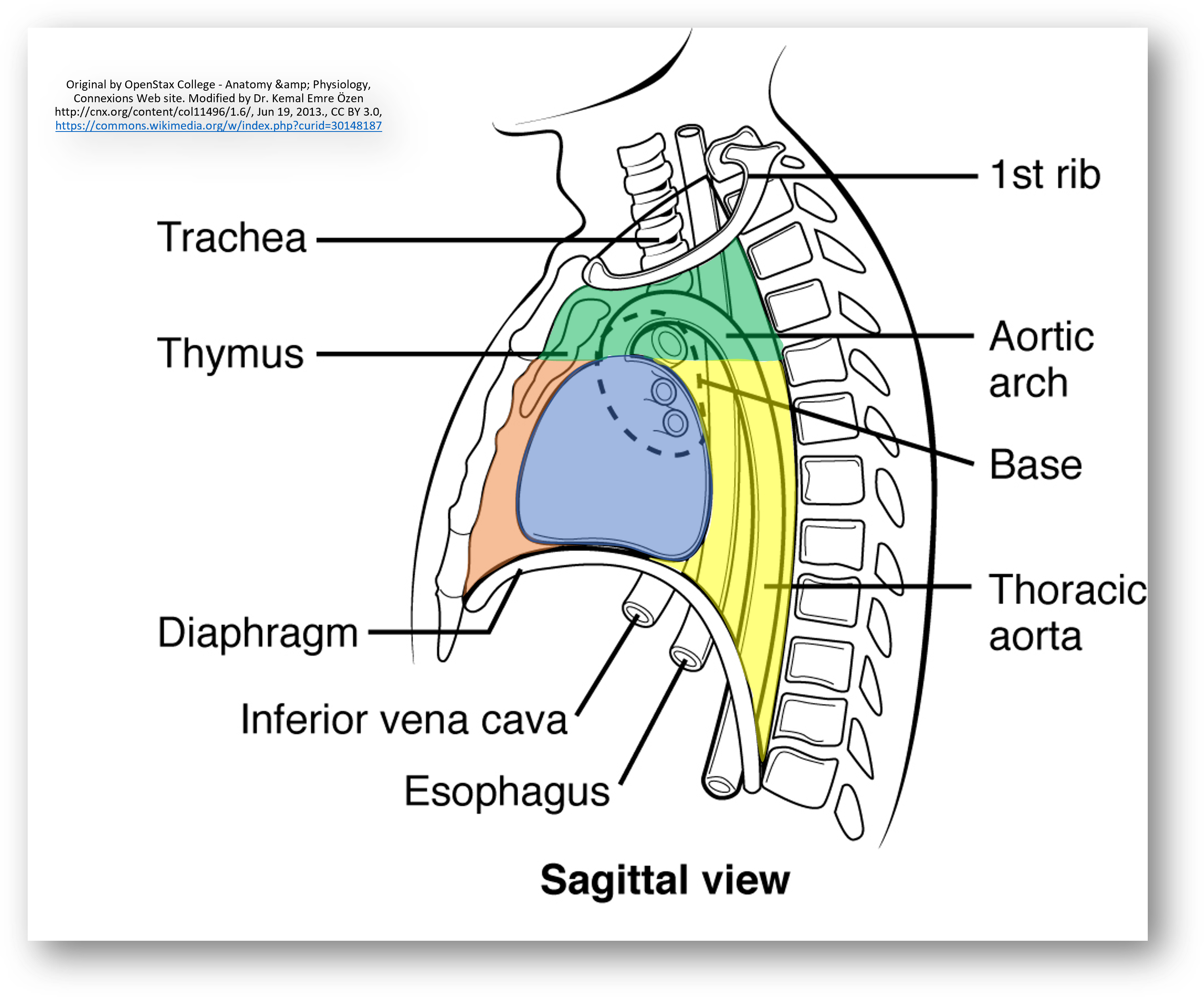
Superior Mediastinum
Thymus, trachea, esophagus, greater vessels
Middle Mediastinum
Heart and pericardium
Posterior Mediastinum
Esophagus and aorta
Heart Layers
Pericardium
Epicardium
Myocardium
Endocardium
Pericardium
Dense irregular + collagen layer that fuses with the central tendon of the diphragm
Parietal
Visceral/Epicardium
Parietal Pericardium
2 layers of the outer heart (fibrous and serous)
Myocardium
Heart muscle
Multiple layers so it squezes upward
Endocardium
Simple squamous inner heart lining that is continuous with the endothelium around vessels
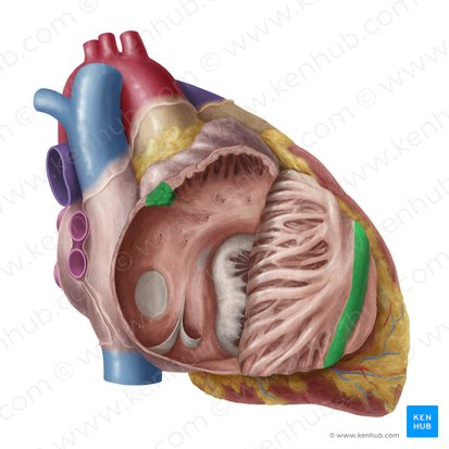
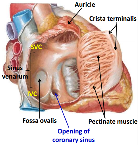
Crista Terminalis
Rudge on the RA endocardium that divides the sinus venarum from the pectinate muscles
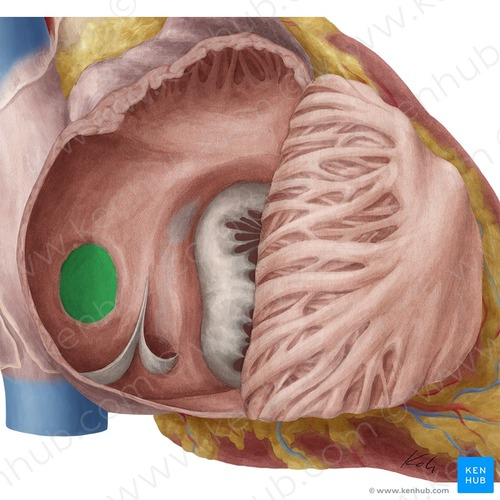
Fossa Ovalis
Depression on the interatrial septum (from embryo foramen ovale)
Sinus Venarum
Smooth wall of posterior RA
Tricuspid Valve
Brings blood from RA to RV

Pectinate Muscle
Ridges on the interior atria for strength and contraction

Sternocostal Surface
Anterior/outer surface of the RV
Conus Arteriosus
Leads to pulmonary valve

Papillary Muscles
Anchor chordae tenineae
3 in RV
2 in LV

Trabeculae Carnae
Bridges in ventricles for contraction
More in LV

Moderator Band
Muscle bridge in lower RV
Shortcut for electrical signal to anterior myocardium
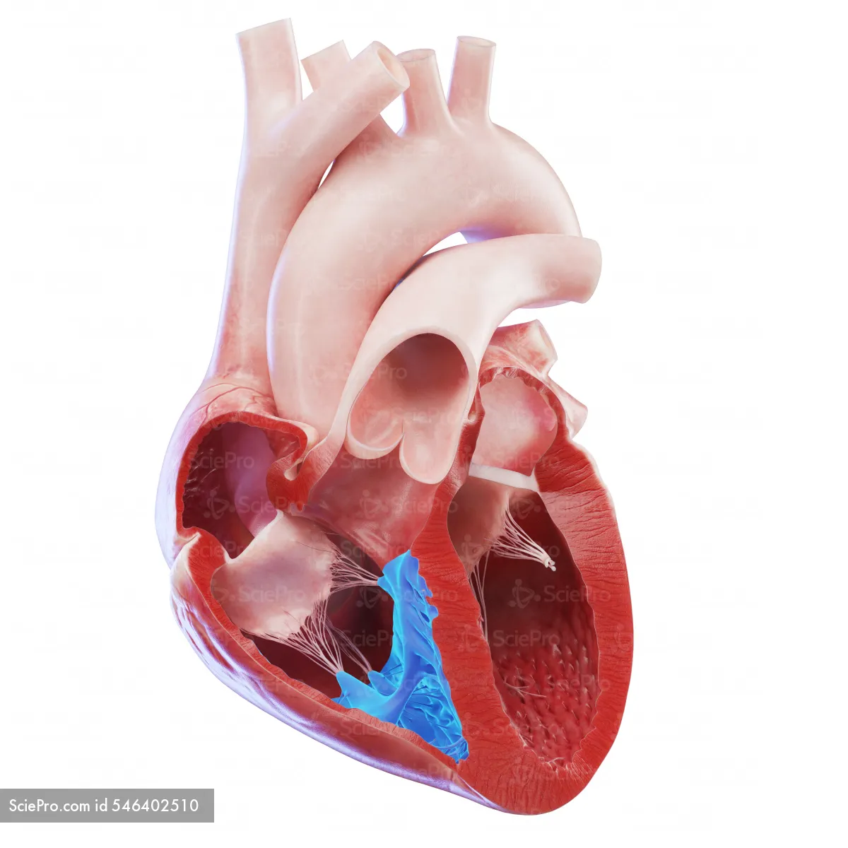
Chordae Tendineae
Connect leaflets to papillary muscles to prevent backflow

Pulmonary Valve
Brings blood to the pulmonary artery
Semilunar

Interventricular Septum
Wall between LV and RV

Bicuspid (Mitral) Valve
Brings blood to LV

Left Aorta
Thicker wall
Smooth with few pectinate muscles
Left Ventricle
No moderator band
More trabeculae carnae
2 papillary muscles
Aortic Valve
Brings blood from the LV to the aorta/body
Semilunar
Atrioventricular Valves
A to V
Tricuspid and bicuspid
Semilunar Valves
V to vessels
Pulmonary valve and aortic valves
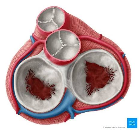
Fibrous Skeleton
Stabilizes heart muscles for contraction and valves
Physically and electrically separates A and V
SA Node
Pacemaker for A
AV Node
Connects to SA node and ventricles
Bundle of His
Nerves that run down the middle of the L/R ventricles

Purkinje Fibers
Nerves that go to the inner ventricle walls

Coronary Arteries
Branch from the aorta to supply the heart with blood
Left and right
Covered by leaflets during contraction
Circumflex Artery
From left main coronary artery and goes to left/back of heartartery
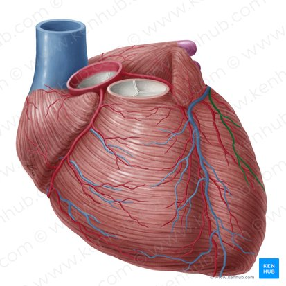
Veinous Drainage
Drains blood to the RA
Coronary Sinus
Largest vein in the heart that brings blood to the RA

Thesbian Veins
Drain RA blood supply directly to the RA without going through the coronary sinus
Ductus Arteriosus
RV to aorta
Fetus
Becomes ligamentum arteriosus
Umbilical Vein
Umbilical cord to liver
Fetus
Becomes ligamentum teres
Ductus Venosus
Liver to IVC
Fetus
Becomes ligamentum venosum
Umbilical Artery
Low O2 fetus blood to umbilical
Becomes medial umbilical ligament
Tunica Intima
Simple squamous inner layer of vessels
Tunica Media
Muscle tissue middle layer of vessels
Tunica Externa
Connective tissue outer layer to anchor vessels
Metarteriole
Direct passage from artery to vein
Thoroughfare canal
Helps determine amount of blood flow through capillaries
Precapillary Sphincters
Alters bloodflow through capillaries by opening and closing
Vaso Vasorum
Blood supply to alrge vessels
Continuous Capillary
Capillary with a continuous tube
Brain, lungs, kidney, skeletal/smooth muscle
Fennestrated Capillary
Capillary with pores in the membrane for protein to pass through
Kidneys
Sinusoid Capillary
Capillary with wide openings for RBC and proteins
Bone marrow, liver, spleen
Aortic Arch Arteries
L/R Coronary
Brachiocephalic
Left Common Carotid
Left Subclavian
Brachiocephalic
Becomes right common carotid and right subclavian arteries

Left Common Carotid Artery
Becomes L/R int/ext carotids

Left Subclavian Artery
Becomes axillary

Coartations
Development of a narrower aorta/artery
Head Arteries
Carotid (Int/Ext)
Verterbrals
Internal Carotid Arteries
Supply brain and orbital

External Carotid Arteries
Supply superficial muscle/structures

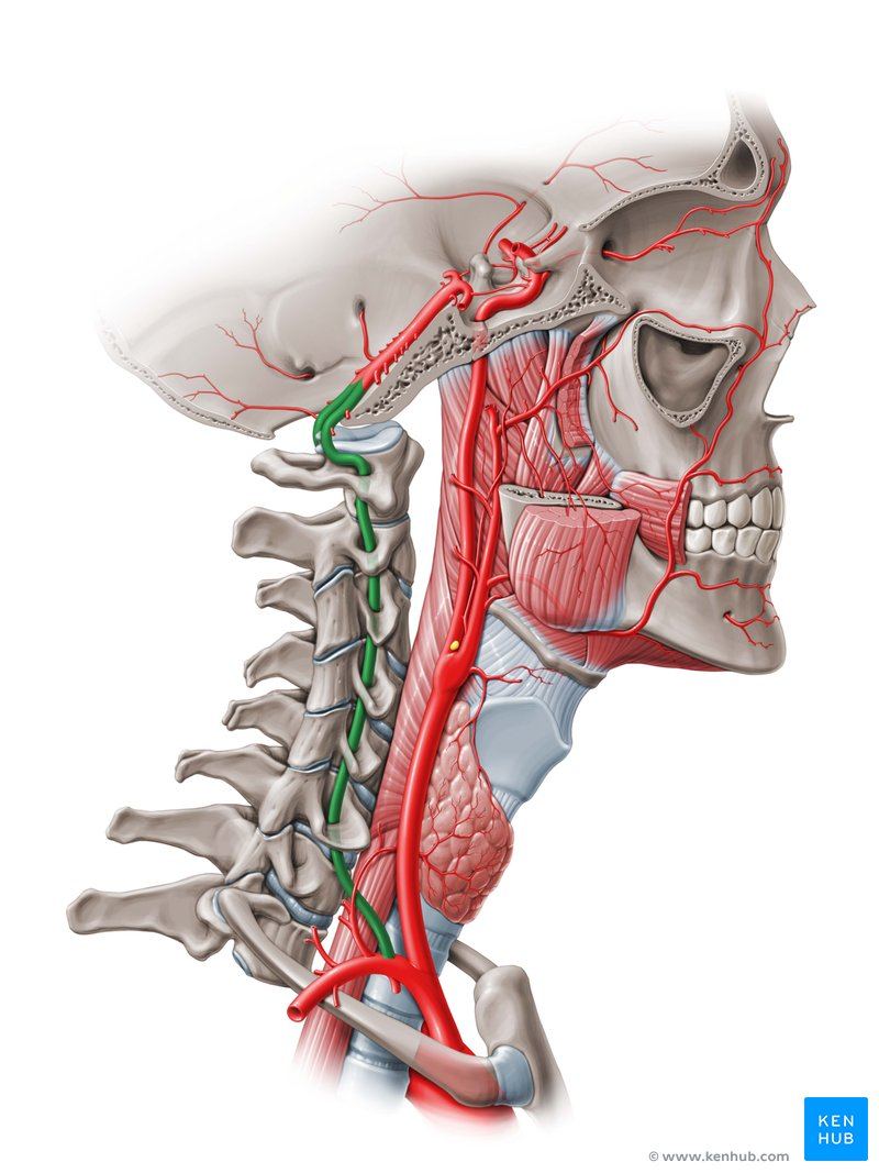
Vertebral Arteries
Comes from the subclavian and supplies the brain
Arm Arteries
Subclavian
Axillary
Brachial
Radial and Ulnar
Palmar and Digital
Thoracic Aorta Arteries
Intercostals
Bronchial: Lungs
Esophageal
Superior Phrenic: Superior diaphragm
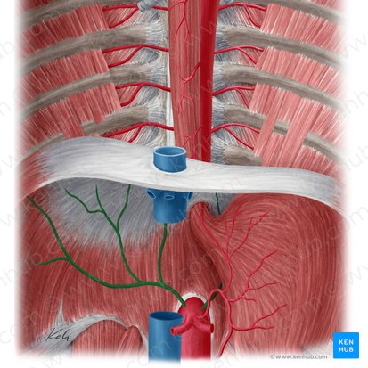
Inferior Phrenic Artery
Supplies diaphragm
Abdominal Aorta
Celiac Trunk
Common Hepatic
Splenic
Left Gastric

Common Hepatic Artery
Liver
Abdominal Aorta
Splenic Artery
Spleen
Abdominal Aorta
Left Gastric Artery
Lesser curve of the stomach
Abdominal Aorta
Suprarenals
Kidneys
Abdominal Aorta

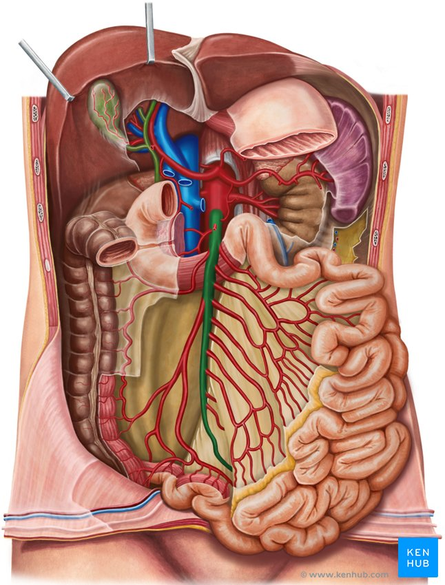
Superior Mesenteric Artery
All small intestines
Abdominal Aorta
Renal Artery
Bigger arteries supplying the kindeys
Abdominal Aorta

Gonadal Arteries
Ovaries and testes
Abdominal Aorta

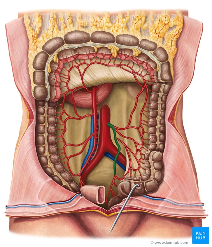
Inferior Mesenteric Artery
Descending and sigmoid colon
Abdominal Aorta
Iliacs Bifurcation
Branches that supply the anus, bladder, uterus
Abdominal Aorta

Sacral Artery
Supply lower spine, sacrum, coccyx, and parts of the pelvis and rectum
Abdominal Aorta
