PSIO 201 - Unit 2
 Call Kai
 Learn
Learn Practice Test
 Spaced Repetition
 Match
Match Flashcards
 Knowt Play
1/195

There's no tags or description
Looks like no tags are added yet.
Name | Mastery | Learn | Test | Matching | Spaced | Call with Kai | Chat |
|---|
No analytics yet
Send a link to your students to track their progress
196 Terms
each bone is an
organ
what does a bone contain that classifies it as an organ?
bone CT, blood vessels and nerves, lymphatic vessels, cartilage CT, and connective tissue coverings
skeletal system functions
support, protection, leverage, mineral homeostasis (storage and release), triglyceride storage, hemopoiesis
Hemopoiesis
red and white blood cell formation. found in red marrow!
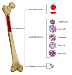
where is triglyceride stored in bones?
yellow marrow

importance of mineral homeostasis?
critical for keeping blood calcium levels regulated.

axial skeleton

apendicular skeleton

how many bones are in the human body?
206
long bones
longer than they are wide

short bones
cube shaped bones. equal in l and w

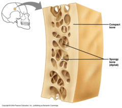
flat bones
thin, flattened, and usually curved
irregular bones
vertebrae and facial bones

seasmoid bones
round bone typically around a joint

sutural bones
tiny bones between cranial bones
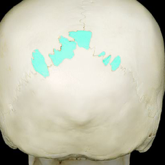
pneumatized bones
a bone that is hollow or contains many air cells

Diaphysis of long bone
shaft

epiphysis of long bone

Metaphysis of long bone

Periosteum (long bone)
outer lining of bone

Endosteum (long bone)
inner lining of bone

medullary cavity
cavity within the shaft of the long bones filled with bone marrow

compact bone
dense bone connective tissue on the outside (hard covering)
spongy bone
porous bone on the inside in the epiphysis

what two components are important for bone growth and remodeling?
endosteum and periosteum
Articulation
joint. point of contact of two bones together, bones and cartilage, and bones and teeth.

study of joints
arthrology
kinesiology
study of body movement
the more stable the joint is the ____ mobility (and vice versa)
less mobility
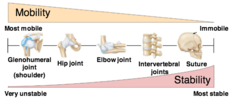
joint classifications via anatomical structure
fibrous, cartilaginous, synovial
joint classifications via function
synarthrosis, amphiarthrosis, and diarthrosis.
Synarthrosis
immovable joint

Amphiarthrosis
slightly movable joint

Diarthrosis
freely movable joint

true or false: all diarthrosis joints are synovial?
true

synovial joints
Freely movable joints with synovial fluid.
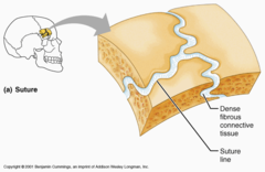
fibrous joints
generally immovable
cartilaginous joints
bones held together by cartilage

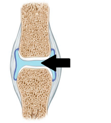
synovial fluid does what?
lubricates joint, provides nutrients for cartilage, and absorbs shocks
osteoarthritis (OA)
overuse (bone on bone contact) - degenerative disease

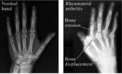
rheumatoid arthritis
Chronic autoimmune disease which attacks the healthy joint tissue causing inflamed joints
The shoulder is an example of which type of synovial joint?
the most free moving = least stable
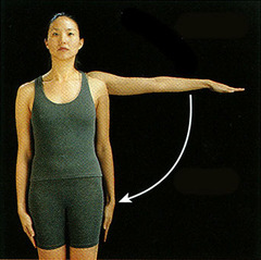
how many muscles does the shoulder rely on to keep it stable?
4
what cushions the joint in the knee?
medial and lateral menisci

name the 2 intracapsular ligaments of the knee
anterior cruciate ligament (ACL) and posterior cruciate ligament (PCL)
ACL
anterior cruciate ligament of the knee; prevents forward movement

PCL
posterior cruciate ligament of knee; prevents backward movement

what test can be done to see if you've injured your ACL or PCL?
drawer test!

Osteology
study of bone tissue
what makes up connective tissue?
cells and extracellular matrix
cells within bone connective tissue
osteogenic cells, osteoblasts, osteocytes, and osteoclasts.
osteogenic cell

what does an osteogenic cell do?
develops into an osteoblast
osteoblast
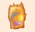
what does an osteoblast do?
builds new bone (bone formation), builds matrix (collagen fibers), initiates calcification (taking calcium from blood and depositing it in bone).
osteocyte

what does an osteocyte do?
maintains bone health, found in lacunae.
osteoclast
from myeloid tissue
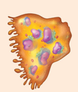
what do osteoclasts do?
break down damage (resorption)
sealing zone (SZ) of osteoclast
blue portion - seals itself to the bone

ruffled border (RB) of osteoclast
red portion - "teeth" which increase surface area and releases enzymes and acids (for breakdown)
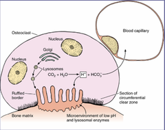
functional secretory domain (FSD) of osteoblast
green portion - where trancytosis occurs, recycling of nutrients from the cell pushing it back out into the blood stream.

goals of bone remodeling
strength for loading, lightness for mobility
in class example of bone remodeling
tennis player! repeated movement, increased loading, bones must be strengthened for their function.
steps of bone remodeling
1. Activation
2. Resorption
3. Reversal
4. Formation
5. Quiescence
extracellular marix of bone connective tissue contains what two components?
ground substance and fibers (collagen)
ground substance contains
glycoproteins (polysaccharide + protein)
GAG's are negatively charged, so what does it do?
traps water! like in hyaluronic acid skincare.
collagen fibers
helical form, provide strength, resistant to forces/tension
what is type 1 collagen made of?
polypeptide chains, using 3, creating a tropocollagen chain.
what do tropocollagens make?
microfibrils, which then create fibrils, and then fibers

inorganic compounds of the ECM
water (25%) and hydroxyapatite (calcium minerals secreted by osteoblasts)
why is the organization of organic and inorganic compounds important in bones?
it gives structure. mineralized fibrils are extremely strong, and collagen fibers contain lots of mineralized fibrils.
collagen provides
flexibility
minerals provide
firmness

collagen: organic or inorganic?
organic
minerals: organic or inorganic?
inorganic
rickets
inorganic compound deficiency, calcium deficiency due to lack of vitamin d. bowed legs.

scurvy
organic compound deficiency, lack of vitamin c, brittle bones that can fracture easily.
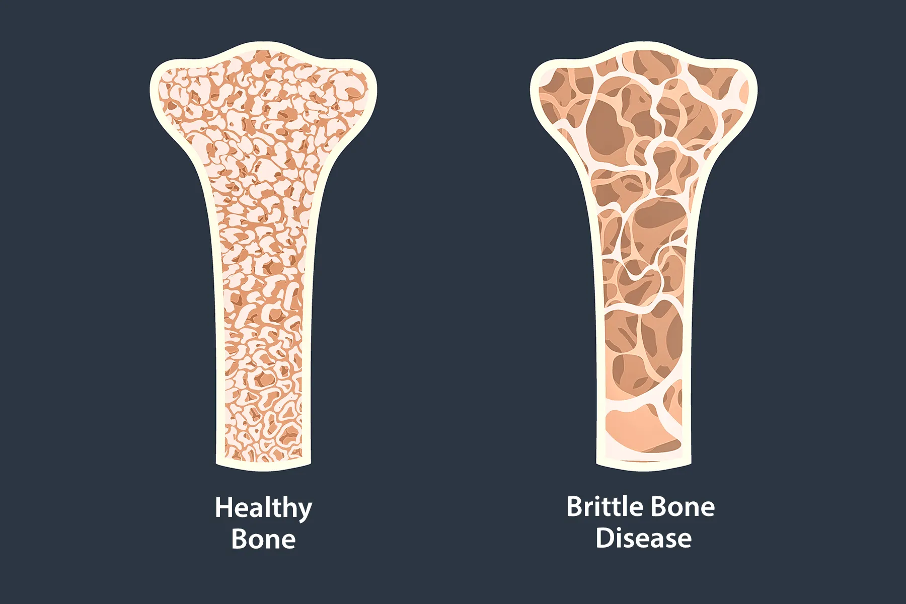
spongy bone and compact bone are similar in their
chemical composition
spongy bone organization
irregular lattice called trabeculae, osteocytes in lacunae

locations of spongy bone
epiphysis of long bones, medullary cavity (where marrow is), insides of smaller bones (packing)

functions of spongy bone
withstand forces in many directions, lightens skeletal system, contains red marrow for hemopoiesis

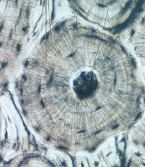
organization of compact bone
solid network in rings called osteons

location of compact bone
externally around all bones, diaphysis of long bones

function of compact bone
gives long bones the ability to withstand forces

osteon
A

central canal
where vessels and nerves run through to provide nutrients
lamellae
layers surrounding the osteon

canaliculi
small canals surrounding osteocyte

why are osteons arranged with each lamallae having alternating collagen fibers?
allows for the bone to withstand pressure, and have twisting force.

soft spot bones in infants include which of the following?
all the above
ossification
replacement of connective tissue by bone, continuous in development before and after birth.
intramembranous ossification
direct replacement of tissue (mesenchyme) to bone, done within the membrane.
what bones form via intramembrane ossification?
crahead, face, sternum, clavicles.
heterotropic bone formation (intramembrane ossification)
bones that feel stress which stimulates its formation. including sesamoid bones like the kneecaps
step 1 of intramembranous ossification
development of ossification center: osteoblasts secrete organic material

step 2 of intramembranous ossification
calcification: osteoblasts secrete calcium which hardens the ECM

step 3 of intramembranous ossification
formation of trabeculae: ECM develops trabeculae and it fuses making spongy bone

step 4 of intramembranous ossification
development of periosteum: spongy bone turns into compact bone
