unit5 anat
 Call Kai
 Learn
 Practice Test
 Spaced Repetition
 Match
 Flashcards
 Knowt Play
1/112

There's no tags or description
Looks like no tags are added yet.
113 Terms
What are the characteristics of a RBC (erthrocyte)?
Transports oxygen and carbon dioxide.
Unique biconcave shape.
Not true cells because they lack a nucleus and organelles.
What is the key characteristic of each white blood cell?
Neutrophil: Fights infections by internalizing microorganisms for destruction; increased in people with bacterial infections.
Eosinophil: Attempts to suppress inflammatory tissue reactions; increased in people with parasitic infections and allergies.
Basophil: Plays a role in acute and delayed allergic reactions; least numerous of WBC’s.
Monocyte: Most important phagocytic cell.
Lymphocyte: Regulates the immune system; plays a role in antibody production.
What are platelets? And what is their function(s)?
Plays a role in blood coagulation.
No nucleus.
What supports the one way bloodflow in the heart?
The 4 heart valves: Tricuspid valve, bicuspid valve, pulmonary valve, and aortic valve.
Why is the wall of the left ventricle thicker than the right ventricle?
Because it’s a stronger contraction/needs high pressure since it needs to pump blood to the rest of the organs in the body.
What prevents blood from moving between the atria?
Interatrial septum.
Is blood in the pulmonary veins deoxygenated? If so, why?
No, the blood in the pulmonary veins deoxygenated.
Because it just passed through the capillaries of the lungs, so it’s now filled with oxygenated blood.
What is the most abundant to least abundant WBC?
Neutrophil → Lymphocyte → Monocyte → Eosinophil → Basophil
Mnemonic: “Never Let Monkeys Eat Bananas”
Neutrophils
Nucleus is multi-lobed (as many as 5 lobes).
Cytoplasm contains neutral or pale, distinct, granules (when stained).
Lymphocytes
Round or slightly indented nucleus (fills the cell in smaller lymphocytes).
Nucleus is usually darkly stained.
Thin rim of cytoplasm surrounds nucleus.
Monocytes
Kidney-shaped or C-shaped nucleus.
Nucleus is generally pale staining.
Abundant cytoplasm around nucleus.
Eosinophils
Nucleus is bilobed.
Cytoplasm contains reddish or pink-orange granules (when stained)
Basophils
Nucleus is bilobed.
Cytoplasm contains deep blue-violet granules (when stained.)
What classes are the 5 leukocytes divided into?
Granulocytes and Agranulocytes.
Granulocytes
WBC’s that contain clearly visible, enzyme-filled granules in their cytoplasm under a microscope.
Agranulocytes
WBC’s that lack obvious, visible granules in their cytoplasm (appearing smooth).
Hemopoesis
Production of formed elements.
Myeloid line: Forms erthrocytes, megakaryocytes, and all leukocytes except lymphocytes.
Lymphoid line: Forms lymphocytes.

What are the three classes of blood vessels?
Arteries, capillaries, and veins.
Arteries
Carry blood away from the heart and become progressively smaller as they branch and finally results in capillaries.
Veins
Return blood to the heart and become progressively larger as they merge and are closer to the heart.
What are the three types of arteries?
Elastic arteries, muscular arteries, and aterioles.

Elastic artery
Largest artery.
Near the heart.
Elastic fibers in all 3 tunics allow these arteries to stretch under the increased pressure generated by blood ejected from the heart.
Branch into muscular arteries.
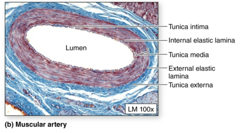
Muscular arteries
Medium-sized arteries
Have thicker tunica media.
Arteriole
Smallest arteries
Sympathetic nervous system: Tightens blood vessels → raises blood pressure
Parasympathetic nervous system: Relaxes blood vessels → lowers blood pressure.
What are the components of blood and their %’s
Erythrocytes (RBC): Makes up 44% of a blood sample
Buffy coat: Middle layer containing leukocytes and platelets; forms about 1% of a blood sample
Plasma: Straw-colored liquid that makes up about 55% of blood.
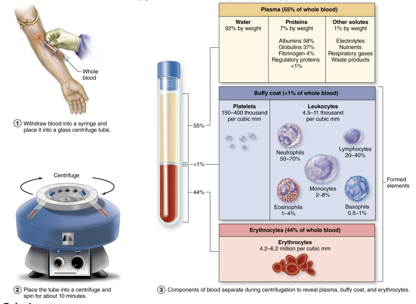
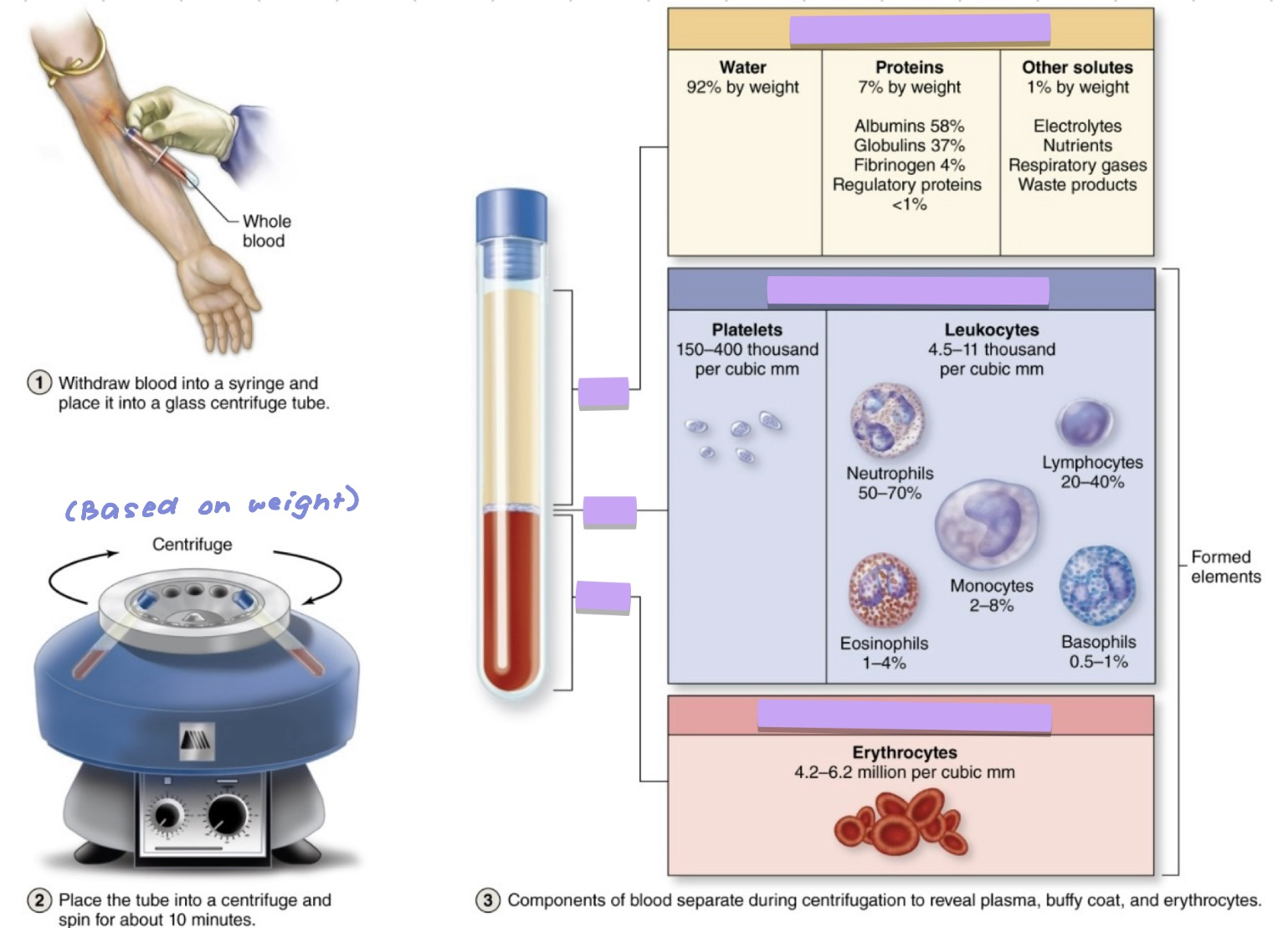
Whole Blood Seperation and Composition

What are the functions of blood?
Transportation (oxygen, carbon dioxide, nutrients, wastes)
Regulation (Temperature, pH, fluid levels)
Protection (Leukocytes guard against infection; blood carries antibodies)
What is hematocrit?
A blood test that measures the percentage of the volume of all formed elements in the blood.
Can vary with age and gender.
Can be viewed by preparing a blood smear.
As erythrocytes pass through small blood vessels, they line up in a single file termed a…?
Rouleau

Every erythrocyte contains 280 million molecules of a red-pigmented protein called…?
Hemoglobin
Hemoglobins
Capable of reversibly transporting oxygen and carbon dioxide in the blood.
Consists of four globin protein molecules:
Two alpha (a) chains
Two beta (β) chains
Each of the four globins possesses a nonprotein heme group containing an iron (Fe²+) molecule.
Each hemoglobin molecule can bind a combination of four oxygen molecules.

Recycling the components of aged or damaged erythrocytes.

What are leukocytes (WBC’s)?
Unlike RBCs, WBCs are true cells because they have a nucleus and organelles.
Help initiate an immune response and defend the body against pathogens.
Capable of diapedesis and chemotaxis

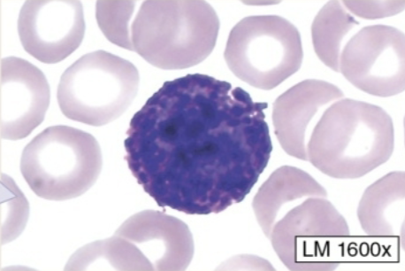
Lymphocyte
Round or slightly indented nucleus.
Nucleus is usually darkly stained.
The rim of the cytoplasm surrounds the nucleus.


Monocyte
Kidney shaped or C-shaped nucleus.
Nucleus is usually pale staining
Abundant cytoplasm around nucleus.


Neutrophil
Nucleus is multilobed (as many as five lobes)
Cytoplasm contains neutral or pale, distinct granules (when stained)


Eospinphil
Nucleus is bilobed
Cytoplasm contains reddish or pink-orange granules (when stained.)


Basophil
Nucleus is bilobed
Cytoplasm contains deep blue-violet granules (when stained)
Where do the arteries carry blood to?
Arteries carry blood away from the heart
Where do veins carry the blood to?
Veins carry blood back to the heart.
What are the two circulations that make up the cardiovascular system?
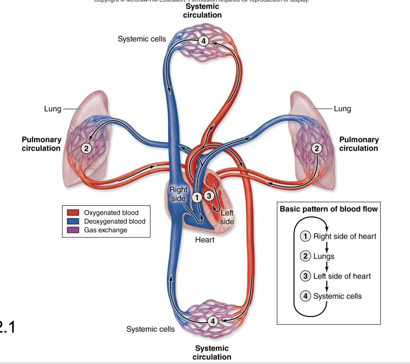
Pulmonary: Right side of the heart and the pulmonary arteries and veins; conveys blood to the lungs and the back left side of the heart.
Systematic: Left side of the heart and arteries and veins; conveys blood to most body tissues and back to the right side of the heart.
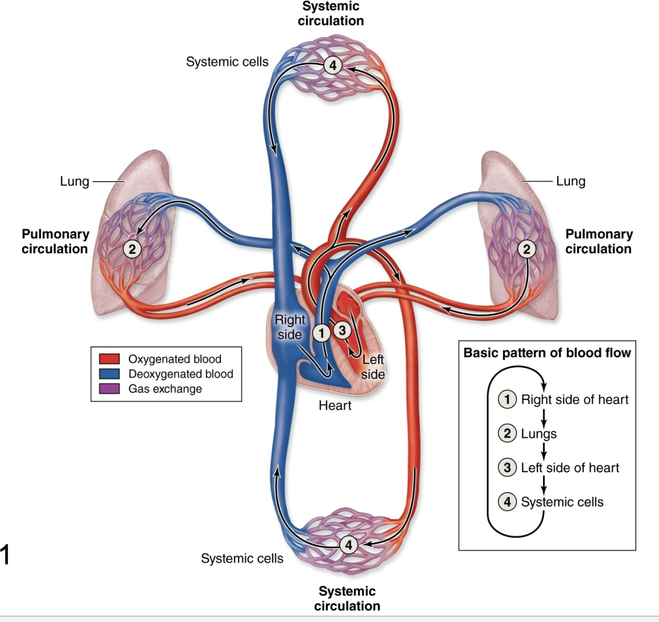
The heart is enclosed within a tough sac called the…?
Pericardium
^ Restricts heart movements so that it moves only slightly within the thorax
What two parts is the pericardium composed of?
Fibrous pericardium: Tough outer sac
Serous pericardium: Composed of parietal and visceral layers; forms the pericardial cavity.
What are the walls of the heart?
Three layers (from superficial to deep):
Epicardium: Consists of the visceral layer of the serous pericardium and areolar connective tissue
Myocardium: Cardiac muscle; thickest of the three layers
Endocardium: Internal surface of the heart chambers and external surface of the heart valves.
What are the heart’s four chambers?
Right atrium, right ventricle, left atrium, and left ventricle.
What are the heart’s four valves?
Right atrioventricular, pulmonary semilunar, left atrioventricular, aortic semilunar.
Contraction of heart muscle
Cardiac muscle fibers contract as a unified whole because low-resistance gap junctions within shared intercalated discs let electrical impulses spread instantly and synchronously across the heart wall.

Autorhythmicity
The heart exhibits autorhythmicity, which means it is capable of initiating its own heartbeat independent of external nerves.
The electrical impulse that initiates the heartbeat comes from…?
Specialized cardiac muscle cells called sinoatrial (SA) nodes/ pacemaker
The heart’s conducting system
SA Nodes sends electrical impulses through the atria of the atrioventricular (AV) Nodes. AV Nodes receives the impulses and passes it to the AV Bundles (Bundle of His).
Bundle of His carries the impulse down into the wall between ventricles (interventricular septum). The Bundle of His splits into two pathways, into the left and right ventricles.
SA Nodes → AV Nodes → Bundle of His → Right and left ventricles
Purkinje fibers
Purkinje fibers receive electrical impulses from the right and left bundle branches.
Carry electrical signal upward from the bottom (apex) of the heart.
The signal spreads throughout the ventricular walls, causing the ventricles to contract and pump blood.
Purkinje fibers = carry electrical signals from the bottom of the heart upward, causing the ventricles to contract.
What is the basic pattern of blood flow?
Right side of heart → lungs → left side of heart → systematic cells
Mediastinum
The central compartment of the thoracic cavity located between the left and right pleural cavities.

Position of the heart
Slightly left of the midline, deep to the sternum, and in the mediastinum

The great vessels
The arteries and veins entering and leaving the heart are called great vessels.
Base of the heart
The posterosuperior surface of the heart is mainly the left atrium and is called the base of the heart

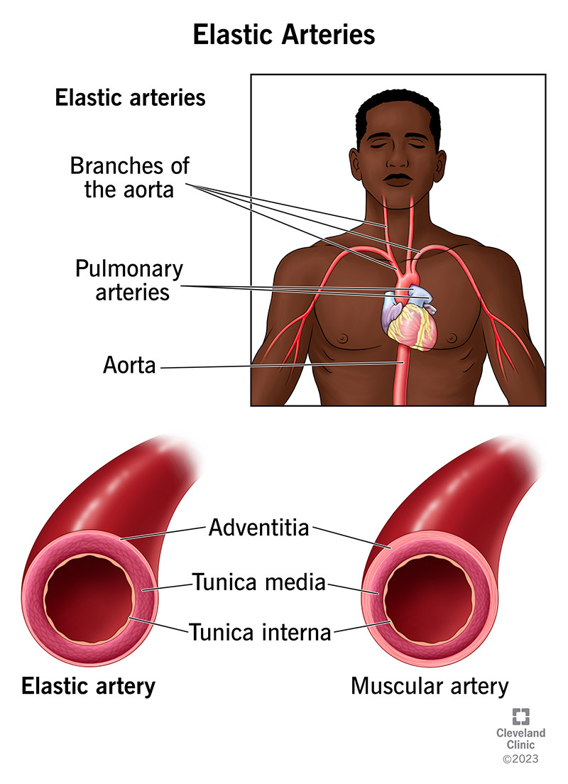
What are the layers of the vein and artery walls?
Inner to outer:
Tunica intima
Tunica media
Tunica externa

What are tissues present in tunica intima?
Endothelium
Subendothelial layer
Internal elastic lamina

What are the tissues present in tunic media?
Smooth muscle cells
Elastic and collagen fibers
External elastic lamina

What are the tissues present in tunica externa?
Dense irregular/loose connective tissue
Vaso vasorum and nervi vasorum

What are the three types of arteries?
Elastic arteries
Muscular arteries
Areterioles
Elastic arteries
Largest arteries
Mostly near the heart
Are able to stretch under the increased pressure generated by blood ejected from the heart
Branch into muscular arteries
Muscular arteries
Medium-sized arteries
Has elastic fibers between the three tunicas:
Internal elastic lamina
External elastic lamina
Has a proportionately thicker tunica media


Aterioles
Smallest arteries
Have less than six cell layers of smooth muscle in their tunica media
Sympathetic effect: Nerves tighten the muscle layer (vasoconstriction), which raises blood pressure
Parasympathetic effect: Nerves relax the muscle layer (vasodilation), which lowers blood pressure.

Capillaries
Smallest blood vessels
Solely consists of the tunica intima (single layer of endothelial cells)
Only type of blood vessel where metabolic exchange can occur between blood and tissues
Capillary beds
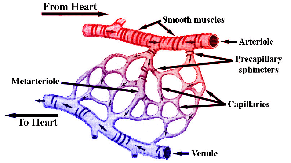
Each bed fed by a metarteriole
A thoroughfare channel connects to a postcapillary venule
Branches from the metarteriole that begin with a ring of smooth muscle on their walls are called true capillaries.
What are the three types of capillaries?
Continuous
Fenestrated
Sinusoids

Venules
Smallest veins
Companion vessels with aterioles
Smallest are the postcapillary venules
Diapedesis occurs through the walls of the postcapillary venules
Venules merge to form veins
What are the functions of the lymphatic system?
Returns interstitial fluid back to the blood stream
Transports lipids and lipid-soluble vitamins into the bloodstream
Production and maturation of lymphocytes
Generates an immune response against antigens in the interstital fluid
Lymph and lymph vessels
Extra fluid and solutes from body tissue enter tiny lymph vessels; after entering vessels they become lymphs
The lymph vessels move through a network of increasingly larger vessels
The vessels empty the lymphs back into the bloodstream
What are lymphatic capillaries?
Close-ended tubes that are found interspersed among most blood capillary beds.

What specialized lymph capillaries are found in the gastrointestinal tract?
Lacteals: Collect interstitial fluid as well as lipids and lipid-soluble vitamins.
Chyle
Lymph collected from the gastrointestinal system.
What are lymphatic vessels?
Lymphatic capillaries merge to form lymphatic vessels
Afferent lymphatic vessels
Bring lymph to a lymph node
Efferent lymphatic vessels
Transport filtered lymph away from the lymph node
Lymphatic trunks
Left and right lymphatic trunks merge form from merging lymphatic vessels
Each trunk drains lymph from a specific region of the body, as follows:
Jugular trunks
Subclavian trunks
Bronchomediastinal trunks
Intestinal trunks
Lumbar trunk
Lymphatic ducts
Are formed from the fusion of lymphatic trunks
What are the 3 types of lymphocytes?
T-lymphocytes (T-cells)
B-lymphocytes (B-cells)
Natural killers (NK cells)
All 3 migrate through the lymphatic system and search for harmful antigens.
T-Lymphocytes
70-80% of the body
Express a plasma membrane coreceptor (CD) that can recognize a particular antigen
Two main types of T-cells:
Helper T-cells
Cytotoxic T-cells
B-Lymphocytes
15-30% of body
Contain antigen receptors to only one antigen and produce immunoglobulins or antibodies against the same antigen
Most B-cells become plasma cells that produce and secrete large amounts of antibodies
Some activated B-lymphocytes become memory B-lymphocytes and confer immunity to certain antigens for many years or a lifetime
NK Cells
Unlike B and T-cells, can kill a wide variety of infected cells and some cancerous cells.
Lymphatic nodules
Small, unencapsulated clusters of immune cells that filter and attack antigens
Germinal Center (center): packed with rapidly dividing B-cells and macrophages
T-cells sit just outside the germinal center
MALT (Mucosa-Associated Lymphatic Tissue)
Nodules embedded in the mucous membrane of the digestive, respiratory, urinary, and genital tracts
Acts as an early-warning system against foreign invaders entering these passages
Peyer patches: Large prominent clusters of these nodules found specifically in the small intestine (ileum)
What are the three types of tonsils?
Pharyngeal
Palatine
Lingual
What are the main lymphatic organs?
Thymus, lymph nodes, spleen
Thymus
Bilobed organ, superficial to the heart
Consists of two fused thymic lobes, which are divided into lobes
Each lobe has an outer cortex and an inner medulla
Continue to grows until puberty and then begins to regress in size and function
Thymus and T-cells
Cortex contains immature T-cells
Medulla contains mature T-cells
Lymph nodes
Filter antigens from the lymph and initiate an immune response
Surrounded by a tough connective tissue capsule
Internal extensions of the capsule, trabeculae, project into the node
Most apparent lymph nodes occur as:
Axillary
Inguinal
Cervical
Lymph node structure
Divided into outer cortex and inner medulla
Cortex: consists of nodules and sinuses (cortical sinuses)
Medulla: contains medullary cords and medullary sinuses
Afferent vessels deliver lymph to the node
Lymph exits nodes via efferent vessels at the indentation of the node (hilum)
Functions of the spleen
Initiates an immune response when antigens are found in blood
Serves as a reservoir for erythrocytes and platelets
Phagocytizes old, defective erythrocytes and platelets
Phagocytizes bacteria and other foreign material
Spleen
Largest lymphatic organ, lateral to the kidney
Splenic artery and splenic vein enter/leave via its hilum
Surrounded by dense irregular connective tissue capsule, which sends extensions called trabeculae into the organ
Trabeculae vessels
(Branches of splenic arteries and veins) extends within the trabeculae
Cells around the trabeculae are subdivided into white pulp and red pulp
Red pulp surrounds each cluster of white pulp
Red and white pulp
White pulp (immune center): Surrounds the arteries, hold immune cells (T-cells, B-cells, macrophages), and fights off infection
Red pulp (blood filter): Connects to the veins, holds open leaky capillaries (sinusoids), cleans out old blood cells, platelets, and wastes
What are the main functions of the respiratory system?
Breathing (ventilation)
Involves inhalation and exhalation
Other functions include:
Gas conditioning
Sound production
Olfaction
defense
What is the respiratory system divided into?
Upper and lower respiratory tracts.
What is the upper respiratory tract divided into?
Nose and nasal cavities
Paranasal sinuses
Pharynx
What are the paranasal sinuses?
Frontal
ethmoidal
sphenoidal
maxillary
The pharynx is divided into…?
Nasopharynx
Oropharynx
Laryngopharynx