SLHS 3560 final quiz questions
 Call Kai
 Learn
 Practice Test
 Spaced Repetition
 Match
 Flashcards
 Knowt Play
1/180

There's no tags or description
Looks like no tags are added yet.
181 Terms
a. Association fibers
These white matter fibers stay within the same hemisphere.
a. Association fibers
b. Projection fibers
c. Limbic fibers
d. Commissural fibers
d. Broca's area
Which of the following areas is represented by the region above?
a. Wernicke's area
b. Precentral gyrus
c. Auditory cortex
d. Broca's area
e. Postcentral gyrus

b. They establish a connection between the neuron and another neuron or the cells of a muscle or gland
What is the function of boutons?
a. They insulate the axon
b. They establish a connection between the neuron and another neuron or the cells of a muscle or gland
c. They transmit a neural impulse more quickly along the axon.
d. They conduct a neural impulse toward the neuron.
b. corpus callosum.
The cerebral hemispheres are connected by the
a. encephalon
b. corpus callosum.
c. basal ganglion
d. gray matter.
c. Commissural fibers
These white matter fibers connect the left and right hemispheres.
a. Limbic fibers
b. Association fibers
c. Commissural fibers
d. Projection fibers
b. Sagittal
What anatomical plane is the brain above shown in?
a. Axial
b. Sagittal
c. Coronal
d. Transverse
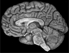
d. Left
Which hemisphere of the brain contains the major neurologic mechanisms for speech and language?
a. Right
b. Neither; speech and language centers are found primarily in the brainstem.
c. Speech and language centers are equally divided between the two hemispheres. d. Left
c. Schwann cells
Which of the following is NOT a type of neuroglial cell in the CNS?
a. Ependyma
b. Astrocytes
c. Schwann cells
d. Microglia
d. ventral
Structures lying at the base of the brain may be anatomically described as being
a. dorsal.
b. rostral
c. cephalic
d. ventral
b. motor
The precentral gyrus makes up the primary
a. None of these
b. motor
c. sensory
d. visual
c. dendrites
The processes specialized to receive the impulses moving toward the cell are the
a. axons
b. nuclei
c. dendrites
d. Golgi apparatus.
a. myelinated axons
The presence of ______ is what gives white matter its white appearance and name.
a. myelinated axons
b. synaptic junctions
c. specialized ependymal cells
d. unmyelinated axons
a. gyri or d. sulci
The enfolding of the cortex of the brain during development forms the
a. gyri
b. cerebral spinal fluid
c. lobes
d. sulci
c. Projection fibers
These white matter fibers connect parts of the brain to the spinal cord.
a. Limbic fibers
b. Commissural fibers
c. Projection fibers
d. Association fibers
h. Dendrites
Gray matter within the brain is made up of?
a. Myelin sheath
b. Postcentral gyrus
c. Blood
d. Wernicke's area
e. Precentral gyrus
f. Cerebrospinal fluid
g. Broca's area
h. Dendrites
b. neurons.
Nerve cells are called
a. neuroglial cells.
b. neurons.
c. dendrites.
d. axons.
false
The motor cortex is housed in the postcentral gyrus.
True
False
a. Axon
Which part of the neuron is critical to communication within the nervous system?
a. Axon
b. Cytoskeleton
c. Perikaryon
d. Soma
d. the frontal, parietal, temporal, and occipital lobes.
The four lobes of each hemisphere of the brain are
a. the occipital, temporal, nasal, and parietal lobes.
b. the parietal, sulcus, gyrus, and frontal lobes.
c. the frontal, dorsal, temporal, and occipital lobes.
d. the frontal, parietal, temporal, and occipital lobes.
d. Oligodendrocytes
Which type of neuroglial cell forms and maintains myelin?
a. Astrocytes
b. Ependyma
c. Microglia
d. Oligodendrocytes
a. an neurologic speech disorder.
A dysarthria is
a. an neurologic speech disorder.
b. a group of fibers connecting areas or centers of the brain.
c. a disorder of executing motor acts as a result of a brain lesion.
d. a disorder of cortical sensory recognition.
b. the brain and spinal cord
The central nervous system consists of
a. the brain only.
b. the brain and spinal cord
c. the spinal cord and cranial nerves.
d. the brain and cranial nerves.
b. myelin sheath
White matter within the brain is made up of?
a. blood
b. myelin sheath
c. dendrites
d. postcentral cortex
e. cerebrospinal fluid
f. precentral cortex
g. Broca's area
h. Wernicke's area
e. Wernicke's area
Which of the following areas is represented by the region above?
a. Visual cortex
b. Broca's area
c. Postcentral gyrus
d. Precentral gyrus
e. Wernicke's area

d. brocas area
Which of the following areas is represented by the region above?
a. precentral gyrus
b. auditory cortex
c. postcentral gyrus
d. brocas area

a. superior
b. posterior
c. dorsal
d. rostral
a.
b.
c.
d.

e. rostral
f. ventral
g. dorsal
e.
f.
g.

1. spinal cord
2. spinal nerves
The CNS includes the brain and the [answer1] while the PNS includes the cranial nerves and [answer2].
c. Thyroid gland
Which of the following structures is NOT a part of the diencephalon?
a. Pituitary gland
b. Thalamus.
c. Thyroid gland
d. Hypothalamus
b. Hypothalamus
Which of the following exerts neural control over the pituitary gland?
a. Thalamus
b. Hypothalamus
c. Subthalamus
d. Subthalamus Epithalamus
b. Dysarthria
Damage to the cerebellum may cause what type of speech problem?
a. None
b. Dysarthria
c. Aphasia
c. left
which hemisphere is the dominant hemisphere in the most persons, meaning that it is the language functions are controlled?
a. right
b. neither
c. left
a. astrocytes
which neuroglial cells may actively participate in promoting brain plasticity through action at the synapse?
a. astrocytes
b. ependyma
c. microglia
d. oligodendrocytes
c. limbic system
the area that brocas considered to be the 5th lobe of the brain & that contains the most primitive portions of the brain is the:
a. hippocampus
b. pons
c. limbic system
d. corpus callosum
c. nucleus
which organelle contains the DNA of a cell?
a. endoplasmic reticulum (ER)
b. lysomones
c. nucleus
d. bilipid membrane
a. dura mater
b. arachnoid mater
c. pia mater
d. subdural space
e. subarachnoid space
a.
b.
c.
d.
e.

c. Periosteal and Meningeal
What are the two layers that make up the dura mater?
a. Periosteal and Arachnoid
b. Arachnoid and Pia
c. Periosteal and Meningeal
d. None of these
b. Leptomeninges
The arachnoid and pia mater are collectively referred to as the ___
a. Falx cerebri
b. Leptomeninges
c. Falx cerebelli
d. Periosteal
d. CS diffuses into the bloodstream in the choroid plexus
Which of the following is NOT a characteristic of cerebrospinal fluid?
a. CSF removes waste products from the central nervous system.
b. CS provides a cushion to protect the brain against trauma.
c. CS is produced in the ventricles.
d. CS diffuses into the bloodstream in the choroid plexus
e. CS provides nutrients to neural tissue.
c. Plays a direct role in repair of neuronal injury
Cerebrospinal fluid (CSF) serves each of the following functions EXCEPT:
a. Buffer between brain and skull
b. Plays a role in regulation of extracellular fluid
c. Plays a direct role in repair of neuronal injury
d. Plays a role in moving neuroactive hormones throughout the nervous system
d. Arachnoid trabeculae
What are the structures that connect the arachnoid and pia mater?
a. All are correct
b. Arachnoid villus
c. Arachnoid granulation
d. Arachnoid trabeculae
c. choroid plexus
The ______ produces cerebrospinal fluid
a. pia mate
b. ependyma
c. choroid plexus
d. arachnoid granulation
e. subarachnoid space
d. pia mater
The meningeal layer that is nearest the brain is the
a. arachnoid mater
b. periosteal dura mater
c. meningeal dura mater
d. pia mater
a. dura mater
The falx cerebri is created by folds in the
a. dura mater
b. arachnoid mater
c. tentorium cerebelli
d. subarachnoid space
e. arachnoid granulations
Hydrocephalus
A disorder caused by the dissociation between production and absorption rate of the CSF is:
b. Ependymal cells
Which type of neuroglial cells line the ventricles?
a. Oligodendrocytes
b. Ependymal cells
c. Microglia cells
d. Schwann cells
a. lateral ventricle
b. third ventricle
c. fourth ventricle
a.
b.
c.

a. allows collateral circulation if blood flow is reduced in one area.
The circle of Willis:
a. allows collateral circulation if blood flow is reduced in one area.
b. is formed by the anastomosis of the internal carotid arteries with the basilar arteries.
c. restricts blood flow to one hemisphere if there is an occlusion.
d. allows a mixing of vertebral and internal carotid arterial blood because of a pressure differential.
False
The posterior cerebral arteries branch from the vertebral arteries.
True
False
d. PCA
These arteries supply the occipital lobe of the brain.
a. PICA
b. AICA
c. MCA
d. PCA
b. face
The external carotid artery supplies the..
a. All of these are correct
b. face
c. ears
d. shoulders, neck, head
a. Left MCA
Aphasia will typically result from a pathology in which artery?
a. Left MCA
b. Right ACA
c. Left ACA
d. Right MCA
b. Pontine arteries
These arteries supply the pons.
a. Lenticulostriate arteries
b. Pontine arteries
c. None of these
d. Circle of Willis
d. Lenticulostriate
This artery supplies the basal ganglia:
a. Pontine
b. Vertebral
c. Anterior Cerebral
d. Lenticulostriate
c. anterior cerebral
The hypothalamus _______ artery supplies the medial surface of the cortex, including
a. basilar
b. middle cerebral
c. anterior cerebral
d. superior cerebellar
e. Vertebral
a. anterior communicating
---c. basilar
The anterior inferior cerebellar arteries are branches of the ____ arteries
a. anterior communicating
b. vertebral
c. basilar
d. internal carotid
e. middle cerebral
b. Basilar artery
--c. Middle cerebral artery
Which of the following arteries is NOT part of the circle of Willis?
a. Posterior cerebral artery
b. Basilar artery
c. Middle cerebral artery
d. Anterior communicating artery
e. Anterior cerebral artery
1. arteries
2. capillary beds
3. venules
4. veins
Oxygenated blood flows through the (1)______ into the arterioles, which lead to the (2) ______. This is where oxygen exchange takes place. Deoxygenated blood then travels to the (3) ____ and into the (4) _____
a. Rhombencephalon
Which of these vesicles undergoes further division?
a. Rhombencephalon
b. None of these vesicles further divides
c. All of these vesicles further divides
d. Mesencephalon
b. muscles and bone.
Somites develop into:
a. the neural crest.
b. muscles and bone.
c. the spinal cord.
d. the jaw.
e. the neural tube.
d. diencephalon
The thalamus.develops from the:
a. neopallium
b. telencephalon
c. rhombencephalon
d. diencephalon
d. rooting
Side-to-side head turning is a characteristic of the reflex:
a. swallowing
b. suckling
c. tongue
d. rooting
a. metencephalon
The pons and cerebellum develop from the:
a. metencephalon
b. myelencephalon
c. prosencephalon
d. mesencephalon
c. 9 to 38 weeks
The fetal period of embryonic development is between which weeks of development?
a. 6 to 39 weeks
b. 8 to 40 weeks
c. 9 to 38 weeks
d. 1 to 16 weeks
a. basal plate
The structure associated with efferent functions of the spinal cord is the:
a. basal plate
b. alar plate.
c. sulcus limitans.
d. conus medullaris.
d. Moro
This reflex is present in almost all newborns.
a. None of these
b. Galant
c. Tonic labryinthine
d. Moro
b. ectoderm
In the embryo, the ______ will develop into the central nervous system.
a. endoderm
b. ectoderm
c. notochord
d. Mesoderm
d. are useful in early diagnosis of cerebral palsy
Persistent primitive reflexes:
a. influence oral and pharyngeal reflexes.
b. are more useful than conventional neurologic signs of muscle tone to detect cerebral injury.
c. can be used to diagnose cerebral injury in children older than 18 months.
d. are useful in early diagnosis of cerebral palsy
c. Moro
This reflex is present in almost all'newborns:
a. None of these
b. Tonic labryinthine
c. Moro
d. Galant
a. when persistent, is associated with cerebral palsy
The Moro reflex:
a. when persistent, is associated with cerebral palsy
b. peaks at approximately 4 months of age.
c. when persistent, is the most significant sign of cerebral injury.
d. tends to reinforce pharyngeal reflexes in children with cerebral palsy.
a. 3 and 16 weeks
The developing brain is most vulnerable to teratogens between
a. 3 and 16 weeks
b. 1 and 3 weeks
c. 4 and 5 months
d. 16 and 24 weeks
c. neuronal migration
Somal translocation and glial-guided locomotion are the two primary types of..
a. reelin expression.
b. vesicle derivation.
c. neuronal migration
d. functional transition.
c. conus medullaris.
The caudal end of the spinal cord is the
a. sulcus limitans.
b. basal plate.
c. conus medullaris.
d. alar plate.
e. cauda equinus.
a. laborious, jerky, and slow movements
A child with spastic cerebral palsy would exhibit:
a. laborious, jerky, and slow movements
b. involuntary writhing movements.
c. poor balance.
d. an incoordination of upper and lower extremities
c. somites.
In the mesoderm, longitudinal columns develop that then divide into paired cubelike formations called:
a. branchial arches.
b. nodes.
c. somites.
d. dermatomes.
b. neuroprogenitor cells
Cells called _____ areas of the brain.
a. neurosynthesis cells
b. neuroprogenitor cells
c. ancillary cells
d. blastocytes
e. stem cells
a. may delay the development of speech and language
Childhood apraxia of speech:
a. may delay the development of speech and language
b. presents with symptoms resembling dysarthria.
c. is associated with specific brain lesions.
d. is a clear-cut syndrome with well-defined patterns of treatment..
b. neural crest.
The dorsal root ganglia develop directly from the:
a. notochord.
b. neural crest.
c. somites.
d. neural tube.
e. central alexia.
b. Dopamine
A patient with Parkinson s disease has a deficiency in which neurotransmitter?
a. GABA
b. Dopamine
c. L-Dopa
d. Glutamate
d. depolarize the cell.
An adequate stimulus to a neuron may act to:
a. close ion channels.
b. make the electrical charge inside the cell more negative than that outside of the cell.
c. prohibit movement of ions across the cell membrane.
d. depolarize the cell.
a. terminal bouton
The point of contact from one axon to another neuron is the:
a. terminal bouton
b. axoplasm
c. endoplasmic reticulum
d. cytoplasm
c. The Axon Hillock
Which of the following is NGT a component of the synapse?
a. The presynaptic terminal
b. The synaptic cleft
c. The axon hillock
d. The postsynaptic terminal
a. Sodium
_______ is/are found in high concentration outside the cell
a. Sodium
b. Protein anions
c. Potassium
d. Calcium
b. may be voltage- or chemically gated
Channels in the cell membrane:
a. are selective to more than one ion per channel
b. may be voltage- or chemically gated
c. do not serve a function
d. are always gated
c. GABA
The major inhibitory neurotransmitter is:
a. dopamine
b. epinephrine
c. GABA
d. glutamate
a. their shape
Neurons are classified according to:
a. their shape
b. the number of dendrites in each
c. the number of ribosomes they contain
d. the length of their axons
c. action
A(n) ______ potential is propagated from the axon hillock to the terminal bouton
a. resting
b. graded
c. action
d. depolarized
d. the difference in potential across the cell membrane is 55 mV.
Depolarization occurs when:
a. there is a discharge of sodium ions into the extracellular space.
b. the neuron has reached its resting potential.
c. the difference in potential across the cell membrane is 70 mV.
d. the difference in potential across the cell membrane is 55 mV.
b. the ionic difference across the membrane is at a steady state.
During resting potential:
a. the cell is actively responding to impulses from other cells.
b. the ionic difference across the membrane is at a steady state.
c. the extracellular fluid is 70 mV more negative than the cell's cytoplasm
d. the cell is firing an impulse.
d. chemical
A neural impulse is transmitted across the synaptic cleft primarily through ____ means
a. electrical
b. thermal
c. metabolic
d. chemical
a. neural information is signaled by the amplitude of the action potential
All of the following are characteristic of an action potential EXCEPT:
a. neural information is signaled by the amplitude of the action potential
b. it begins at the axon hillock.
c. it functions in an all-or-nothing manner.
d. it is propagated at a constant velocity.
b. Myasthenia gravis
______ results from an inability to transmit acetylcholine across the synapse to muscle fibers.
a. Muscular dystrophy
b. Myasthenia gravis
c. Seizure
d. Parkinson s disease
b. vary greatly in diameter.
Axons of neurons
a. vary greatly in number.
b. vary greatly in diameter.
c. are relatively uniform in regard to conduction velocity.
d. conduct nerve impulses toward the neuron s cell body.
c. Sodium-potassium pumps
_________ allow some ions to move against their gradient to provide extra energy to the cell.
a. Chemical gradients C. Sodium-potassium pumps Correct D. b. Metabolic activities
c. Sodium-potassium pumps
d. Voltage gradients
a. protein ions in the intracellular fluid can permeate the neural cell membrane.
Regarding the concentration gradient, all of the following are true EXCEPT:
a. protein ions in the intracellular fluid can permeate the neural cell membrane.
b. fluids across a membrane move from a higher to lower concentration.
c. sodium ions do not diffuse across the cell membrane.
d. interstitial fluid contains, a higher concentration of water than intracellular fluid.
false
A neuron with an axon of small diameter will conduct neural impulses faster than a neuron with an axon of a large diameter.
True
false
a. dendrite
b. soma/cellbody
c. axon
d. myelin sheath
a.
b.
c.
d.

e. snyapse
f. axon hillock
g. terminal bouton
e.
f.
g.

d. The lemniscus
Which of the following structures would contain a second-order neuron in the somatic sensory pathway?
a. The dorsolateral fasciculus
b. A spinal ganglion
c. A spinal nerve
d. The lemniscus
d. are part of the posterior root of the spinal nerves
---b. are located in the posterior root ganglion.
The sensory fibers of the spinal nerves
a. are efferent fibers
b. are located in the posterior root ganglion.
c. are also called anterior horn cells.
d. are part of the posterior root of the spinal nerves