Animal Physiology 301 Final
 Call Kai
 Learn
 Practice Test
 Spaced Repetition
 Match
 Flashcards
 Knowt Play
1/59

There's no tags or description
Looks like no tags are added yet.
60 Terms
Repolarization
The return of membrane potential toward its resting negative value after depolarization. In a typical neuronal action potential, voltage-gated Na⁺ channels inactivate and voltage-gated K⁺ channels open. K⁺ exits, carrying positive charge out and making the interior more negative.
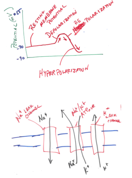
Hyperpolarization
A change in membrane potential to a value more negative than the resting potential. Following an action potential, delayed closure of voltage-gated K⁺ channels allows continued K⁺ efflux and produces afterhyperpolarization. Inhibitory inputs can also hyperpolarize a membrane.
Dura mater
The tough, fibrous outermost meningeal covering around the brain and spinal cord. It lies superficial to the arachnoid mater and helps protect the central nervous system.

Arachnoid mater
The middle meningeal layer, between dura mater and pia mater. The subarachnoid space between arachnoid and pia contains cerebrospinal fluid and blood vessels that cushion and support the CNS.

Efferent nerve
A nerve pathway carrying information away from the CNS toward peripheral effectors. Somatic motor pathways supply skeletal muscle. Autonomic efferent pathways supply cardiac muscle, smooth muscle and glands.
Afferent nerve
A nerve pathway carrying sensory information from receptors in the periphery toward the CNS for processing. Afferent input can report the external environment, the internal environment or body position.
Cholinergic receptor
A receptor that binds acetylcholine. The two major classes are nicotinic receptors, which are ligand-gated ion channels, and muscarinic receptors, which signal through G proteins. Nicotinic receptors occur in autonomic ganglia and at skeletal neuromuscular junctions. Muscarinic receptors mediate many parasympathetic effects at target organs.
Compare and contrast (i.e., similarities and differences) the divisions of the autonomic nervous system.
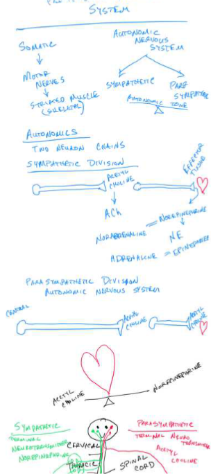
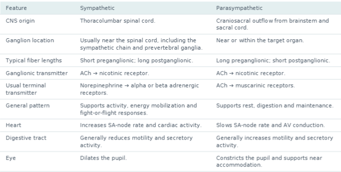
The sympathetic and parasympathetic divisions are involuntary visceral motor divisions of the peripheral nervous system. Both regulate smooth muscle, cardiac muscle and glands, helping maintain homeostasis. Their usual efferent pathway contains a preganglionic neuron with its cell body in the CNS and a postganglionic neuron with its cell body in a peripheral autonomic ganglion. In both divisions, the preganglionic neuron releases ACh onto nicotinic receptors on the postganglionic neuron. Their activity can vary continuously as autonomic tone.
Many organs receive both divisions, with the final response reflecting their relative activity and the receptors present. The divisions can have opposing or coordinated effects. The adrenal medulla is a specialized sympathetic pathway. Preganglionic ACh stimulates nicotinic receptors on chromaffin cells, which release epinephrine and norepinephrine into the blood. An exception to the usual sympathetic terminal transmitter is ACh release onto muscarinic receptors at many mammalian sweat glands, with species differences in sweating control.
Describe how the resting membrane potential is generated. Be sure to include the important membrane proteins, ions involved and indicate why the resting membrane potential has the value that it does.
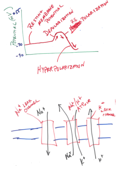
Resting membrane potential is the voltage difference across the membrane of an unstimulated cell, measured as inside relative to outside. In the neuron example used in lecture it is about −70 mV. This results from unequal ion distributions and selective membrane permeability.
The Na⁺/K⁺ ATPase uses ATP to pump three Na⁺ out and two K⁺ in per cycle. It maintains high intracellular K⁺ and high extracellular Na⁺ against their concentration gradients. Its unequal exchange also moves one net positive charge outward, making a small direct electrogenic contribution. Large intracellular anions, including proteins, remain inside and contribute to the intracellular ionic environment.
At rest there are more open K⁺ leak pathways than Na⁺ leak pathways, so the membrane is much more permeable to K⁺. K⁺ diffuses outward down its concentration gradient. As positive charge leaves, the inner membrane surface becomes negative relative to the outer surface. That electrical gradient attracts K⁺ inward and opposes the outward concentration gradient. If K⁺ were the only permeant ion, the two forces would balance at its equilibrium potential, approximately −90 mV in the lecture example.
A smaller amount of Na⁺ enters through Na⁺ leak channels. Both the Na⁺ concentration gradient and the negative interior favor inward Na⁺ movement. This inward positive current shifts the voltage toward the Na⁺ equilibrium potential, approximately +55 mV in the lecture. Because resting K⁺ permeability is much greater, the actual resting voltage lies much closer to −90 mV, at approximately −70 mV. The resting voltage is therefore determined by the ion gradients and relative permeabilities. Individual Na⁺ and K⁺ fluxes continue at rest, while the pump maintains their concentration gradients over time. Only a tiny separation of charge near the membrane is needed to establish the voltage.

Inhibitory post-synaptic potential
Change in the membrane potential of a neuron as a result of input that makes the cell membrane more negative
Excitatory post-synaptic potential
Change in the membrane potential of a neuron as a result of input that makes the cell membrane more positive
Ventral horn
Part of the spinal cord where efferent fibers exit the cord to the periphery
Dorsal horn
Part of the spinal cord where afferent fibers enter the cord from the periphery
Schwann cell
The cell that enwraps some peripheral neurons, insulating them and speeding propagation
Oligodendrocyte
The cell that enwraps multiple central neurons, insulating them and speeding propagation.
Dendrites
Extensions of the neuron cell body
Axon Hillock
Junction between neuron cell body and axon where action potentials are initiated
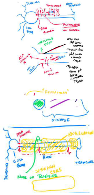
What is ‘propagation’ in an excitable cell and why does it occur? How is the process accelerated in higher vertebrates?
Propagation is the spread of an action potential along an excitable membrane through regeneration of the action potential in successive regions. In a neuron, depolarizing inputs are integrated at the trigger zone near the axon hillock and initial segment. If the membrane reaches threshold, approximately −55 mV in lecture, voltage-gated Na⁺ channels open rapidly. Na⁺ enters down its electrochemical gradient, making the inside more positive. Depolarization opens additional Na⁺ channels through positive feedback and generates the rising phase of an all-or-none action potential.
The entering positive charge produces local circuit current that spreads along the axon interior and returns through the extracellular fluid. This depolarizes adjacent resting membrane. When that region reaches threshold, its own voltage-gated Na⁺ channels open, fresh Na⁺ enters across its membrane,
and another action potential is generated. Repetition carries the signal along the axon. Because each new region regenerates the action potential, its amplitude is maintained along a healthy axon. A stronger suprathreshold stimulus can increase firing frequency rather than the size of each action potential.
Behind the advancing signal, Na⁺ channels inactivate and delayed voltage-gated K⁺ channels open. K⁺ exits, repolarizing the membrane. Continued K⁺ conductance can produce afterhyperpolarization. During the absolute refractory period, inactivated Na⁺ channels cannot immediately reopen, which
prevents the recently active region from firing again and favors forward propagation in normal signaling. During the relative refractory period, partial Na⁺-channel recovery and elevated K⁺ conductance mean a stronger stimulus is needed.
Myelination accelerates conduction in vertebrates. Schwann cells produce myelin in the PNS and oligodendrocytes in the CNS. Myelin increases membrane resistance, reducing current leakage, and decreases membrane capacitance, reducing the charge needed to change voltage. Local depolarizing current therefore spreads rapidly and farther beneath the myelin. Voltage-gated Na⁺ channels are concentrated at the nodes of Ranvier, where the membrane is exposed and the action potential is regenerated. This node-to-node regeneration is saltatory conduction. Larger axon diameter also speeds conduction by reducing internal resistance to current flow.
Accommodation
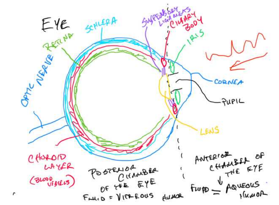
Adjustment of lens shape to focus light from objects at different distances onto the retina. For near vision, parasympathetic ACh stimulates ciliary muscle contraction, reducing tension in the suspensory ligaments. The elastic lens becomes rounder, increasing refractive power. For distant vision, ciliary muscle relaxation increases ligament tension and flattens the lens, reducing refractive power. The lecture associates distant accommodation with sympathetic activity.

Proprioception
The sense of body position and movement, including limb position, muscle length and tension. Proprioceptors in muscles, tendons and joints provide information used for posture, balance and coordinated movement.
Rod cells
Retinal photoreceptors specialized for dim-light vision. They contain rhodopsin, have high light sensitivity and provide low visual acuity with little color discrimination. Signals pass through retinal circuits to ganglion cells, whose axons form the optic nerve.
Zona fasciculata
The middle layer of the adrenal cortex. It produces glucocorticoids such as cortisol, which participate in metabolism and the stress response. ACTH from the anterior pituitary stimulates its activity.

Islets of Langerhans
Clusters of endocrine cells within the pancreas. Alpha cells secrete glucagon, beta cells secrete insulin, and delta cells secrete somatostatin. These hormones participate in regulation of nutrient metabolism and blood glucose.

Z-line
The protein-rich boundary of a sarcomere, where thin actin filaments are anchored. A sarcomere extends from one Z-line to the next. During shortening, adjacent Z-lines move closer together as thin filaments slide toward the sarcomere center.

Vitreous humor
The transparent gel occupying the large vitreous chamber behind the lens and in front of the retina. It transmits light and helps maintain the globe’s shape and support the retina. The anterior chambers contain aqueous humor.
Describe what happens within the cochlea following vibration of the oval window by the stapes.
The stapes pushes on the oval window and generates pressure waves in the perilymph of the scala vestibuli. These waves deform the cochlear partition and produce a traveling wave along the basilar membrane. The cochlear duct, or scala media, contains endolymph and houses the organ of Corti resting on the basilar membrane. Motion of the basilar membrane relative to the tectorial membrane creates shearing that bends the hair-cell stereocilia.
Bending the stereocilia toward their tallest members increases tension on tip links and opens mechanically gated channels. K⁺ from the K⁺-rich endolymph enters the hair cell and depolarizes it. Depolarization opens voltage-gated Ca²⁺ channels near the basal synapse. Ca²⁺ entry promotes neurotransmitter release onto sensory neurons of the spiral ganglion, which generate action potentials
carried in the cochlear branch of cranial nerve VIII to the brain. Bending the stereocilia in the opposite direction closes channels, hyperpolarizes the hair cell and reduces transmitter release. Hair cells produce graded receptor potentials.
Pressure is transmitted through cochlear fluids to the scala tympani and is relieved by movement of the round window. The round window bulges as the oval window is displaced inward, allowing movement within the fluid-filled cochlea. This pressure-release step is specifically called out in the instructor’s correction.
The location of maximal basilar-membrane displacement encodes sound frequency. The narrow, stiff base near the oval window responds best to high frequencies. The wider, more flexible apex responds best to low frequencies. Sound intensity is associated with displacement amplitude and the resulting pattern and rate of sensory nerve activity.

Describe the relationship between the hypothalamus and both the anterior and posterior pituitary.
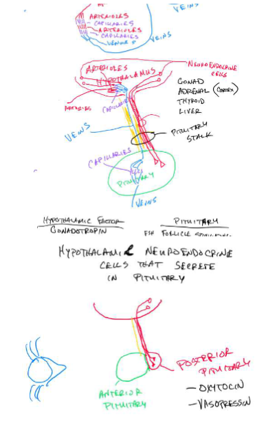
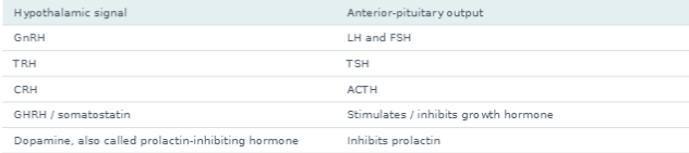
The hypothalamus links nervous-system information to endocrine output. It controls both pituitary lobes, using a vascular hormonal connection to the anterior lobe and a direct axonal connection to the posterior lobe.
Anterior pituitary
Hypothalamic neuroendocrine neurons synthesize releasing or inhibiting hormones and secrete them into a primary capillary plexus near the median eminence. Hypothalamic-hypophyseal portal vessels carry them directly down the pituitary stalk to a secondary capillary plexus in the anterior pituitary. A
portal system links two capillary beds in series. It delivers hypothalamic signals in relatively high concentrations before they are diluted in the general circulation. The factors regulate glandular anterior-pituitary endocrine cells, which synthesize and release their own hormones into the systemic blood.
Tropic hormones such as TSH, ACTH, LH and FSH regulate other endocrine tissues. Their downstream hormones commonly provide negative feedback to the anterior pituitary and hypothalamus. For example, CRH stimulates ACTH, ACTH stimulates adrenal glucocorticoid secretion, and glucocorticoids
inhibit further CRH and ACTH release.
Posterior pituitary
The posterior pituitary is neural tissue continuous with the hypothalamus. Large hypothalamic neurosecretory neurons synthesize oxytocin and vasopressin (ADH), then transport them down their axons through the stalk to terminals in the posterior lobe. Action potentials trigger Ca²⁺-dependent secretion from those terminals into systemic blood. Thus, the posterior lobe stores and releases hormones synthesized in hypothalamic neurons. Oxytocin participates in uterine contraction and milk ejection. ADH increases renal water retention.
H-zone
Portion of the sarcomere that contains only myosin.
Alpha cells
Cells in the pancreas that produce and secrete glucagon.
Beta cells
Cells in the pancreas that produce and secrete insulin.
Sclera
Outer reflective layer of the posterior chamber of the eye.
Ciliary body
Smooth muscle that is responsible for allowing for very near field vision.
Zona Glomerulosa
Region of the adrenal cortex that produces mineralocorticoids.
Thyroxin
Hormone produced in the thyroid that has four iodine atoms bound.
I-band
Portion of the sarcomere that contains no myosin.
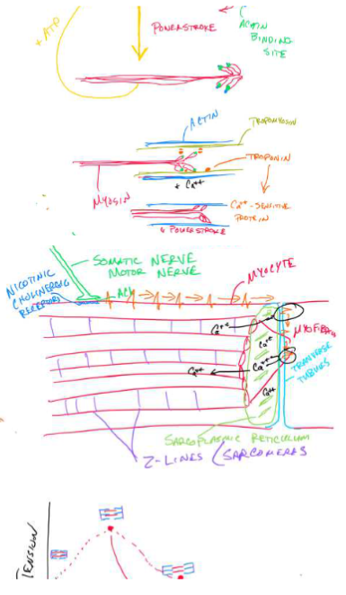
Beginning with the initiation of an action potential in a motor nerve, describe the events that lead to skeletal muscle contraction.
A motor neuron reaches threshold at its trigger zone, opening voltage-gated Na⁺ channels and initiating an action potential. Local current regenerates the action potential along the motor axon, reaching the terminal at the neuromuscular junction. Terminal depolarization opens voltage-gated Ca²⁺ channels.
Ca²⁺ enters and triggers synaptic vesicles to fuse with the presynaptic membrane, releasing ACh by exocytosis into the synaptic cleft.
ACh diffuses across the cleft and binds nicotinic cholinergic receptors on the motor end plate of the muscle fiber. These ligand-gated cation channels open. Na⁺ influx exceeds K⁺ efflux at the resting voltage, producing a local end-plate depolarization. When adjacent sarcolemma reaches threshold, voltage-gated Na⁺ channels open and generate a muscle action potential. It propagates along the sarcolemma and down the transverse, or T-, tubules.
T-tubule depolarization activates voltage sensors that are coupled to Ca²⁺-release channels in the sarcoplasmic reticulum. In skeletal muscle, the named proteins are the dihydropyridine receptor voltage sensor (DHPR/Caᵥ1.1) and ryanodine receptor (RyR1). Their coupling opens SR release channels, raising cytosolic Ca²⁺. The lecture drawing shows the required T-tubule-to-SR-to-Ca²⁺ sequence. Naming DHPR and RyR1 supplies the molecular connection if that level of detail was discussed in class.
Ca²⁺ binds troponin C on the thin filament, changing the troponin complex’s conformation and shifting tropomyosin away from myosin-binding sites on actin. Energized myosin heads carrying ADP and inorganic phosphate can then attach to exposed actin sites and form cross bridges. Phosphate release is coupled to strong binding and the power stroke, in which the myosin head pivots and pulls the thin filament toward the sarcomere center. ADP is released. A new ATP binds the myosin head and causes detachment from actin. Hydrolysis of that ATP to ADP and phosphate re-energizes and repositions the head for another cycle. Cross-bridge cycling continues while cytosolic Ca²⁺ remains elevated and ATP is available.
Thin and thick filaments slide past each other. The sarcomere shortens, Z-lines move closer together, and the I-band and H-zone narrow. The A-band remains the same length because the thick filaments retain their length. Individual actin and myosin filaments retain their lengths during this sliding process.
When stimulation ends, acetylcholinesterase breaks down ACh in the cleft, and ATP-dependent SERCA pumps return Ca²⁺ to the SR. Ca²⁺ dissociates from troponin, tropomyosin re-covers the binding sites, and cross-bridge activity declines, allowing relaxation. ATP is required for myosin detachment, head re-energization and Ca²⁺ reuptake. Without ATP, attached cross-bridges cannot detach normally, contributing to rigor.
Diastole
The relaxation phase of a cardiac chamber’s cycle. During ventricular diastole, ventricular pressure falls and the ventricles fill when atrial pressure exceeds ventricular pressure and the AV valves open. Diastole begins with isovolumetric relaxation before filling.
Systole
The contraction phase of a cardiac chamber’s cycle. Ventricular systole includes isovolumetric contraction and ejection. Ejection starts when ventricular pressure exceeds the pressure in the aorta or pulmonary artery and opens the corresponding semilunar valve.
End diastolic volume
The volume of blood in a ventricle at the end of filling, immediately before ventricular contraction. EDV is the maximum ventricular volume in that cycle and is related to preload.

Stroke volume
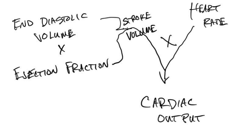
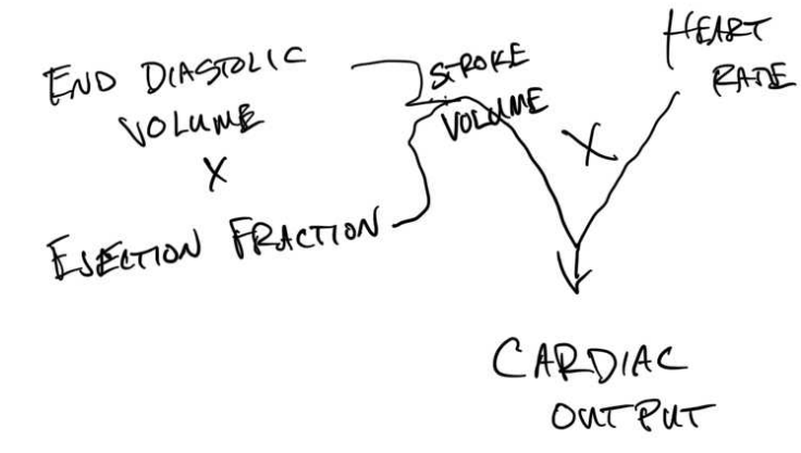
The volume of blood ejected by one ventricle in a single beat. SV = EDV − ESV, where ESV is the volume remaining after ventricular ejection.
Ejection fraction
The fraction of end-diastolic ventricular volume ejected in one beat. EF = SV/EDV, or (EDV − ESV)/EDV. Multiply by 100 to express it as a percentage.
Cardiac output
The volume pumped by one ventricle per unit time. CO = heart rate × stroke volume. Equivalently, CO = HR × EDV × EF when EF is a fraction. Typical units are mL/min or L/min.

Surfactant
A surface-active mixture, rich in phospholipids and proteins, secreted by type II alveolar epithelial cells. It reduces surface tension at the alveolar air-liquid interface, increases compliance, reduces the work needed for inflation and helps stabilize alveoli against collapse.

Describe how the partial pressure of oxygen changes in plasma as blood circulates from the right ventricle back to the right atrium.
Blood in the right ventricle is relatively low in oxygen after returning from systemic tissues. It passes through the pulmonary semilunar valve into the pulmonary trunk and pulmonary arteries, then into pulmonary capillaries surrounding the alveoli. The lecture diagrams give a range of roughly 40–80 mmHg for returning venous PO₂, depending on tissue extraction. About 40 mmHg is a common resting mixed-venous example.
Alveolar PO₂ is approximately 100 mmHg in the lecture example, higher than the incoming pulmonary-capillary plasma PO₂. Dissolved O₂ diffuses down this partial-pressure gradient across the thin alveolar-capillary barrier into the plasma and then into red blood cells. O₂ binds hemoglobin inside
the RBCs. Binding removes O₂ from the dissolved pool and permits continued diffusion into the blood while hemoglobin loads. As oxygenation proceeds, plasma PO₂ rises toward alveolar PO₂, approximately 100 mmHg.
Oxygenated blood leaves through the pulmonary veins, enters the left atrium, crosses the mitral/bicuspid valve into the left ventricle and is ejected through the aortic semilunar valve into the aorta. Plasma PO₂ stays relatively high through these chambers and the systemic arteries because
major gas exchange occurs at capillaries.
At systemic capillaries, tissue PO₂ is lower because cells continually consume O₂ in metabolism. Dissolved O₂ diffuses from plasma into interstitial fluid and cells, lowering plasma PO₂. Hemoglobin then releases O₂, replenishing dissolved plasma O₂ and supporting continued diffusion to the tissues. Both total blood oxygen content and plasma PO₂ fall as oxygen is delivered. The amount of extraction depends on tissue activity and blood flow.
Blood then travels through systemic venules and veins into the cranial and caudal venae cavae and back to the right atrium, with relatively low PO₂. It crosses the tricuspid valve to return to the right ventricle and repeat the cycle. PO₂ reflects dissolved O₂. Hemoglobin-bound O₂ contributes strongly to total oxygen content but does not directly exert the plasma partial pressure.

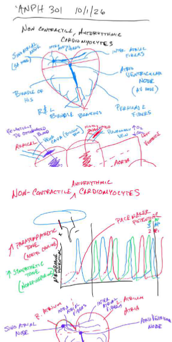
Describe the system of noncontractile, autorhythmic cardiomyocytes in initiating contraction of the heart and how heart rate is modulated by autonomic inputs to these cells.
Specialized noncontractile, autorhythmic cardiomyocytes spontaneously depolarize and organize the heart’s electrical activity. The sinoatrial (SA) node in the right atrial wall normally has the fastest intrinsic rhythm and acts as the pacemaker. Its cells have an unstable pacemaker potential. Slow inward currents gradually depolarize the membrane to threshold, Ca²⁺ entry generates the nodal action-potential upstroke, and K⁺ efflux repolarizes the cells. The cycle then repeats. Cardiac rhythm is myogenic, originating in cardiac cells, and is modulated by autonomic input.
The impulse spreads through atrial conduction pathways and neighboring atrial myocardium, linked by gap junctions, causing the atria to depolarize and contract. It reaches the atrioventricular (AV) node, where slow conduction produces a delay that allows atrial emptying and ventricular filling before ventricular contraction. The signal then travels through the AV bundle, or bundle of His, the right and left bundle branches in the interventricular septum, and the Purkinje fibers. Rapid Purkinje conduction distributes the excitation through the ventricles, coordinating contraction from the apex toward the outflow regions. The fibrous skeleton electrically insulates the atria from the ventricles, with the normal conduction route passing through the AV system.
Sympathetic postganglionic nerves release norepinephrine, which activates β₁-adrenergic receptors on nodal cells. Circulating epinephrine can act similarly. This increases inward pacemaker and Ca²⁺ currents, steepening pacemaker depolarization so threshold is reached sooner. SA-node firing frequency and heart rate increase, and AV conduction is accelerated. Sympathetic activity also
increases contractility of working myocardium.
Parasympathetic vagal nerves release ACh, which activates M₂ muscarinic receptors. Increased K⁺ conductance and reduced inward pacemaker/Ca²⁺ currents hyperpolarize nodal cells and slow their approach to threshold. SA-node firing and heart rate decrease, and AV conduction slows. The net heart rate reflects the intrinsic rhythm and the balance of autonomic tone. The transmitter here is acetylcholine, abbreviated ACh.
Protoporphyrin ring
Common structure contributing to respiratory pigments coordinated with Fe or Cu.
Single unit s