Genetics Exam 2
 Call Kai
 Learn
Learn Practice Test
 Spaced Repetition
 Match
Match Flashcards
 Knowt Play
1/50

There's no tags or description
Looks like no tags are added yet.
Name | Mastery | Learn | Test | Matching | Spaced | Call with Kai | Chat |
|---|
No analytics yet
Send a link to your students to track their progress
51 Terms
Genetic Material
contains information that is expressed to determine all traits & functions of an organism
is stable, but must also have the ability to change (variation in many traits without lethality to organism)
is carried on chromosomes (contains proteins & DNA)
Order of Experiments to Disprove “Genes are mad of protein” hypothesis
Griffith (1928) ; Avery et al (1944) → streptococcus pneumoniae strains & transformation
Hershey & Chase (1952) → bacteriophage T2 & radioactive labeling
Griffith & Avery et al Experiments
Streptococcus pneumoniae = bacteria that causes pneumonia in humans & lethal in mice
2 strains
smooth (S)
virulent (infectious)
wild-type
cells enclosed in polysccharide capsule
rough (R)
nonvirulent (easy to engulf & destroy)
mutant
no polysaccharide capsule
Griffith:
transformation: conversion of rough → smooth
living cells necessary for virulence

Avery, Macleod, and McCarty continued Griffith’s work
they knew genetic material must be either protein, RNA, or DNA
concluded transforming principle is made of DNA
transformed bacteria remained virulent in subsequent generations, so they concluded that transformed traits = heritable

Hershey & Chase
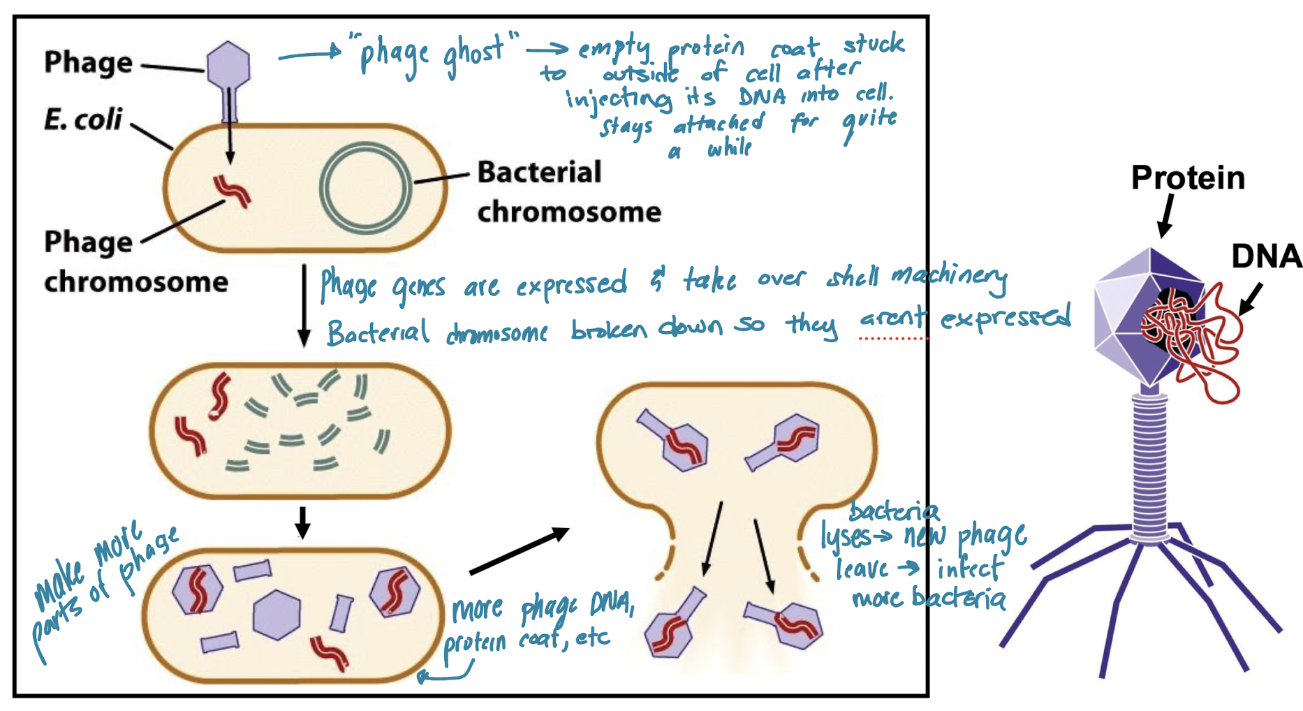
bacteriophage T2 = virus that infects bacteria ; DNA core surrounded by protein coat
they knew T2 is ~50% protein and 50% DNA
infection steps:
adsorption - phage attaches to bacteria via tail fibers
phage material enters bacteria - production of new viruses within bacterial cell
phage material must be the genetic material of the phage because it is necessary to make a new phage (molecule of inheritance!)
bacterial lysis - new phage progeny is released
radioactive labeling experiment steps
preparation
grow sample of phage in presence of E. coli
separate into two samples
add 35S (radioactive sulfur) into one media
add 32P (radioactive phosphorus) into the other
results in two labeled phages (phosphorus - DNA labeled & sulfur - protein labeled)

infection
infect two separate fresh E. coli samples (each with one of the labeled phages)
blend - shears protein coats & anything that did not enter the bacterial cell
spin - infected bacteria will pellet (concentrate & collect on bottom & separate from media)
found that the phage with DNA labeled had radioactive pellets and the other didnt → DNA is genetic material

Components of DNA (base unit & what that’s made of)
basic building block of DNA = nucleotide

deoxyribose - 5C sugar (one less O than ribose: ribose has OH on 2’ and deoxyribose has H)

Base Pairs & Chargaff’s Rule
Purines (nitrogenous base with two rings)
adenine (A)
guanine (G)
Pyrimidines (nitrogenous base with one ring)
thymine (T)
cytosine (C)
Chargaff’s Rule:
total pyrimidine (T+C) = total purine (A+G)
T = A & G = C
A + T is not equal to G + C
Pairings:
A-T
G-C
Rosalind Franklin & Maurice Wilkins Findings
DNA is made of 2 parallel parts
DNA is helical
Helix contains 10 nucleotides per turn
measured helix diameter
Watson & Crick Findings
figured out DNA is a double helix
two strands of DNA wound around each other
each strand is one chain of nucleotides
5’ phosphate of one nucleotide is linked to 3’ OH of another (antiparallel)
two strands of DNA of double helix held together by H bonds between complementary bases (A + T & G + C)
recognized DNA structure is important for replication
complementary strands (each strand can be a template for the other)
specific base-pairing (only one sequence is possible from a template)
Three Models for Mechanism of DNA Replication
add to this???

DNA Backbone
made of sugar & phosphate (covalently bonded)
phosphate has a (-) net charge, giving DNA a negative charge since backbones is on the outside of the helix
Major vs Minor Grooves
Major: area between turns of double helix
Minor: distance between 2 back bones of anti-parallel strands

Three Forms of DNA
A Form
less hydrated
more compact (10.9 base pairs per turn)
right-handed
typically seen in vitro
in vitro = in controlled lab conditions (for this situation: high salt conditions)
B Form (most common)
more hydrated
less compact (10 base pairs per turn)
right-handed
typically found in vivo (in living organism/cell)
proposed by Watson & Crick
Z form
left-handed
zig-zag phosphate backbone
seen in short DNA pieces with alternating purine/pyrimidine
seen in small localized regions (ex: regions of big chromosome)

Oligonucleotide
short chain of nucleotides
single-stranded DNA
Denaturing
separating 2 strands of DNA
aka “melting”
can be done by heating DNA, which disrupts H bonds between strands
G/C-rich DNA has higher melting temp than A/T-rich DNA because G/C has 3 H-bonds & A/T has 2 H-bonds
Annealing
when 2 complementary single strands of DNA bind back together via H-bonds
DNA vs RNA
DNA
deoxyribose (2’ C has an H)
A, G, C, T
double-stranded
more stable (relative to RNA)
RNA
ribose (2’ C has an OH)
A, G, C, U
single-stranded
less stable (relative to DNA)

Special Structures found in Single-Stranded Nucleic Acids
Hairpin
sequences on same strand are inverted complements
sequence between inverted repeats forms loops

Stem
hairpin without loop

Complexity of RNA
molecules fold into complex 2˚ & 3˚ structures
RNA is synthesized as single stranded nucleic acid
hairpins, bulges, stems, loops, etc fold onto each other (these structures typically are binding sites for proteins)

3 Models for Mechanism of DNA Replication
Conservative: template strands create new strands - template strands recombine & new strands combine
Semi-Conserative: template strand binds with the new strand it made
Dispersive: pieces of template strand synthesize new strands - recombination is random, results in mixed strands
Meselson-Stahl Experiment
proved semi-conservative model for prokaryotes
E. coli - circular chromosome
15N = heavy isotope of nitrogen that labels DNA
14N = “normal” nitrogen
used density gradient centrifugation at different points in time
time = 0 → 15N-containing ds DNA is extracted and goes through gradient centrifugation → result = one band of heavy DNA
time = 1 (after E. coli is exposed to 14N & one replication occurs - all new dna = 14N) → DNA extracted, gradient centrifugation = 1 intermediate band between light and heavy (supports semi-conservative OR dispersive)
time = 2 (another cycle of replication in 14N) → DNA extracted, gradient centrifugation = 1 band of all light & 1 band of intermediate (consistent with semi-conservative)

Density Gradient Centrifugation
DNA is extracted form cells, cut into pieces & added to a gradient of Cesium Chloride (CsCl) - higher density towards bottom
samples are centrifuged at high speed
at equilibrium, DNA concentrates within the gradient where its density is equivalent to that of CsCl
See DNA by staining with ethidium bromide & shining UV light

Taylor Experiment
proved semi-conservative model in eukaryotes
studied bean root tips - followed cells through 2 rounds of mitosis - labeled DNA with tritriated (3H) thymidine (radioactive)
started with unlabeled chromosome in G1 & added 3H-thymidine → chromosome goes through S, G2, and prophase → autoradiography at metaphase = both sisters are radiolabeled (consistent with semi-conservative OR dispersive)
anaphase occurs & they follow one sister through the next round of mitosis
sister in G1 → unlabeled thymidine is added, sister goes through s-phase, g2, and prophase → autoradiography in metaphase = only one chromatid is labeld (consistent with semi-conservative! if dispersive, then both sisters = labeled)

Autoradiography
Method Taylor used in his experiment that proved eukaryotic DNA replication is through the semi-conservative method
done during metaphase of mitosis - exposes DNA to film, dark areas show radioactivity
picture shows DNA molecules with both sister chromatids labeled

Things needed for DNA Synthesis
template (ssDNA) - single stranded DNA
substrate (dNTPs) - deoxyribonucleotide triphosphates (A, G, C, T)
cellular machinery (enzymes, etc) to read template and synthesize new DNA strands
DNA polymerase
DNA Pol I & III
initiator protein
helicase
SSBs
topoisomerase (DNA gyrase)
RNA polymerase (primase)
DNA ligase
Telomerase
Initiator Protein
starts process binds to oriC & begins melting DNA at A/T rich regions (easier to melt than G-C)
Origin of Replication (OriC)
where replication begins
eukaryotic chromosomes have many
only 1 in E. Coli - OriC
sequence is recognized by initiator proteins
Helicase
continues to melt DNA (breaks down H-Bond)
DNA Polymerase
lots of types in eukaryotes
5 types in prokaryotes (only need to know DNApol I & III)
makes new DNA strand 5’ → 3’
needs -OH group to begin
DNApol I
3 activities
5’ → 3’ exonuclease activity - destroys RNA primer
5’ → 3’ polymerase activity - replaces RNA primer with DNA
3’ → 5’ exonuclease activity - allows proofreading
DNApol III
prokaryotic enzyme
does most of DNA synthesis
elongates RNA primer by adding new DNA (5’ → 3’ polymerase activity)
very fast (~1,000 nucleotides/sec)
3’ → 5’ exonuclease activity to correct mistakes
corrects mistakes for the nucleotide that was just added to new DNA strand - destroys incorrect nucleotide & corrects mistake
RNA polymerase
prokaryotic enzyme
type of RNApol - primase
doesn’t need free 3'‘-OH group to work
makes RNA primer using ssDNA as template
RNA primer provides free 3’-OH for DNApol to add onto
Single-Strand-Binding Proteins
keeps DNA melted
non-specifically binds to ss DNA to stop H-bonds from reforming
Topoisomerase
DNA Gyrase - type of isomerase for prokaryotes
works ahead of replication fork to reduce strain due to helix undwinding
relieves supercoiling by cleaving dsDNA of sugar-phosphate backbone, allowing it to unwind & recreates those covalent bonds before helicase gets there to melt the DNA
because as DNA helicase unwinds DNA @ fork, it sets up supercoiling
topoisomerase can either undo supercoiling or cause it
DNA Ligase
binds okazaki fragments together
creates covalent bond between 5’ phosphate & 3’ OH to join 2 nucleotides in newly synthesized DNA
Replication Licensing Factor (RLF)
controls timing of replication initation in eukaryotes
keeps track of where replication already happened & where it needs to happen because eukaryotes have multiple OriCs that open at different times
contains several proteins that bind to OriC during G1 phase of cell cycle
marks an OriC as ready for DNA replication
once replication begins, different proteins will bind to mark that OriC as already replicated
Telomerase Enzyme
extends DNA at chromosome ends
synthesizes highly repetitive DNA in telomeres
has protein & RNA components
protein part - synthesizes new DNA
RNA part - acts as template for DNA synthesis
-telomerase moves down & continues extending 3’ overhang
-primase makes primer on 5’ prime end
DNA polymerase fills in , ligase seals nick
primer is removed
Telomerase is expressed in germ cells (give rise to gametes) & stem cells (immortal cells that provides new cells for processes like wound healing)
Telomeres & Replication
Telomeres contain condensed highly repetitive DNA that do not code for anything - impossible to replicate end of lagging strand - each time you go through replication (without telomerase) the chromosome should be getting smaller because end does not replicate (typically not good because you lose the info at the end of the strand)
repeat sequence depends on sequence of telomerase RNA (all eukaryotes have a version of telomerases, but different RNA sequences)
2 Types of DNA Replication in Prokaryotes
circular chromosome replication: theta replication
plasmid replication: rolling circle replication

Theta Replication in Prokaryotes
starting at oriC, DNA is melted so single strands act as templates
DNA synthesis happens simultaneously in both directions
1 replication bubble made of 2 replication forks
product = 2 circular chromosomes (each with 1 old & 1 new DNA strand)

Leading Strand
since DNApol makes DNA 5’ → 3’, each fork will have only 1 strand made continuously
strands coming straight out from oriC

Lagging Strand
made discontinuously
short pieces = okazaki fragments
strands going toward oriC

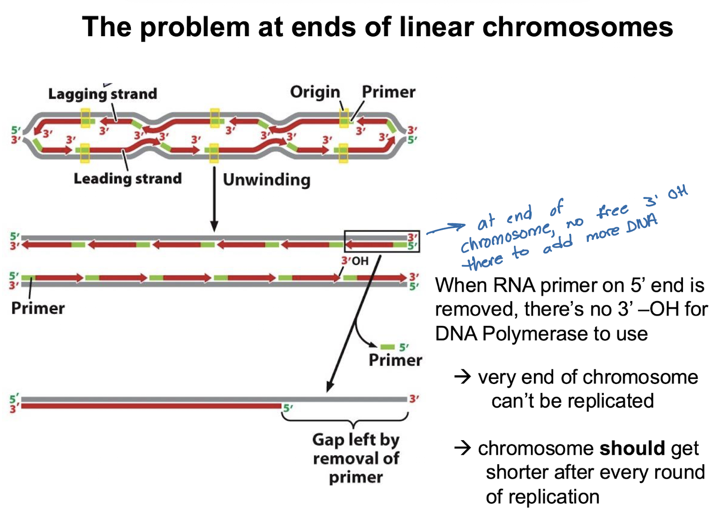
The problem at ends of linear chromosomes & what fixes them
When RNA primer 5’ end is removed at end of DNA strand, there’s no 3’-OH for DNApol to use
very end of chromosome cannot be replicated (end of lagging strand)
chromosome should get shorter after every round of replication, but telomerase prevents this
Replication in Eukaryotes
eukaryotic chromosomes have many DNA packaging proteins bound to DNA
histones
must be synthesized during DNA replication
many replicons (point of origin & area of DNA replicated)
slower than prokaryotes
many types of DNApol
1 bubble (2 forks) from each origin
bidirectional DNA synthesis
final product = 2 linear chromosomes (1 old + 1 new DNA strand)

Two Levels of DNA Packaging in Prokaryotes
formation of loop domains
each loop is held in place by DNA binding proteins
keeps chromosomes organized - most of the time, related genes are present near each other, so expressing these types of genes only needs one loop to be turned on (ex: genes that code for a biochemical pathway all in one loop)
supercoiling
happens within loop domains
condenses DNA even further

Levels of DNA Packaging In Eukaryotes (Simplest to Complex)
DNA double helix (2nm) → nucleosome / chromatosome (11nm) → 30nm → 300nm → 250-nm-wide fiber (700nm) → chromosome (1400nm)
chromatin = DNA + proteins
chromosome = chromatin that is highly condensed
Two Types of Chromatin in Eukaryotes
Heterochromatin
remains highly condensed (tightly packed) throughout cell cycle
no gene expression here
major state of human Y chromosome
can be constitutive (always condensed ; telomeres/centromeres) or facultative (condensed at certain times ; x-inactivation)
Euchromatin
packaging changes during cell cycle (based on whether or not gene needs to be expressed & where in the cell cycle we are in)
contains expressed genes
major state of most genes
only this type found in prokaryotes
Centromeres
made up of heterochromatin
part of chromosome that attaches to spindle fibers - acts as a protein binding sit
required for proper segregation of chromatids
no genes present here; DNA sequence is highly repetitive (# of repeats on each chromosome varies)
DNA remains tightly packaged throughout cell cycle
Differences in Replication in Eukaryotes vs Prokaryotes
prokaryotic replication
A continuous process that can happen rapidly, even while the cell is dividing
eukaryotic replication
Strictly restricted to the S phase (synthesis phase) of the cell cycle

General Process of DNA Replication (one oriC in view)
SEMI-CONSERVATIVE
Initiation
initiator proteins bind to origin of replication & begin process of melting DNA into two ss DNA
2 DNA helicases continue to melt DNA away from origin of replication & towards forks (two diff directions)
SSBs bind to single strands to prevent re-annealing (rebinding/reforming H bonds)
topoisomerase relieves the strain of supercoiling ahead of replication fork by cutting & resealing the phosphate backbone
elongation
primase synthesizes RNA primer to provide free 3’-OH
DNA polymerase attaches to free 3’-OH & reads template strand from 3’ to 5’ direction to build new strand in 5’ to 3’ direction
leading strand: synthesized continuously in same direction that replication fork opens
lagging strand: synthesized discontinuously in short segments away from the opening fork (okazaki fragments)
termination & processing
primer removal & replace with DNA (prokaryotes = DNApol I)
DNA ligase seals nicks in phosphodiester backbones of okazaki fragments (turns them into a continuous strand)
EUKARYOTES ONLY: telomere maintenance - telomerase uses internal RNA template to replicate ends of chromosomes to prevent gene loss

Telomeres & Single Stranded Overhang
made up of heterochromatin & also is a protein binding site
POT (protection of telomere) proteins bind single stranded overhand (3’ overhang due to end of chromosome’s inability to be replicated)
provides stability
prevents ends from degrading and/or sticking together
overhang = single stranded nucleic acid that will get destroyed by nucleases in cell if no proteins bind to it for protection or if no other protection mechanisms occur (destruction = shorter chromosome & sometimes loss of vital information)
sometimes folds over to form triple-stranded structure (SEE PICTURE)
DNA remains tightly packaged throughout cell cycle
Packaging of Euchromatin
dynamic! level of packaging changes throughout cell cycle
FINISH SLIDE 275!!!!