8 - Clinical assessment, classification and grading of AMD
 Call Kai
 Learn
Learn Practice Test
 Spaced Repetition
 Match
Match Flashcards
 Knowt Play
1/48

Earn XP
Description and Tags
Block 2
Name | Mastery | Learn | Test | Matching | Spaced | Call with Kai | Chat |
|---|
No analytics yet
Send a link to your students to track their progress
49 Terms
What are the two types of AMD?
Wet aka active
Dry aka inactive
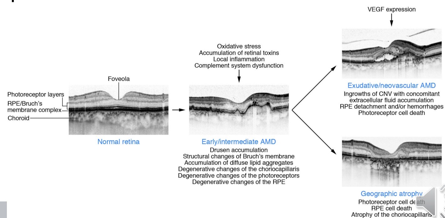
What is AMD and what are its 2 main types?
AMD is a leading cause of visual impairment in people aged ≥50 in developed countries; wet/active AMD and dry/inactive AMD
What are the risk factors for AMD?
Increasing age; genetics; gender; lifestyle factors: smoking/diet/hypertension; myopia; CSR; cataract surgery; diabetes; inflammation
What does fundus autofluorescence (FAF) assess?
Lipofuscin fluoresces and indicates the presence of RPE; excess lipofuscin may occur when cells overlap due to early degeneration; oxidative stress = hyperfluorescence; RPE cell death = hypofluorescence
What is fluorescein angiography used for in AMD?
Investigates retinal leakage; the pattern of leakage can be used to classify the lesion
What is indocyanine green angiography used for?
Imaging the choroidal vasculature
What is OCT angiography (OCTA)?
Non-invasive imaging that detects blood flow and abnormal patterns of vascular flow
How does NICE classify normal/no AMD?
No signs of AMD or only small hard drusen <63 µm
What is NICE low-risk early AMD?
Medium drusen ≥63 to <125 µm OR pigmentary abnormalities
What is NICE medium-risk early AMD?
Large drusen ≥125 µm OR reticular drusen OR medium drusen with pigmentary abnormalities
What is NICE high-risk early AMD?
Large drusen ≥125 µm with pigmentary abnormalities; reticular drusen with pigmentary abnormalities; vitelliform lesion without significant visual loss (BCVA better than 6/18); OR atrophy <175 µm not involving the fovea
What is NICE late AMD (indeterminate)?
RPE degeneration/dysfunction with degenerative AMD changes and subretinal or intraretinal fluid without neovascularisation; OR serous PED without neovascularisation
What is NICE late AMD (wet active)?
Classic CNV; occult CNV including fibrovascular PED/serous PED with neovascularisation; mixed CNV; retinal angiomatous proliferation (RAP); polypoidal choroidal vasculopathy (PCV)
What is NICE late AMD (dry)?
Geographic atrophy without neovascular AMD; OR significant visual loss of 6/18 or worse associated with dense/confluent drusen; advanced pigmentary changes/atrophy; or a vitelliform lesion
What is NICE late AMD (wet inactive)?
Fibrous scar; subfoveal atrophy/fibrosis secondary to RPE tear; atrophy with absent/thinned RPE and/or retina; cystic degeneration with persistent intraretinal fluid/tubulations unresponsive to treatment
What are drusen?
Extracellular deposits between the RPE and Bruch's membrane due to incomplete processing of photoreceptor outer segments; largely composed of lipids; act as a barrier and are a precursor to atrophy and CNV
How does drusen prevalence change with age?
≈2% in ages 45–55; increases to ≈25% in those aged >75
What are the features of hard; intermediate and soft drusen?
Hard:
What are reticular pseudodrusen (RPD) and how can they be imaged?
Reticular pseudodrusen can be demonstrated using colour photography; IR imaging; FAF; and SD-OCT
What does the conversion-to-large-drusen graph show?
Risk of developing large drusen increases most when medium drusen are present in both eyes; lower with medium drusen in one eye; lower still with small/no drusen
What are the AREDS 5-year risk factors used to estimate AMD progression?
Large drusen and pigmentary abnormalities in each eye are assigned risk factors; additional risk factors are assigned when one eye already has late AMD
What is atrophy in AMD?
Associated with collapse of drusen; irreversible tissue loss; appears as a hypopigmented/depigmented area where choroidal vasculature becomes visible; geographic atrophy is end-stage
What is cORA?
Complete outer retinal atrophy: intermittent hypertransmission; continuous absence of ellipsoid/interdigitation zones with corresponding outer nuclear layer loss but intact RPE; usually after prolonged SRF or regression of subretinal deposit
What is iORA?
Incomplete outer retinal atrophy: detectable ellipsoid layer disruption and outer retinal loss; RPE remains intact; no hypertransmission
What is cRORA?
Complete RPE and outer retinal atrophy: hypertransmission with an area of RPE disruption >250 µm and evidence of overlying photoreceptor disruption
What is iRORA?
Incomplete RPE and outer retinal atrophy: hypertransmission with corresponding RPE disruption ± basal laminar deposits and evidence of overlying photoreceptor disruption
What does NICE recommend for referral of early AMD?
Confirm early AMD using slit-lamp biomicroscopic fundus examination alone; do not refer asymptomatic early AMD to hospital eye services for further diagnostic tests
When should late dry AMD be referred according to NICE?
Only for sight impairment certification; if hospital referral is the local route to low-vision services; if new symptoms suggest wet active AMD; or to enable participation in research into new dry AMD treatments
What OCT features should be assessed in AMD?
Subretinal fluid; intraretinal fluid; PED; hyper-reflective dots (HRD); subretinal hyper-reflective material (SHRM); double-layer sign (DLS); outer retinal tubulation (ORT)
What types of pigment epithelial detachment (PED) may occur?
Serous; drusenoid; fibrovascular; haemorrhagic
What are hyper-reflective dots (HRD)?
Possibly inflammatory/disrupted cells; can occur in all retinal layers; usually located around fluid
What can subretinal hyper-reflective material (SHRM) represent?
Haemorrhage; lipid; pigment; subretinal fibrosis; CNV itself can also be hyperreflective
What is the double-layer sign (DLS)?
Irregular elevation of the RPE from Bruch's membrane; can occur in CSR and AMD subtypes such as PCV and non-exudative CNV
What is outer retinal tubulation (ORT)?
Degenerative photoreceptors and Müller cells forming a hyper-reflective ring with a hypo-reflective centre; indicates chronicity and poor response to treatment
Where are the 3 types of CNV located?
Type 1 = below RPE; Type 2 = above RPE; Type 3 = intraretinal neovascularisation
What is non-exudative/quiescent CNV?
CNV with abnormal vascular flow detectable on OCTA but without the leakage detected by FA in exudative CNV; ≈20% risk of progression to exudative CNV within 1 year
How urgently should suspected wet active AMD be referred according to NICE?
Urgent referral to a macula service whether or not visual impairment is reported; normally within 1 working day; does not require emergency referral
What are the NICE anti-VEGF treatment criteria stated in the lecture?
BCVA 6/96–6/12; no permanent structural damage to the fovea; lesion ≤12 disc areas; features of recent progression
What are the main components of AMD assessment?
History; refraction; examination; Amsler; imaging
What should be established from the history in suspected AMD?
Reduced VA; distortion; whether unilateral or bilateral; duration and progression
How should refraction be approached in AMD?
Objective refraction may be more reliable; use changes appropriate to VA e.g. larger subjective steps if VA is poor; obtain a measurable VA as <6/60 is not ideal
How should the fundus be examined when assessing AMD?
Consider dilation with usual precautions; red-free may help; use low magnification; indirect/oblique illumination; contact lens examination where appropriate; examine both eyes
What is the role of the Amsler grid in AMD assessment?
Standard Amsler may not always be easy to administer; it can be useful for patient self-monitoring
What is the role of imaging in AMD assessment?
It complements clinical examination and helps identify AMD features
What happened in Case 1?
77-year-old woman with dry AMD for 10 years and progressive worsening; VA R 6/9-2 and L 6/12; developed stable right-eye distortion over several weeks; referred to macular clinic for assessment
What were the key details of Case 2?
Previously treated with anti-VEGF; last injection 4 weeks ago; VA stable at 6/48; fellow eye dry AMD with VA 6/7.5; no new symptoms; attending routine review
What OCT findings were considered in Case 2 and what were the management options?
New/recurrent intraretinal oedema/cysts with HRD versus degenerative cysts associated with damaged/defective RPE; options were re-inject; observe; or perform angiography
What are the key take-home points for assessment and referral of AMD?
AMD terminology/classification has evolved; dry AMD affects the majority but can convert; imaging is a useful adjunct; full evaluation is needed to determine diagnosis and management