Lt 14 Degenerative and other joint diseases
 Call Kai
 Learn
 Practice Test
 Spaced Repetition
 Match
 Flashcards
 Knowt Play
1/49

There's no tags or description
Looks like no tags are added yet.
50 Terms

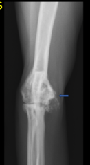
What is the arrow pointing to in this image?
Osteophyte

What is an Osteophyte?
New bone formation at the Osteochondral junctions (around periphery of articular cartilage)
What is an Enthesophyte?
New bone that develops at an Enthesis (the point of insertion of a tendon, ligament, joint capsule or fascia onto bone)

Radiographic signs of Joint Disease
- Increased synovial volume.
- Decreased or increased subchondral bone opacity.
- Soft tissue mineralization.
- Osteophytes.
- Enthesophytes.
Degenerative joint disease is also known as _____
Osteoarthrosis
Degenerative joint disease has a _____ appearance
Non-aggressive
Degenerative Joint Disease is a slow progressive disease that leads to _____
Synovial effusion and Cartilage degradation
Degenerative Joint Disease frequently occurs where?
Weight-bearing joints in large-breed dogs and cats
Degenerative Joint Disease can be caused by ______
Aging,
Developmental disorders,
Acquired disorder
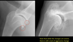
Why is this DJD and NOT neoplasia?
Because there is proliferation WITHOUT cortical destruction

What are the circles highlighting in this image?
Osteophytes associated with Elbow DJD

What is being highlighted in this image?
Coxofemoral DJD

What is being highlighted in this image?
Stifle DJD

What is being highlighted in this image?
Tarsus DJD

What are signs of Cruciate Ligament Disease?
Synovial effusion,
Osteophytes,
Cranial displacement of the tibia,
Distal displacement of the popliteal sesamoid bone,
Cranial displacement of the interpatellar fat pad.

Hip dysplasia is indicated by _____
< 50% coverage of femoral heads by acetabulum.
Osteophytes.
Widened joint space/subluxation.
What are the 2 methods used to determine Hip Dysplasia?
OFA and PennHip
What is an OFA?
Extended ventrodorsal projection hip radiographs.
Patient must be at least 2 YEARS OLD.

What is a PennHip?
3 different views of the hip that are sent to a certified radiologist.
Can predict osteoarthritis in dogs 16 WEEKS or OLDER.

What is being highlighted in this skyline view of the shoulder?
Bicipital tendon mineralization or partial avulsion

1-year-old male neutered Labrador.
• Bilateral forelimb lameness, worse on the left
Normal or Abnormal?
If abnormal, what abnormalities do you see?
Osteophyte and mineralization; Osteochondrosis
Degenerative Joint Disease in CATS is primarily caused by _____
Age-related,
Osteochondral dysplasia of Scottish fold cats,
Mucopolysaccharidosis (MPS),
Hypervitaminosis A
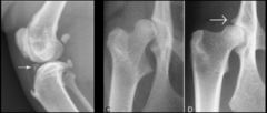
Radiographic findings associated with DJD in CATS
• Periarticular osteophytes.
• Subchondral bone changes.
• Intra-/periarticular calcification and ossification.
What are the MOST COMMON causes of Polyarthropathies in Dogs and Cats?
Septic arthritis,
Inflammatory arthritides,
Immune-mediated polyarthritis (IMPA)
Which joints are common affected for Immune-mediated polyarthritis (IMPA)
Distal joints (Carpus and Tarsus)
Is IMPA erosive or non-erosive?
NON-erosive
Causes of IMPA
Idiopathic,
Breed associated (Greyhound, Shar-pei, Akita),
Systemic Lupus Erythematous,
Inflammatory/reactive (Lyme disease)

Radiographic signs associated with non-erosive IMPA
ONLY synovial/intra-articular swelling
Is septic arthritis erosive or non-erosive?
EROSIVE
What is septic arthritis?
Microorganisms penetrate the synovial membrane and joint; Iatrogenic or systemic disease
Where does septic arthritis commonly occurs where?
Larger proximal joints
What are the initial signs of septic arthritis?
Acute lameness and painful joints.
Soft tissue swelling and joint effusion.

In 7-10 days, what radiographic signs can be seen for septic arthritis?
- Subchondral bone destruction and irregularity.
- Adjacent sclerosis.
- Periosteal reaction.
_______ causes secondary polyarthropathy in CATS
Infectious diseases

For EROSIVE Polyarthropathies, the _____ are the MOST affected and appear radiographically aggressive.
Carpus and Tarsus

What are the 2 most common causes of EROSIVE Polyarthropathies?
Rheumatoid arthritis and Periosteal proliferative polyarthritis
Feline noninfectious NON-erosive Polyarthritis in CATS is caused by _____
Idiopathic or primary IMPA or Systemic Lupus Erythematous
Feline noninfectious EROSIVE Polyarthritis in CATS is caused by _____
Feline Periosteal Proliferative polyarthritis and Feline Rheumatoid arthritis

Radiographic signs of EROSIVE Polyarthropathies
- Severe soft tissue swelling centered around the carpal joint.
- Severe moth eaten lysis of carpal bones.
- Aggressive osteal changes.
Osteochondrodysplasia is common for which breed of CATS?
Scottish folds
What is Osteochondrodysplasia?
Defective cartilage maturation affecting the joints of the DISTAL LIMBS —> ANKYLOSIS of Metatarsal and Metacarpals and malformed bones (Shorter and Fatter)

Radiographic signs of Osteochondrodysplasia
Ankylosis (bridging and fusion of the joints) with smoothly marginated periosteal proliferation
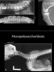
What is Mucopolysaccharidosis?
Polyarthropathy due to faulty cartilage formation from tissue accumulation of glycosaminoglycans; Most common in CATS;
Associated with Dwarfism, Facial dysmorphism, Short and square vertebral bodies

Radiographic signs of Mucopolysaccharidosis
- Generalized epiphyseal dysplasia.
- Short and square vertebral bodies.
Causes and clinical signs of Hypervitaminosis A
Cats fed large amounts of raw liver; Cats are obtunded, apprehensive, lame.
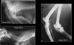
Radiographic signs of Hypervitaminosis A
- Ankylosing arthropathy (proximal joints) and spondylopathy (vertebrae).
- Ankylosis that does NOT resolve.

Once Ankylosis of the proximal joints occur for Hypervitaminosis A, what happens?
The joints can NOT bend and extend
Most common joints affected by neoplasia
Stifle and elbow
What are the most common types of neoplasias that affect the joints?
Histiocytic sarcoma and synovial cell sarcoma

Radiographic signs of neoplasia of the joints
- Soft tissue swelling*
- Progress to subchondral lysis
- Cortical lysis (Permeative and moth eaten)
- Amorphous periosteal proliferation