BDS1.CP.06 Action Potential
 Call Kai
 Learn
Learn Practice Test
 Spaced Repetition
 Match
Match Flashcards
 Knowt Play
1/35

Earn XP
Description and Tags
• Characterise parameters of biological electricity and outline electrophysiological approaches • Describe changes in cellular electricity (Depolarization and hyperpolarization) • Define action potential and characterise ion permeability of cell membrane during its development • Identify key components and main characteristics of action potential in different excitable tissues
Name | Mastery | Learn | Test | Matching | Spaced | Call with Kai | Chat |
|---|
No analytics yet
Send a link to your students to track their progress
36 Terms
·
What is biological electricity?
Electrical activity produced by living cells. It is essential for communication between nerve cells, muscle contraction and the normal functioning of the heart and brain.
The basic principle is that ions carry electrical charge, and their movement across cell membranes creates electrical signals.

What are the important ions involved in biological electricity? What roles do each typically play?
Na+: Concentration more out>in. Moving into cell depolarises cell and makes it more positive.
K+: Concentration more in> Out. Moving out of cell makes cell more negative – REPOLARISATION.
Ca2+: Concentration more out>in. Vital in cell signalling and MUSCLE contraction.
Cl-: Concentration more out >in. Involved in membrane voltage and neuronal inhibition.
What is the resting potential within outside a cell, inside a cell and of a neuron at rest?
Neuron at rest = electrically charged
Outside = positive
Inside = negative

3x examples of biological electrical activity being tested clinically?
ECG- electrical activity in heart
EEG- electrical activity in brain, using electrodes
EMG- electrical activity in muscles
None of the above measures the membrane voltage of one individual cell directly instead the surface electrodes generally record voltage differences generated by populations of cells.
How do we measure electrical activity?
Electrophysiology is the study and measurement of electrical properties in biological systems, ranging from individual ion channels to whole organs.
What are 6 important electrical parameters involved in electrophysiology? and their units?
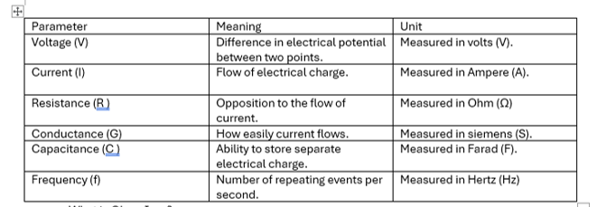
Voltage (V) – difference in electrical potential between two points. Measured in volts (V).
Current (I) – flow of electrical charge. Measured in Ampere (A).
Resistance (R ) – Opposition to the flow of current. Measured in Ohm (Ω)
Conductance (G). How easily current flows. Measured in siemens (S).
Capacitance (C ). Ability to store separate electrical charge. Measured in Farad (F).
Frequency (f). number of repeating events per second. Measured in Hertz (Hz)

Fill in the gaps
What is Ohms Law?
Describes the relationship between voltage, current and resistance:
V = IxR. Which is Voltage = Current x Resistance
Or easier to remember RVI R= V/I
Ion channels provide pathways for ions to cross membranes. When channels open, membrane conductance changes, allowing electrical currents to flow. Biological membranes are more complex than a simple resistor, but Ohm's law is still a useful foundation.

In a formula how do Conductance and resistance related?
Conductance is the reciprocal of resistance: because how easily current flows is the opposite of how opposed it is.
o Therefore G = 1/R.
o OR G = I/ V
How do both intracellular and extracellular electrophysiological recording techniques work?
Extracellular recording: Electrodes are positioned outside cells. They detect voltage changes produced by nearby cellular activity. Can record activity from multiple neurons – can study patterns of neuronal firing. Does not directly measure the membrane voltage inside one cell.
Intracellular recording / patch-clamp: a fine glass pipette accesses the cell membrane, allowing researchers to measure membrane voltage or ionic currents.
Voltage-clamp: holds the membrane voltage at a chosen level and measures the current needed to maintain it.
Current-clamp: injects or controls current and measures the resulting change in membrane voltage.
An easy way to remember the difference:
Voltage-clamp: control voltage, measure current. Current-clamp: control current, measure voltage.
Membrane potential: why is the inside of a resting neuron negative?
Membrane potential (Vm) is a sum of equilibrium potentials (Eion) of all contributing ions. Membrane potential is a voltage difference across the plasma membrane
A typical resting neuron has a membrane potential of approximately −70 mV.
Ion transport: sodium-potassium pump maintains ion gradients by moving three Na⁺ ions out of the cell for every two K⁺ ions moved in, using ATP.
Selective permeability = K+ has a substantial electrochemical gradient and the resting membrane is highly permeable to K⁺. Meaning K⁺ has a major influence on resting Vm.

What is Depolarisation vs hyperpolarisation? Which ions are usually responsible?
AND which for each is the neuron MORE or LESS excitable?
Depolarisation (reduction in charge): membrane potential becomes less negative (moves towards zero or becomes positive). Often caused by Na⁺ entering the cell through open sodium channels. Depolarisation INCREASES neuronal excitability by bringing the membrane closer to the action potential threshold.
Hyperpolarisation: membrane potential becomes more negative than its starting value. Often caused by K⁺ leaving the cell, or by the movement of other ions depending on the cell and its channels. Effect: generally, decreases neuronal excitability because a larger depolarisation is needed to reach threshold.

How do dental anaesthetics use the principles of Depolarisation vs hyperpolarisation to block pain?
Nerve excitability is fundamental for pain transmission. Local anaesthetics block voltage-gated sodium channels, preventing normal action potential initiation and propagation in nerves.
What is an action potential?
e.g. If a Typical neuronal action potential starts from a resting potential of around −70 mV. If sufficient depolarisation brings the membrane to a threshold of approximately −40 mV (example in lecture), an action potential can be triggered.
§ The action potential is like a wave then triggered - rapid, temporary change in membrane voltage in an excitable cell, such as a neuron or muscle cell. The electrical signal allows a neuron to transmit information along its axon or a muscle cell to initiate contraction.

What are the following spike characteristics of an action potential? (1) Amplitude (2) Overshoot?
Amplitude is = Spike height, combination of the overshoot and afterhiperpolarisation
Overshoot = period where the cell membrane is +ve

With neuronal action potentials they follow the all or nothing principle – what is this?
Once threshold is reached, a typical neuronal action potential is generated as a full event. A stronger stimulus does not normally produce a proportionally taller action potential.
Instead, stronger stimulation can increase the frequency of action potentials.
Below threshold: no full action potential is generated.
Which cells have Hyperpolarization and Cyclic Nucleotide (HCN) sensitive channels?
Cells that can produce spontaneous action potentials e.g. cardiac cells.
They are found in the sinoatrial node and play a key role in action potential initiation AND repolarisation.
With neuronal action potentials they follow the all or nothing principle – what is this?
Once threshold is reached, a typical neuronal action potential is generated as a full event. A stronger stimulus does not normally produce a proportionally taller action potential.
Instead, stronger stimulation can increase the frequency of action potentials.
Below threshold: no full action potential is generated.
What are the five phases of a neuronal action potential?
Phase 0 — Resting potential and slow depolarisation
Phase 1 — Fast depolarisation
Phase 2 — Repolarisation
Phase 3 — Afterhyperpolarisation
Phase 4 — Return to resting conditions
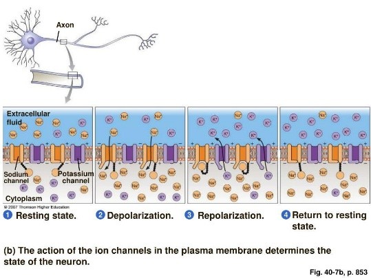
Within Phase 0 — Resting potential and slow depolarisation. What is happening within a neuronal cell?
The neuron starts near its resting membrane potential. Ion gradients are maintained by transporters and pumps, while leak channels influence the voltage.
Hyperpolarisation-activated cyclic nucleotide-gated (HCN) channels can contribute to slow depolarisation in certain neurons and pacemaker cells.
Voltage gated sodium channels are CLOSED
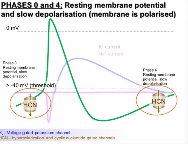
Phase 1 — Fast depolarisation. What is happening within a neuronal cell?
Once threshold is reached, voltage-gated sodium (Na⁺) channels open. Na⁺ enters the cell down its electrochemical gradient, causing the membrane potential to rise rapidly and may make the inside positive. RAPID DEPOLARISATION.

Phase 2 — Repolarisation. What is happening within a neuronal cell?
Sodium channels inactivated, reducing Na⁺ entry.
While voltage-gated potassium (K⁺) channels open. K⁺ leaves the cell, making the inside more negative again. REPOLARISATION.

Phase 3 — Afterhyperpolarisation. What is happening within a neuronal cell?
Some K⁺ channels close slowly, so K⁺ continues to leave the cell.
The membrane potential can temporarily become more negative than the resting potential before returning towards baseline.

Phase 4 — Return to resting conditions. What is happening within a neuronal cell?
The membrane returns towards its resting potential as ion-channel activity and membrane conductances return towards baseline.
The Na⁺/K⁺ ATPase maintains the Na⁺ and K⁺ concentration gradients over time.

What is the refractory period?
Period during which an excitable cell (neuron or muscular cell) is incapable to response to the further stimulation. Recharge of the membrane capacitator (Na+ channels cannot immediately reopen).
What is the difference between absolute and relative refractory period?
Absolute refractory period phase when excitable cell does not respond for stimulation no matter how great stimulus is applied. This is mainly because many voltage-gated Na⁺ channels are already open or inactivated and cannot immediately reopen.
Relative refractory period phase when excitable cell could respond for the stimulation that is greater than normal stimulus. Some Na⁺ channels have recovered, but the membrane may still be hyperpolarised and K⁺ conductance may remain elevated.
Why are refractory periods important? (3)
They limit how rapidly a neuron can fire.
They help prevent immediate re-excitation of the same membrane region.
They support forward propagation of action potentials along an axon

Fill in the gaps

Why do action potentials differ between tissues?
Different tissues use different ion channels, so the shape and duration of their action potentials differ.


Fill in

How does the action potential in cardiac cells differ? What Ions are involved?
Cardiac ventricular action potentials last much longer because they contain a plateau phase. The longer action potential and refractory period help prevent sustained, repeated contractions that would interfere with the heart's pumping cycle.
DEPOLARISATION: Fast Na⁺ entry causes rapid depolarisation. Ca²⁺ entry through L-type calcium channels helps maintain the plateau.
REPOLARISATION: K⁺ currents contribute to repolarisation.
Why is the plateau seen in cardiac action potentials so crucial?
The ventricular action potential lasts much longer than a neuronal action potential because of its calcium-dependent plateau. This prolonged electrical activity supports coordinated contraction and gives the heart muscle a long refractory period.

Which cells have Hyperpolarization and Cyclic Nucleotide (HCN) sensitive channels?
Cells that can produce spontaneous action potentials e.g. cardiac cells.
They are found in the sinoatrial node and play a key role in action potential initiation AND repolarisation.

What does the ECG record?
Electrical activity from the heart as a whole. A ventricular action potential, by contrast, measures the voltage change across the membrane of an individual cardiac cell.
How does the action potential in smooth muscle cells differ? What Ions are involved?
E.g. Blood vessels, GI tract, respiratory system.
Its action potentials can depend strongly on Ca²⁺ entry through voltage-gated calcium channels. Calcium both contributes to the electrical signal and helps initiate contraction.

How does the action potential in skeletal muscle cells differ? What Ions are involved?
Skeletal muscle action potentials are also brief and depend mainly on voltage-gated Na⁺ channels for their rising phase.
The electrical signal triggers calcium release from the sarcoplasmic reticulum, which enables muscle contraction.
