laboratory testing in hematology
 Call Kai
 Learn
Learn Practice Test
 Spaced Repetition
 Match
Match Flashcards
 Knowt Play
1/65

Earn XP
Description and Tags
status: unfinished
Name | Mastery | Learn | Test | Matching | Spaced | Call with Kai | Chat |
|---|
No analytics yet
Send a link to your students to track their progress
66 Terms
RBC reference range for males
4.6 – 6.2 x10¹²/L or x 106/mm3 or /μL (millions)
RBC reference range for females
4.2 – 5.4 x10¹²/L or x 106/mm3 or /μL (millions)
WBC counting area for neubauer hemacytometer
4 corners counting area: #’s 1, 3, 7, 9. contains 16 smaller squares. the remaining large squares (2, 4, 6, 8) contain 20 squares.

RBC & platelet counting area for neubauer hemacytometer
center large square: #5 counting area. contains 25 squares and 16 smaller squares

RBC thoma pipette (for RBC counts)
101 total units - 1 unit in stem = 100 units.
blood to 0.5 units + fluid to 101 units = 1:200.
dilution is saline

RBC thoma pipette (for platelet counts)
blood to 1.0 units + fluid to 101 units = 1:100.
dilution in 1% oxalate

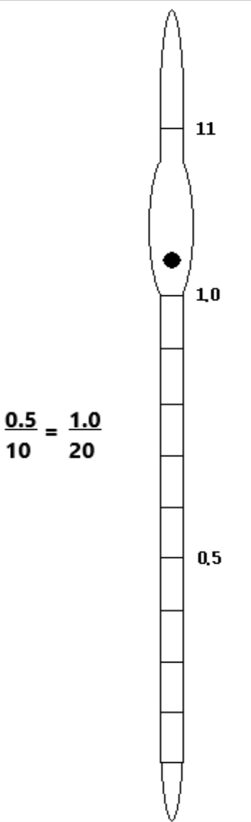
WBC thoma pipette
11 total units - 1 unit in stem = 10 total units.
blood to 0.5 units + fluid to 11 units = 1:20.
diluent is 2% acetic acid
prefabricated dilution systems
RBC count (ery-tic) = 1:200 dilution in NaCl (saline).
WBC count (leuko-tik) = 1:20 dilution in 2% acetic acid (methylene blue).
plt count (thrombo-tik): 1:100 dilution in 1% ammonium oxalate.
hemacytometer calculations
average # counted per side x dilution x depth x or / depending what area was counted.
if more than 1mm2 then divide, if less then multiply. goal is to reach 1mm2. report as # x 1012/L or x 106/uL or x 106/mm3 (millions)
typical RBC calculation (ery-tic)
average # x 200 × 10 × 5 = # cells/mm3 or µL. 200 is standard dilution, 10 used to compensate for depth, 5 converts area (25 total squares/5 squares counted = 5). report as X.Xx1012/L. 5 small squares counted within large center square.
shortcut = average # x 10,000 = # cells/ mm3 or uL (millions).
typical WBC calculation (leuko-tik)
average # x 20 × 10/4 = #cells/mm3 or µL. 20 is dilution, 10 used to compensate for depth, 4 converts 4 large corner squares. report as X.Xx109/L.
shortcut = average # x 50 = # cells/mm3 or µL (thousands).
parts of coulter/sysmex/cell-dyne
cell bath/chamber = container with saline & cells. + electrode in glass tune, - electrode in diluted blood, both connected to power supply. aperture is small hole in glass tube that allows fluid communication b/t electrodes. saline maintains intact cells & electrical conductivity b/t electrodes through aperture. data of average cell # and size are fed into computer that makes more calculations: measures count (#) & MCV (size); calculates HCT, MCH, & MCHC. automatic dilution of blood sample before counting (dilutor). automated differential: electrical impedance, electromagnetic, laser light scatter.

what does the current do in coulter/sysmex/cell-dyne?
small stable current measured b/t electrodes. vacuum draws diluted cells toward + electrode. as nonconductive cell enters aperture, conductive fluid is displaced & detector records a current drop. each current drop is recorded as a cell & contributes to total cell count. the larger the cell, the larger the fluid displacement & the larger the amplitude of current drop. this is a measure of cell size (MCV)
coincidence error in coulter/sysmex/cell-dyne
counting 2 or more cells as 1 when more than 1 cell pass through aperture simultaneously. either mathematically corrected (coulter) or reversed prior to entering aperture using a stream of fluid forcing cells into single file called hydrodynamic focusing (cell dyne & sysmex)
hgb reference range for males
14 - 18 g/dL
hgb reference range for females
12 – 16 g/dL
hgb reference ranges for anemia
slight = 10-12g/dL
moderate = 8-10g/dL
severe = <8g/dL
copper sulfate
method of hgb quantitation. add one drop of blood to appropriate container. if Hb is denser than solution, drop will sink in 15 sec. used by red cross to screen blood donors.
male: 1.055 sp. gr. = 13.5g/dL Hb or greater.
female: 1.053 sp. gr. = 12.5 g/dL Hb or greater.
cyanmethemoglobin (drabkin’s reagent)
method of hgb quantitation. detergent (saponin) hemolyses RBCs, releasing Hb.
Hb + potassium ferricyanide → (ox) methemoglobin + potassium cyanide → cyanmethemoglobin → spec at 540nm.
common before 2000s, not used anymore b/c cyanide is toxic.
azidemethemoglobin method
method of hgb quantitation. sodium deoxycholate is lysing agent. sodium nitrite → (ox) methemoglobin. sodium azide → azidemethemoglobin → 570nm. most common method after 2000s, azide less toxic than cyanide.
reflectance (point of care)
method of hgb quantitation. handheld instrument with strips that hemolyze RBCs, oxidize Hb, & Hb stains strip. measures Hb as light reflects off the color of Hb.
use of standards for hgb measurement
not necessary for copper sulfate or reflectance technology, used for cyan or azidemethemoglobin. plot standard curve: x axis = standard hb concentrations, y axis = absorbance reading. determine unknown by reading absorbance from standard curve

hemoglobin electrophoresis
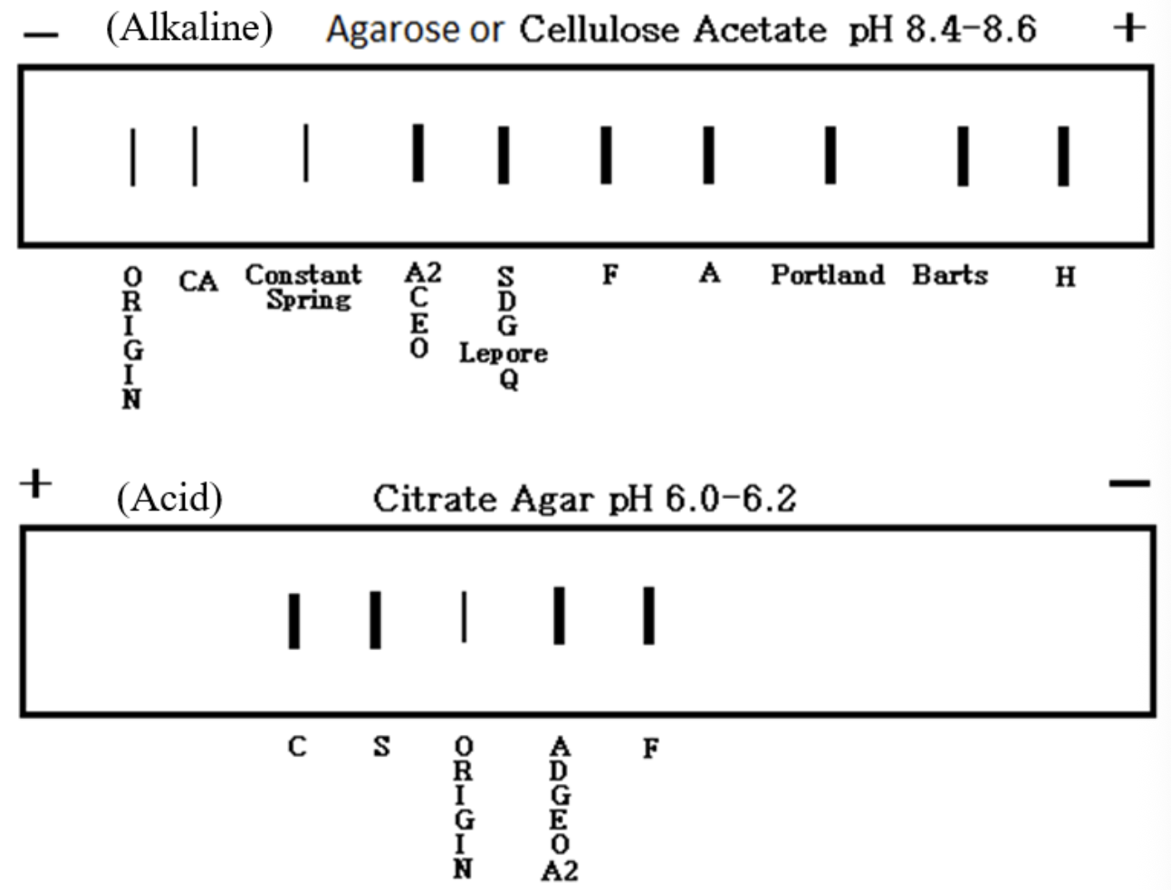
determine types of Hb (A, A2 F, S, C) present in a blood sample. any charged particle or molecule will move in an electric field toward pole of opposite charge at a rate dependent on the net charge of the particle or molecule and its size & shape. Hb molecules have the same shape & similar size but different charges
hemoglobin electrophoresis steps/parts
detergent used to free Hb from RBC. wells hold buffer of precise pH, ± electrodes immersed in each well & connected to power. agar w/ tiny pores molecules will navigate through to migrate toward pole of opposite charge. wicks: filter paper partially immersed in buffer, placed on each end of gel, fluid connection b/t electrodes & gel to complete circuit. glass cover placed over gel. prepare cellulose acetate/agarose plates: label & wet plates, load patients & controls into holes cut in a row at one end of gel. electrophorese at 550 volts for 15 min.

how is hemoglobin electrophoresis read?
staining bands (ponceau S or acid blue) allows Hb bands to be visualized to determine which types are present. scan plate on a densitometer that is used to measure density of stained protein bands and quantitate amount of protein present. CA = carbonic anhydrase, enzyme found in high concentrations within all RBCs
alkaline Hb electrophoresis vs acid Hb electrophoresis
alkaline uses cellulose acetate or agarose. pH = 8.4 - 8.6. normal adult = Hgb A, A2 and F. all others are abnormal. HbC=Crawl, HbS=Slow, HbF=Fast, HbA=Accelerated.
acid uses citrate agar. pH = 6.0 - 6.2
hematocrit reference range for males
42-52%
hematocrit reference range for females
37-47%
microhematocrit
manual (measured) method to measure HCT. HCT determined by spinning blood-filled capillary tube in centrifuge.

automated (calculated) method to measure HCT
electrical impedance cell counter instrument: MCV x RBC count/10 = HCT
RBC indices
values calculated from RBC count, Hgb, & HCT, which describe RBC size & Hgb content mathematically. MCV, MCH, MCHC. MCV & MCHC primarily used to determine classification of anemia
mean corpuscular volume (MCV)
average volume or amount of space occupied by the red blood cell. expressed in femtoliters (fL=10‑15 L). Hct (%) x 10 fL/RBC (x 1012) = MCV fL.
reference range: 80‑100 fL: < 80 fL = microcytic RBCs, >100 fL = macrocytic RBCs.
used to determine average RBC size & classify anemias.
mean corpuscular hemoglobin (MCH)
average weight of hemoglobin in the red blood cell. expressed in picograms (10‑12 g). [Hb (g/dL) x 10 pg]/RBC (x 1012) = MCH pg.
reference range: 27‑31 pg: < 27 pg – micro or normocytic/hypo or normochromic RBCs, > 31 pg ‑ macrocytic RBCs or occasionally spherocytes.
used to determine the average amount of Hb by weight in the RBCs & classify anemias.
mean corpuscular hemoglobin concentration (MCHC)
average concentration of hemoglobin in a RBC. expressed in grams per deciliter (g/dL). Hb (g/dL) x 100/Hct (%) = MCHC g/dL.
reference range: 32‑36 g/dL: < 32 g/dL ‑ hypochromic, > 36 g/dL – hyperchromic (occasionally spherocytosis).
used to determine the concentration of Hgb in RBC & classify anemias
reticulocyte count
used to determine the % of RBCs that are immature by detecting cytoplasmic RNA. uses supravital staining: new methylene blue. count the # retics/1000 RBC. report as % retics by dividing by 10
absolute retic count (actual # of retics/unit volume not % of total) = % retic x RBC count/100. done by automated instrument based on fluorescent staining of RNA.
reference range: 60,000/mm3 μL or 60 x 109/L, 23 – 80 x 109/L.
reticulocyte reference range for adults
0.5 – 1.5%
reticulocyte reference range for newborns
2.5 – 6.0% for 2 weeks after birth
corrected retic count
used to adjust a falsely elevated retic % due to a low hematocrit. when a standard number of retics (bone marrow output) are placed in a low number of circulating RBCs (low crit), the % of retics (retic count) is a falsely elevated representation of bone marrow output.
retic (%) x Patients Hct/Normal Hct.
male: 47%, Female: 42%
reticulocyte production index (RPI)
used to correct for both a low hematocrit (as the corrected retic count) and shift reticulocytes which are very young retics. when bone marrow in under pressure to release RBCs quickly, it can release young retics that are larger and contain excess reticulum (shift retics), which require longer to differentiate into mature RBCs. this slowed maturation results in an accumulation of retics in circulation, which also gives a falsely elevated retic count. must see shift retics.
corrected Retc (%)/maturation time in days (from table).
reticulocyte maturation index (RMI)
quantitation of cytoplasmic RNA in reticulocytes, much more accurate representation of retic age and thus actual bone marrow output.
modern Instrument = absolute count with RMI
erythrocyte sedimentation rate (ESR) mechanism
in abnormal conditions where RBC’s form rouleaux, the ESR is ↑ in any inflammatory condition. an increase in plasma proteins is produced & bind to RBC surface, neutralize sialic acid residues, and remove natural repulsion of RBCs. RBCs stack like coins (rouleaux), increasing density and sedimentation rate. (roll of quarters).
in abnormal conditions where plasma viscosity is ↑, the ESR is ↓. increased viscosity retards RBC sedimentation.
wintrobe (landsberg) ESR method
fill tubes to the 0 mark (100 mm long). let stand upright for 1 hr. read in mm at the RBC/plasma interface.
reference ranges:
male: 0‑9 mm/hr
female: 0‑15 mm/hr
westergren (modified for EDTA) ESR method
dilute 1mL [2mL] (4mL) blood with 0.25mL [.5mL] [1mL) saline or citrate (4:1). fill tube to 0 mark (200 mm long). read in mm at RBC/plasma interface.
normal:
male: 0‑15 mm/hr
female: 0‑20 mm/hr
hemoglobin solubility test
used to detect sickle Hgb or few other Hb variants. saponin, a detergent, lyses the RBCs releasing the hemoglobin into solution. dithionite or sodium hydrosulfite reduces the hemoglobin (Fe+3>>Fe+2) converting oxyhemoglobin to deoxyhemoglobin. reduced Hb A, and most other normal and abnormal hemoglobins, are soluble in the concentrated inorganic salts but Hb S and Hb CHarlem & few rare abnormal hemoglobins are insoluble creating a turbid solution.
hemoglobin solubility test method & interpretation
add 50μL [20μL] of blood to 4mL [2mL] of saponin/dithionite/salt solution. invert and allow to incubate for 5 minutes at RT. invert and read for turbidity against a lined card.
clear solution = normal Hb A. turbid solution = abnormal Hb S or others.
positive test can NOT determine zygosity. follow-up testing with Hb electrophoresis testing is required to identify abnormal Hb
![<p><span style="background-color: transparent;">add 50</span><span style="background-color: transparent; font-family: "Noto Sans Symbols", sans-serif;">μ</span><span style="background-color: transparent;">L [</span><span style="background-color: transparent; font-family: "Noto Sans Symbols", sans-serif;">20μ</span><span style="background-color: transparent;">L] of blood to 4mL [2mL] of saponin/dithionite/salt solution. invert and allow to incubate for 5 minutes at RT. invert and read for turbidity against a lined card.</span></p><p><span style="background-color: transparent;">clear solution = normal Hb A. turbid solution = abnormal Hb S or others.</span></p><p><span style="background-color: transparent;">positive test can NOT determine zygosity. follow-up testing with Hb electrophoresis testing is required to identify abnormal Hb</span></p>](https://assets.knowt.com/user-attachments/10e34580-dc8c-479c-abe7-998bd4f561eb.png)
osmotic fragility test
used to validate the presence of spherocytes due to their increased osmotic fragility. a method of evaluating RBC’s susceptibility to hypotonic damage. dilute NaCl: 0.85% - 0.15%. add 50 μL of patient blood or control. let stand at room temperature for ½ hour or 24 hrs at 370 to increase sensitivity. centrifuge and evaluate degree of hemolysis.
RR: 0.45% - 0.35%, salt concentration when normal RBCs lyse.
visual method of measurement for osmotic fragility test
note tube that shows initial hemolysis, note tube that shows complete hemolysis

spectrophotometric method of measurement for osmotic fragility test
measure the supernatant in each tube at 540nm.
calculate % hemolysis: [A (test) – A (0%)] x 100 [A (100%) – A (0%)]
plot curve.
normal fragility = 0.45 - 0.35.
increased fragility (spherocytes): 0.8 - 0.55.
decreased fragility (target cells): 0.45 - 0.25.
![<p><span style="background-color: transparent;">measure the supernatant in each tube at 540nm. </span></p><p><span style="background-color: transparent;">calculate % hemolysis: [<u><sup>A</sup> (test) – <sup>A</sup> (0%)</u>] x 100 [<sup>A</sup> (100%) – <sup>A </sup>(0%)]</span></p><p><span style="background-color: transparent;">plot curve.</span></p><p><span style="background-color: transparent;">normal fragility = 0.45 - 0.35.</span></p><p><span style="background-color: transparent;">increased fragility (spherocytes): 0.8 - 0.55.</span></p><p><span style="background-color: transparent;">decreased fragility (target cells): 0.45 - 0.25.</span></p><p></p>](https://assets.knowt.com/user-attachments/def52a7c-f31e-44ed-899d-bee296754339.png)
kleihauer/betke stain
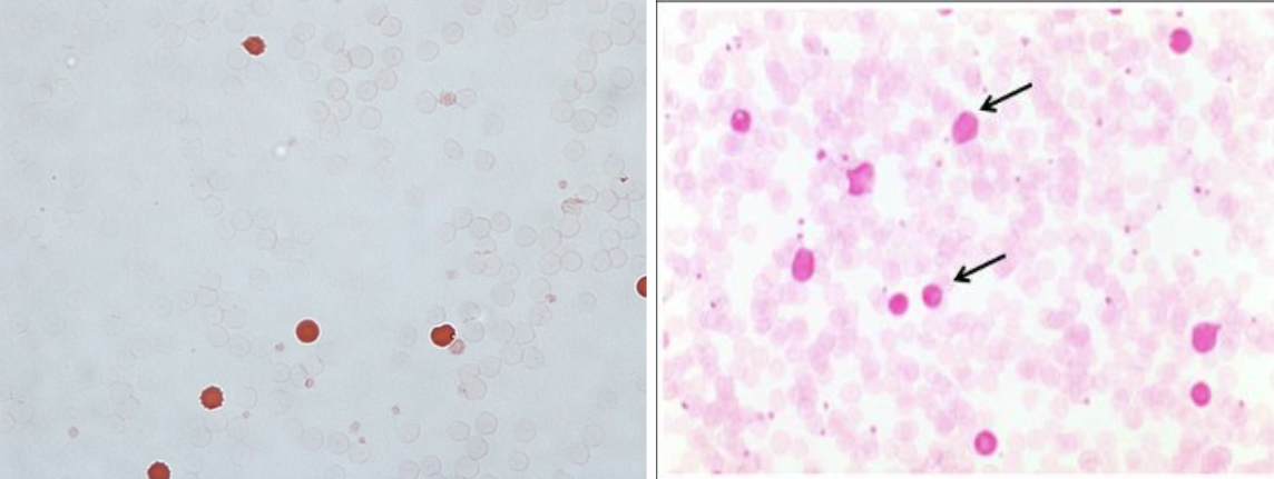
used to look for fetal cells in maternal blood (HDN), high HbF in β-thalassemias, evaluate response to hydroxuria in sickle cell patients. HbF inside RBCs will resist elution by weak acid (citrate). observe for fetal cells that counterstained due to retention of Hb F vs adult cells that did not counterstain due to elution of Hb A.
acidified serum test (ham’s test)
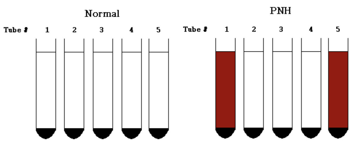
used to diagnose PNH due to the RBCs increased sensitivity to an acid environment. complement will affix to RBCs in a slightly acidic pH from patients with paroxysmal nocturnal hemoglobinuria & will become activated by the alternative pathway and lyse the RBCs. wash patient and control RBCs and make 50% suspension, prepare five tubes, 3 components (RBC – complement – acid), incubate tubes at 37º C for 1 hr, centrifuge & examine for hemolysis.
positive for PNH if tubes 1 and 5 are positive and tubes 2-4 are negative.
sugar water test (sucrose hemolysis)
used to diagnose PNH due to the RBCs increased sensitivity to hypotonicity. sucrose provides a low ionic strength environment that allows complement to bind, which will lyse PNH cells. same method as ham’s but incubate for 30 min.
PNH = lysis in tube 1 only.
invalid = lysis in tube 2.
normal = no lysis.
flow cytometry Analysis: absence of most important markers - CD16, CD55, and CD59

WBC reference range
4.5 – 11.0 x10⁹/L or 4,500 – 11,000/ mm3 or x 103/μL
plt estimate for manual diff
count average # plts/field x 20,000. should agree within 10% of plt count
relative count
the % of particular WBC type with respect to all others
absolute count
total # of a particular WBC x 109/L (or x 103/mm3 or /µL).
WBC count = 9.4 x 10⁹/L or x 103/mm3 or µL and Lymphs = 32%.
absolute estimate = 32 x 9,400 ÷ 100 = 3 x 10⁹/L or 3,008/mm3 or µL.
thrombocyte reference range
150-450 x109/L or 150-450,000/mm3 or µL
manual methods to measure plt count
rees-ecker: stains blue, 1:100 dilution.
brecker-cronkite: ammonium oxalate, 1:100 dilution, phase contrast.
thick smears
spread one drop of blood the size of dime on slide, let dry for 30 min. stain with 4% Giemsa diluted in water for 30 minutes. used to screen for malaria. hemolyze RBCs and release parasites
thin smears
make very thin feathered smear, let dry for 10 min. fix in methanol for 5 seconds & stain with Wright’s stain. used to speciate already identified organisms (malarial parasites).
bone marrow examination: aspirate
liquid marrow aspirated through needle into a syringe. evaluate for spicules to determine specimen is BM. crush/squash, T- prep slides, made by rubbing drop of aspirate between slides. slides made like a peripheral blood smear. slides are stained
bone marrow examination: biopsy
solid marrow that sicks in needle. touch prints are made by blotting specimen several places on a few slides. slides are stained, formalin fixed
bone marrow evaulation
normal ratio of fat to cells = 50:50 or 1:1. 5-10 megakaryocytes/10x field. look for M:E ratio (myeloid:erythroid ratio)
normal M:E ratio = 3:1 to 4:1 (adults), 6:1 (newborns).
increased ratio = 5:1 or greater = ↑ M or ↓ E.
decreased ratio = 2:1 or smaller = ↓ M or ↑ E
flow cytometry uses what 3 primary mechanisms?
laser light scattering, fluorescence detection, cell sorting
laser light scattering
used to count cells. differentiates between cell size & granule contents. cells flow by laser single file. hydrodynamic focusing: laser shines on cells, cell deflects laser to detector
fluorescence detection
monoclonal antibodies are fluorescently labeled. antibodies are specific to cell surface markers (CD). cells are fluorescently labeled & flow past UV lamp. fluorescence is detected. used to ID cell lineage & determine cancer type (leukemias)
cell sorting
labeled cells are given a charge. charged cells are deflected toward an oppositely charged plate. cells are separated based on charge which is based on antibody binding. cells can be collected in a tube and further studied.