A&P Quiz 3
 Call Kai
 Learn
 Practice Test
 Spaced Repetition
 Match
 Flashcards
 Knowt Play
1/83

Earn XP
Description and Tags
ch. 12, 13, 14, 15?
84 Terms
Skeletal Muscle
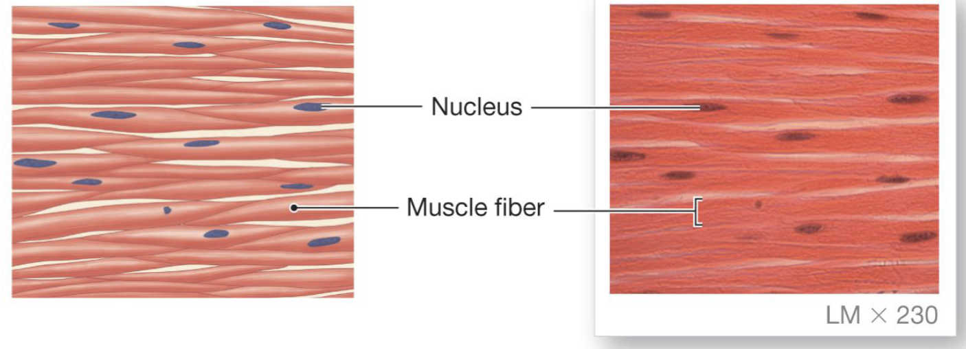
Support voluntary movement. Fibers are large, multinucleate cells that appear striped or striated under microscope. Usually attached to bones by tendons

Cardiac Muscle
Fibers are striated but smaller, branched and uninucleate. Cells are joined in series by junctions called intercalated disks. Cells have one nucleus.

Smooth Muscle
Fibers are small and lack striations. Cells have one nucleus. Muscle in hollow organs and blood vessels
Flexor
Brings bones together
Extensor
Moves bones away
Antagonistic Muscles
Move bones in opposite directions. Flexor-extensor pairs
Muscles fibers/cells
Compose skeletal muscles. Long and cylindrical, fused cells with many nuclei
Satellite cells
Type of stem cells. Differentiate into muscle for growth or repair
Fascicles
Bundled muscle fibers. surrounded by connective tissue sheath.

Connective tissue (in muscles)
Continuous with connective tissue sheath. Holds muscle to bone with tendon
Muscle equivalent for muscle cell
Muscle fiber
Muscle equivalent for cell membrane
Sarcolemma
Muscle equivalent for cytoplasm
Sarcoplasm
Muscle equivalent for modified endoplasmic reticulum
Sarcoplasmic reticulum
Myofibrils
Bundles of contractile and elastic proteins responsible for muscle contraction. Contain actin and myosin
Myosin
forms thick filament of the myofibril that convert chemical bond energy of ATP into motion
Transverse Tubules (T-Tubules)
Extensions of the cell membrane (sarcolemma) that associate with the ends (terminal cisternae) of the sarcoplasmic reticulum

Sarcomere during contraction (image)

What initiates an action potential in muscle fiber?
Acetylcholine (ACh)
Steps of excitation-contraction coupling
Acetylcholine is released from the somatic motor neuron
Acetylcholine initiates an action potential in the muscle fiber
The muscle action potential triggers calcium release from the sarcoplasmic reticulum
Calcium combines with troponin to initiate contraction
Latent period
Short delay between the muscle action potential and beginning of muscle tension development
Anaerobic glycolysis
Produces lactate and acid. Quick, no oxygen required, small amount of energy released
Aerobic Respiration
Citric acid cycle and electron transport chain. Slow, oxygen required, large amount of energy released
Slow twitch fibers
Rely primarily on oxidative phosphorylation. Fatigue-resistant
Fast twitch fibers
Develop tension faster, easily fatigued
Summation
Stronger contraction when the muscle does not relax completely between action potentials
Tetanus
maximal contraction
Isotonic Contractions
contraction where the muscle changes its length while maintaining a constant level of tension or load (bicep curl)
Isometric Contractions
create force without movement
Autonomic Reflexes
Unconscious, involuntary motor response that regulates internal organs, smooth muscle and cardiac muscle
Proprioceptors
Sensory receptors located in skeletal muscle, joint capsules and ligaments. Input signals go to the CNS through sensory neurons
Myotatic Unit
Collection of pathways controlling a single joint
Reciprocal Inhibition
Antagonistic muscles must relax as the prime mover muscles contract
Flexion reflexes
Pulls limbs away from painful stimuli
Reflex movement
least complex, integrated at the spinal cord or brain
Postural reflexes
Help us maintain body position
Voluntary movements
Most complex, integrated in cerebral cortex
Rhythmic movements
intermediate complexity, integrated in spinal cord with higher center input required
Poiseuille’s Law
Resistance is proportional to length and viscosity. Inversely proportional to radius

Pathway of deoxygenated blood
vena cava → right atrium → right ventricle → pulmonary trunk

Pathway of oxygenated blood
pulmonary veins → left atrium → left ventricle → aorta
Atrioventricular valves
Between atria and ventricles (Tricuspid and bicuspid valve)
Semilunar valves
Between ventricles and arteries (aortic and pulmonary valve)
Contractile Cells
Striated fibers organized intro sarcomeres
Autorhythmic cells (Pacemaker cells)
Signal for contraction, smaller and fewer contractile fibers, don’t have organized sarcomeres
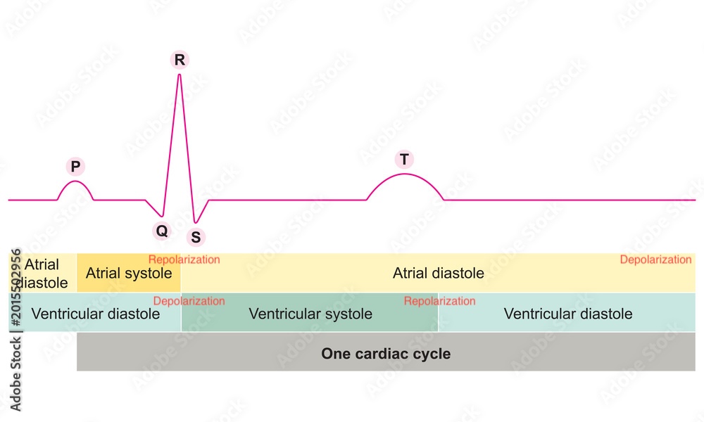
Electrocardiogram
Show the summed electrical activity generated by all cells of the heart
P wave
Depolarization of the atria

QRS complex
wave of ventricular depolarization, and atrial repolarization

T Wave
Repolarization of the ventricle

Tachycardia
Faster than normal heart rate
Bradycardia
Slower than normal heart rate
Diastole
Cardiac muscle relaxes
Systole
Cardiac muscle contracts
Efferent Neurons
Carry commands from CNS to the muscles and glands of the body
Somatic Motor Neurons
Control skeletal muscles, mostly voluntary
Autonomic Neurons
Controls smooth muscle, cardiac muscle, many glands and adipose tissue. Mostly involuntary. Divide into sympathetic (fight-or-flight) and parasympathetic (rest-and-digest)
Neuroeffector junction
Synapse between postganglionic autonomic neuron and target cell
Neuromuscular Junction
Synapse between somatic motor neuron and skeletal muscle fiber
Troponin
Complex of three regulatory proteins associated with tropomyosin that binds calcium ions to help initiate muscle contraction (skeletal and cardiac)
Tropomyosin
Regulatory protein that lies along actin filaments and blocks the myosin binding sites, helping control when muscle contraction can occur
Golgi Tendon Organs
Found in junction of tendons and muscle fibers. Composed of free nerve endings that wind between collagen fibers inside connective tissue capsule
P-R segment
Atrioventricular (AV) nodal delay - slows down electrical impulses as they travel from the atria to the ventricles
T-P segment
ventricular and atrial relaxation

How to find heart rate from ECG
Time between two P waves or two Q waves
Parasympathetic Control
Decreases heart rate, K+ permeability increases
Sympathetic Control
Increases heart rate, Na and Ca permeability increases
Arteries
Act as a pressure reservoir, thick layers of vascular smooth muscles, lots of elastic and fibrous connective tissue
Arterioles
Site of variable resistance, part of the microcirculation, less elastic and more muscular
Metarterioles
Branches of arterioles, partial smooth muscle layer, pre-capillary sphincter's open and close to direct blood flow to capillaries or venous circulation
Pericytes
Contractile cells associated with capillaries, contribute to capillary impermeability, secrete paracrine factors that promote vascular growth and differentiation
Venules
Receive blood from capillaries, thin exchange epithelium, little connective tissue, convergent pattern of flow
Veins
Take blood back to the heart, act as a volume reservoir, thin walls of vascular smooth muscles, contain one-way valves
What causes a striated appearance in muscles?
Organized myofibrils made of actin and myosin arranged into repeating sarcomeres
Contractility
The ability of muscle tissue to shorten fibers, generating movement
Extensibillity
The capacity of muscle tissue to be stretched without damage
Elasticity
The property of muscle tissue to return to its original shape after contraction or extension
I band
Region of skeletal muscle sarcomere that contains only thin filaments

Z line/disk
Set of sarcomere proteins located at the ends of a sarcomere, serving as attachment sites for actin filaments, help define boundaries of each sarcomere

H Zone
Region of a sarcomere that contains only thick filaments and no overlap with thin filaments

A band
Contains the entire length of the thick myosin filaments, plus the overlapping portions of the thin actin filaments

M line
Structural feature of skeletal muscle sarcomeres that divides the A band in half and serves as an attachment site for thick (myosin) filaments

Thick filaments are made of
Myosin
Thin filaments are made of
Actin
Phases of an ECG (depolarization vs. repolarization, diasytole vs. systole)