SECTION 02: CONDUCTING PORTION
 Call Kai
 Learn
 Practice Test
 Spaced Repetition
 Match
 Flashcards
 Knowt Play
1/28

There's no tags or description
Looks like no tags are added yet.
29 Terms
What are the main functions of the conducting portion of the respiratory system?
The conducting portion transports air to and from the lungs without gas exchange. Its main functions are:
Air conditioning: Warms, humidifies, and filters inhaled air.
Defense: Traps particles and microorganisms.
Each structure has unique gross (visible) and microscopic features that support these functions.
What are the paranasal sinuses, and what are their functions?
The paranasal sinuses are air-filled spaces in the skull bones that communicate with the nasal cavity.
Functions: Warm and humidify air, defend against pathogens, and act as resonance chambers for speech.
4 paired sinuses (8 total): Frontal (2), maxillary (2), ethmoid (2), and sphenoid (2).
Lining: Respiratory tract epithelium, which helps with air conditioning and defense.

What are the functions of the nose and nasal cavity?
The nose and nasal cavity act as the first line of defense in the respiratory system.
Coarse hairs and mucus trap pathogens and debris.
Air enters through the nasal vestibules (nostril openings).
Air passes into the nasal cavity, where it is conditioned (warmed, humidified, and filtered).
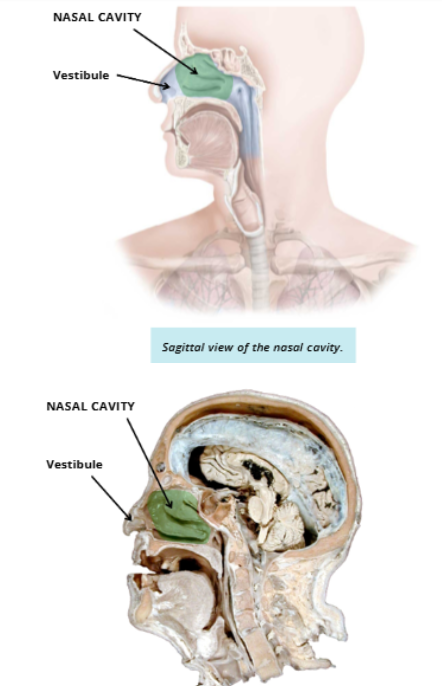
What are the six boundaries of the nasal cavity?
Roof:
Floor:
Medial Wall:
Lateral Wall:
Anterior: .
Posterior:
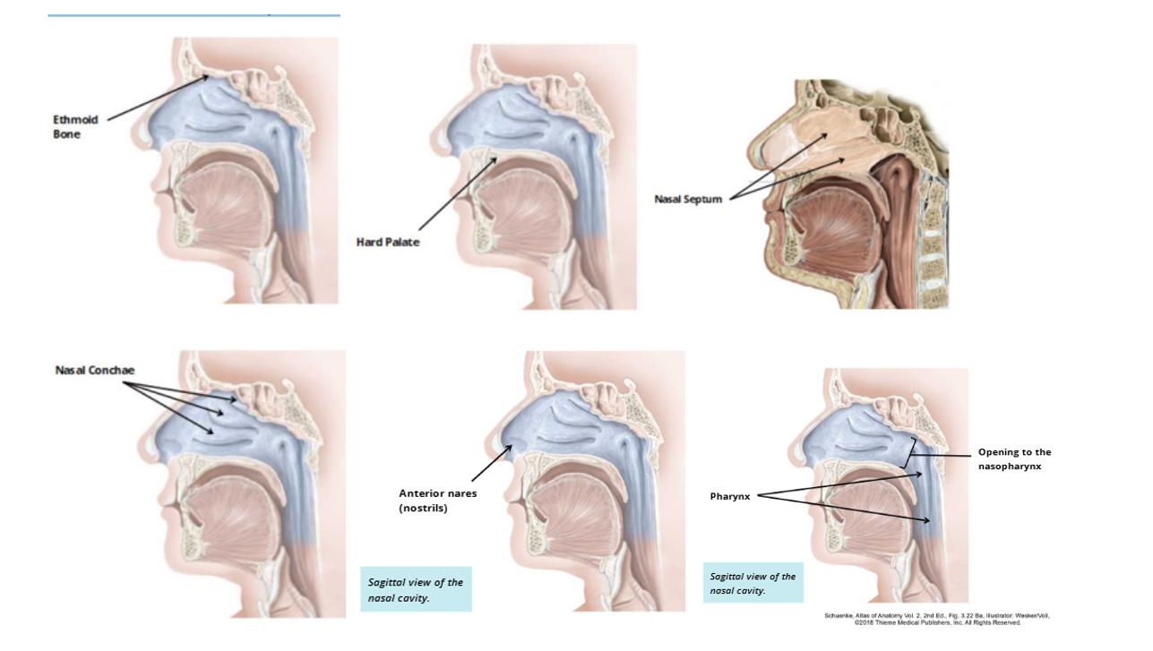
What are the six boundaries of the nasal cavity?
Roof?
The roof of the nasal cavity is a bone in the skull called the ethmoid.
What are the six boundaries of the nasal cavity?
Floor?
The floor is composed of the hard palate (roof of the mouth)
What are the six boundaries of the nasal cavity?
Medial Wall
Medial Wall: The medial wall makes up the nasal septum. The septum is composed of the vertical bones in the skull, and separates the two halves of the nasal cavity.
What are the six boundaries of the nasal cavity?Lateral Wall
Lateral Wall: The lateral walls of the nasal cavity contain structures known as the nasal conchae. These structures create turbulence in the air as it passes through the cavity, allowing for conditioning and catching debris.
What are the six boundaries of the nasal cavity?
Anterior
Anterior: The anterior border of the cavity is made by the nares, which is the opening between the nose and nasal cavity.
What are the six boundaries of the nasal cavity?Posterior
Posterior: The posterior border is the opening to the nasopharynx (choanae), where the nasal cavity connects to the pharynx.
What type of epithelium lines most of the nasal cavity, and why?
Most of the nasal cavity is lined with respiratory tract epithelium (RTE), which:
Protects against airborne debris and microorganisms.
Conditions inhaled air by warming, humidifying, and filtering it.

What is the olfactory epithelium, and where is it located?
The olfactory epithelium lines the roof of the nasal cavity and contains sensory receptors for smell.
Structure: Pseudostratified ciliated columnar epithelium and bipolar olfactory receptor neurons.
Function: Detects airborne molecules responsible for the sense of smell.

What is the pharynx, and what are its functions?
The pharynx is a muscular tube shared by the respiratory and digestive systems.
Respiratory: Connects the nasal cavity to the larynx (air passage).
Digestive: Connects the oral cavity to the esophagus (food passage).

What are the three regions of the pharynx, and what are their functions?
Nasopharynx: Most superior region; passageway for air.
Oropharynx: Middle region; carries air from the nasopharynx and food from the oral cavity into the laryngopharynx.(part of respiratory and digestive)
Laryngopharynx: Most inferior region; directs air toward the respiratory system and food toward the digestive system. (part of respiratory and digestive)

What type of epithelium lines each region of the pharynx, and why?
Nasopharynx: Mostly respiratory tract epithelium (RTE) because its main function is conducting air.
Oropharynx and laryngopharynx: Stratified squamous epithelium for durability and protection against friction when swallowing food.

What is the larynx, and what are its main functions?
The larynx (voice box) is an organ made of cartilages, ligaments, and muscles that produces sound.
Sound production: Produces sound through vocal cord vibrations.
Location: Anterior to the esophagus, connecting the pharynx to the trachea.
Protection: Prevents food from entering the trachea during swallowing.

What is the function of the cartilage in the Larynx?
The larynx is made up of many cartilages that serve to protect, attach, and aid in the function of the vocal cords.
What is the epiglottis, and what is its function?
A large, spoon-shaped elastic cartilage that folds downward during swallowing to cover the airway opening, preventing food from entering the trachea.
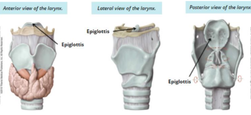
What is the thyroid cartilage, and what is its function?
A shield-shaped hyaline cartilage that provides attachment points for the vocal cords and many muscles of the larynx.

What is the cricoid cartilage, and what is its function?
a complete ring of hyaline cartilage. It is narrow anteriorly and broad posteriorly. This structure functions as an attachment for muscles and the vocal cords.

What are the vocal cords, and how do they function during breathing, speaking, and swallowing?
The vocal cords are small ligaments attached to laryngeal cartilages that vibrate to produce sound when air passes through. Changing their tension creates different sounds.
True vocal cords: Produce sound.
False vocal cords: Protect the true vocal cords.

How do vocal cords function during breathing, speaking, and swallowing?
Breathing: Vocal cords open to allow airflow.
Speaking/Singing: Vocal cords come together and vibrate as air passes through, producing sound.
Swallowing: Epiglottis covers the airway entrance to prevent food and liquids from entering.
Coughing: Removes liquids or particles from the airway.
Voice production: Vibrations create sound that is shaped into speech or singing.
What is the trachea (windpipe), and what is its function?
The trachea is an airway that:
Extends: From the larynx to approximately T4/T5.
Carina: Junction where the trachea splits into the two main bronchi.
Function: Conducts air to the lungs.
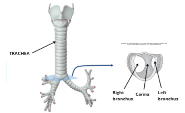
What is the structure of the trachea, and what is its function?
The trachea consists of:
15–20 C-shaped cartilage rings that keep the airway open.
Rings are incomplete posteriorly (at the back).
Trachealis muscle: Connects the open ends of the cartilage rings posteriorly.

Histology of the trachea: what is the Muscosa?
Mucosa: The trachea is lined with RTE in order to clear any debris or pathogens that make it into the lower respiratory tract.

Histology of the trachea: what is the Submucosa?
Submucosa: lies between the mucosa and adventitia. It is made up of loose (areolar) connective tissue, containing larger vessels and nerves, as well as mucus secreting glands.

Histology of the trachea: what is the Adventitia?
Adventitia: The outer connective tissue layer of the trachea that surrounds the C-shaped hyaline cartilage rings, keeping the trachea flexible and strong as the chest moves during breathing.

What are the primary bronchi, and how do the right and left differ?
The trachea divides into two primary bronchi (right and left), which have the same functional and histological features as the trachea.
Location: Each bronchus enters its lung medially at the hilus (where the primary bronchi enter the lung)
Right primary bronchus: Wider, shorter, and more vertical.
Left primary bronchus: Narrower, longer, and more horizontal.

What happens to the airways toward the end of the conducting portion?
The bronchi branch into smaller and smaller tubes, resulting in:
Decreased diameter and thinner walls.
Changes in histology (tissue structure) as the airways branch.
A gradual transition toward the respiratory portion, where gas exchange occurs.
Primary bronchi
Secondary bronchi
Tertiary bronchi
Terminal bronchioles
