Ex. 1: Intro to Microscopy
 Call Kai
 Learn
Learn Practice Test
 Spaced Repetition
 Match
Match Flashcards
 Knowt Play
1/23

There's no tags or description
Looks like no tags are added yet.
Name | Mastery | Learn | Test | Matching | Spaced | Call with Kai | Chat |
|---|
No analytics yet
Send a link to your students to track their progress
24 Terms

How do bright-field and dark-field microscopy differ in appearance?
Bright-field: a dark specimen against a light background
Dark-field: a light specimen against a dark background.

How does a simple microscope differ from a compound microscope?
Simple microscope uses one magnifying lens
Compound microscope uses an objective lens and an ocular lens to magnify the image
How is total microscope magnification calculated?
Total magnification = ocular lens magnification × objective lens magnification
What are the ocular and objective lens magnifications on the lab microscope, and what total magnifications do they produce?
Ocular: 10×
Objectives: 4×, 10×, 40×, and 100×
Corresponding total magnifications: 40×, 100×, 400×, and 1000×.

What does parfocal mean, and which microscope in the slide comparison is parfocal?
The specimen remains approximately in focus when switching objectives, requiring only minor focusing adjustments
Microscope B is parfocal; Microscope A is not.
How does magnification differ from resolution?
Magnification enlarges an image
Resolution allows nearby points to be distinguished as separate

What is the resolution limit, and how does it relate to resolving ability?
The resolution limit is the smallest distance between two points that can still be seen as separate
A smaller minimum distance = better resolving ability.

What microscope is used in this lab, and what are its main optical characteristics?
The LEICA DM 500 is a parfocal compound light microscope with a resolution limit of approximately 0.2 µm.
With a resolution limit of 0.2 µm, can two points separated by 0.1 µm or 0.5 µm be distinguished?
At 0.1 µm, no: they are too close
At 0.5 µm, yes: their separation exceeds the resolution limit.

What is refraction, and why can it reduce microscope image clarity?
Refraction is the bending of light as it passes between materials with different refractive indices.
Light can bend away from the objective, reducing the light collected and image clarity.
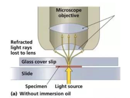
What are the refractive indices of air, glass, and immersion oil?
Air: approximately 1.0
Glass: approximately 1.5
Immersion oil: approximately 1.5.
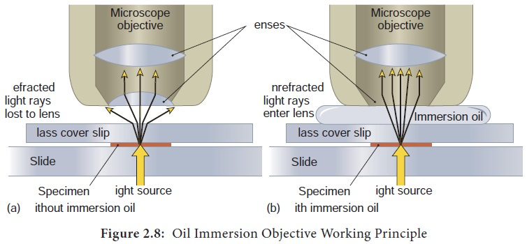
Why does immersion oil improve resolution when using the 100× objective?
Oil replaces the air gap between the slide and objective
Its refractive index closely matches glass, reducing refraction and allowing more light to enter the objective
What is the function of the ocular lens (eyepiece)?
It magnifies the image formed by the objective lens for viewing
The lab microscope has 10× ocular lenses.
What are the functions of the objective lenses and revolving nosepiece?
Objective lenses provide the initial magnification of the specimen
The nosepiece holds them and rotates to change the objective in use
What are the functions of the stage, stage clips, and slide adjustment knobs?
The stage supports the slide
Stage clips hold it in place
Slide adjustment knobs move it horizontally to position the specimen
How do the coarse and fine focus knobs differ?
Coarse focus knob makes large focusing adjustments
Fine focus knob makes small adjustments for precise focus
Best to use fine focus when viewing with the 100× oil-immersion objective
What does the iris diaphragm control, and where is it located?
It is located under the stage and controls the amount of light reaching the objective lens by changing the opening
Should be adjusted to the same level of the objective lens in use
Doesn’t apply to viewing living microorganisms as they require low light
What does the rheostat control, and how should light intensity vary with specimen type?
The rheostat adjusts light-source intensity
Lower intensity is best for living or transparent specimens; higher intensity is best for stained specimens.
What is the purpose of an ocular micrometer, and what does µm mean?
An ocular micrometer is a scale in the eyepiece used to measure specimens after converting ocular units to actual length.
µm means micrometer; 1 µm = 0.001 mm.

What actual length does one ocular micrometer unit represent at each objective magnification in this lab?
4× objective: 25 µm
10×: 10 µm
40×: 2.5 µm
100×: 1 µm.
How do you calculate a cell's actual size from an ocular micrometer reading?
Cell size in µm = number of ocular units spanned × µm per ocular unit for the objective in use.
At the 40× objective, a cell extends from ocular mark 12 to mark 18. What is its length?
It spans 18 − 12 = 6 ocular units
Length = 6 × 2.5 µm = 15 µm.
How do you prepare the slide for oil immersion according to the lab procedure?
Focus with the 10× objective, then rotate the 4× objective into viewing position without moving the slide or adjusting focus
Add one drop of immersion oil directly to the slide
Move back to the 100x objective and use the fine focus to adjust + the iris diaphragm
What are the microscope care and storage steps after use?
Remove oil from the 100× objective with lens paper
Return the 4× objective to viewing position
Lower the stage fully
Wrap the power cord
Return the microscope to the cabinet with the red handle facing out