Biology Chapter 14 review (2)
 Call Kai
 Learn
 Practice Test
 Spaced Repetition
 Match
 Flashcards
 Knowt Play
1/129

Earn XP
Description and Tags
DNA structure and function
130 Terms
Models of DNA replication
Before the correct mechanism was proven, scientists proposed three possible models for how DNA molecules copy themselves:
Possible models of DNA replication:
Conservative replication – parent strands stay intact and two completely new strands are synthesized
Semi-conservative replication– the two strands separate– each is a template for a new complementary strand
Dispersive replication– parent strands are broken up into small pieces and incorporated into new strands

Models of DNA replication
Before the correct mechanism was proven, scientists proposed possible models for how DNA molecules copy themselves:
Possible models of DNA replication:
Conservative replication – parent strands stay together, and two new strands are synthesized
Semi-conservative replication– the two strands separate– each is a template for a new complementary strand

Conservative model of DNA replication
A hypothesized mode where the original parental double-stranded DNA molecule remains completely intact, and an entirely new double-stranded DNA molecule is synthesized from scratch using the parental molecule as a template
Parental molecule conserved: after replication, the original double helix reforms completely so that both parental strands stay together
New molecule created: a separate, brand-new double-stranded DNA molecule is assembled with two completely newly synthesized strands
Disproved by Meselson and Stahl: this model was tested and disproven by the famous Meselson-Stahl experiment, which proved that actual DNA replication is semi-conservative (each new molecule has one old strand and one new strand)
parent strands stay intact and two completely
new strands are synthesized

Conservative model of DNA replication
A hypothesized mode where the original parental double-stranded DNA molecule remains completely intact, and an entirely new double-stranded DNA molecule is synthesized from scratch using the parental molecule as a template
Disproved by Meselson and Stahl: this model was tested and disproven by the famous Meselson-Stahl experiment, which proved that actual DNA replication is semi-conservative (each new molecule has one old strand and one new strand)
parent strands stay intact and two completely
new strands are synthesized
After replication, the two original parental strands come back together and are fully conserved ("saved") as a single double helix
A completely separate second double-stranded molecule is synthesized from two brand-new complementary strands
This model was tested and disproved by the Meselson-Stahl experiment, which proved that real DNA replication is semi-conservative (each new double helix has one old strand and one new strand)

Semi-conservative model of DNA replication
Sates that each newly formed DNA double helix consists of one conserved parental (old) strand and one newly synthesized complementary strand
the two strands separate– each is a template for a new complementary strand
states that when a double-stranded DNA molecule copies itself, each of the two resulting daughter molecules consists of one original (parental) strand and one newly synthesized strand
Template Mechanism: During DNA replication, the two parental strands unwind and separate. Each individual strand serves as a template to build a new, complementary strand using base-pairing rules (A = T, G = C)
Historical Proof (Meselson-Stahl Experiment): Matthew Meselson and Franklin Stahl proved the semi-conservative model in 1958 using heavy (¹⁵N) and light (¹⁴N) nitrogen isotopes in E. coli. After one round of replication in a light medium, the DNA was a "hybrid" density (half heavy, half light), ruling out the conservative model

Semi-conservative model of DNA replication
Sates that each newly formed DNA double helix consists of one conserved parental (old) strand and one newly synthesized complementary strand
the two strands separate– each is a template for a new complementary strand
states that when a double-stranded DNA molecule copies itself, each of the two resulting daughter molecules consists of one original (parental) strand and one newly synthesized strand
Template Mechanism: During DNA replication, the two parental strands unwind and separate. Each individual strand serves as a template to build a new, complementary strand using base-pairing rules (A = T, G = C)
Historical Proof (Meselson-Stahl Experiment): Matthew Meselson and Franklin Stahl proved the semi-conservative model in 1958 using heavy (¹⁵N) and light (¹⁴N) nitrogen isotopes in E. coli. After one round of replication in a light medium, the DNA was a "hybrid" density (half heavy, half light), ruling out the conservative model

Semi-conservative model of DNA replication
Sates that each newly formed DNA double helix consists of one conserved parental (old) strand and one newly synthesized complementary strand
The two strands separate– each is a template for a new complementary strand
Historical Proof (Meselson-Stahl Experiment): Matthew Meselson and Franklin Stahl proved the semi-conservative model in 1958 using heavy (¹⁵N) and light (¹⁴N) nitrogen isotopes in E. coli. After one round of replication in a light medium, the DNA was a "hybrid" density (half heavy, half light), ruling out the conservative model

Dispersive model of DNA replication
Proposes that the parental double helix breaks into multiple segments, and each resulting daughter DNA molecule consists of a "patchwork" or interspersed mixture of old (parental) and newly synthesized DNA strands
Interspersed Segments: Unlike keeping entire strands intact, both strands of the resulting double helices contain alternating blocks of parental and newly made DNA
parent strands are broken up into small pieces and
incorporated into new strands(Meselson-Stahl Experiment): the experiment disproved the dispersive model (and conservative model), proving instead that DNA replication is semi-conservative (each new molecule has one complete old strand and one complete new strand)

Dispersive model of DNA replication
Proposes that the parental double helix breaks into multiple segments, and each resulting daughter DNA molecule consists of a "patchwork" or interspersed mixture of old (parental) and newly synthesized DNA strands
Interspersed Segments: Unlike keeping entire strands intact, both strands of the resulting double helices contain alternating blocks of parental and newly made DNA
parent strands are broken up into small pieces and
incorporated into new strands(Meselson-Stahl Experiment): the experiment disproved the dispersive model (and conservative model), proving instead that DNA replication is semi-conservative (each new molecule has one complete old strand and one complete new strand)

Models of DNA replication
Before the correct mechanism was proven, scientists proposed three possible models for how DNA molecules copy themselves:
Possible models of DNA replication:
Conservative replication – parent strands stay intact and two completely new strands are synthesized
Semi-conservative replication– the two strands separate– each is a template for a new complementary strand
Dispersive replication– parent strands are broken up into small pieces and incorporated into new strands
The Meselson-Stahl experiment proved that DNA replication is semi-conservative, meaning each new DNA double helix contains one original (parental) strand and one newly synthesized strand

Meselson and Stahl’s experiment
The Meselson-Stahl experiment (1958) is the classic study by Matthew Meselson and Franklin Stahl that proved DNA replicates via a semiconservative mechanism
Meselson and Stahl tracked DNA generations using heavy and light nitrogen isotopes via equilibrium density-gradient centrifugation
Labeling with Heavy Nitrogen (¹⁵N): they grew Escherichia coli (E. coli) bacteria in a medium containing the heavy nitrogen isotope (¹⁵N) for many generations so all DNA incorporated heavy nitrogen
Transfer to Light Nitrogen (¹⁴N): they transferred the bacteria to a normal, lighter nitrogen medium (¹⁴N) and allowed the cells to replicate
Centrifugation and Analysis: They extracted the DNA and spun it in a cesium chloride (CsCl) density gradient to separate DNA molecules based on density
After Generation 2 (Second round of replication in ¹⁴N):
The DNA separated into two distinct bands: one intermediate-density band and one light-density band (¹⁴N)
Rule-out: this disproved the dispersive model (which would have continued to show a single blended intermediate band)
Conclusion: proved that each strand separates and acts as a template, confirming semiconservative replication

Meselson and Stahl’s experiment
The Meselson-Stahl experiment (1958) is the classic study by Matthew Meselson and Franklin Stahl that proved DNA replicates via a semiconservative mechanism
Meselson and Stahl tracked DNA generations using heavy and light nitrogen isotopes via equilibrium density-gradient centrifugation
Labeling with Heavy Nitrogen (¹⁵N): they grew Escherichia coli (E. coli) bacteria in a medium containing the heavy nitrogen isotope (¹⁵N) for many generations so all DNA incorporated heavy nitrogen
Transfer to Light Nitrogen (¹⁴N): they transferred the bacteria to a normal, lighter nitrogen medium (¹⁴N) and allowed the cells to replicate
Centrifugation and Analysis: They extracted the DNA and spun it in a cesium chloride (CsCl) density gradient to separate DNA molecules based on density
After Generation 2 (Second round of replication in ¹⁴N):
The DNA separated into two distinct bands: one intermediate-density band and one light-density band (¹⁴N)
Rule-out: this disproved the dispersive model (which would have continued to show a single blended intermediate band)
Conclusion: proved that each strand separates and acts as a template, confirming semiconservative replication
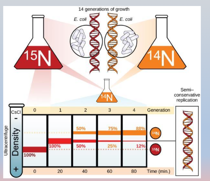
Meselson and Stahl’s experiment procedures
The Meselson-Stahl experiment (1958) is the classic study by Matthew Meselson and Franklin Stahl that proved DNA replicates via a semiconservative mechanism
How the experiment worked:
Meselson and Stahl tracked DNA generations using heavy and light nitrogen isotopes via equilibrium density-gradient centrifugation
Labeling with Heavy Nitrogen (¹⁵N): they grew Escherichia coli (E. coli) bacteria in a medium containing the heavy nitrogen isotope (¹⁵N) for many generations so all DNA incorporated heavy nitrogen
Transfer to Light Nitrogen (¹⁴N): they transferred the bacteria to a normal, lighter nitrogen medium (¹⁴N) and allowed the cells to replicate
Centrifugation and Analysis: They extracted the DNA and spun it in a cesium chloride (CsCl) density gradient to separate DNA molecules based on density
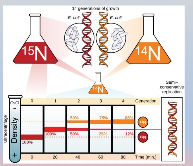
Meselson and Stahl’s experiment procedures
The Meselson-Stahl experiment (1958) is the classic study by Matthew Meselson and Franklin Stahl that proved DNA replicates via a semiconservative mechanism
How the experiment worked:
Meselson and Stahl tracked DNA generations using heavy and light nitrogen isotopes via equilibrium density-gradient centrifugation
Labeling with Heavy Nitrogen (¹⁵N): they grew Escherichia coli (E. coli) bacteria in a medium containing the heavy nitrogen isotope (¹⁵N) for many generations so all DNA incorporated heavy nitrogen
Transfer to Light Nitrogen (¹⁴N): they transferred the bacteria to a normal, lighter nitrogen medium (¹⁴N) and allowed the cells to replicate
Centrifugation and Analysis: They extracted the DNA and spun it in a cesium chloride (CsCl) density gradient to separate DNA molecules based on density

Meselson and Stahl’s experiment - key results
The Meselson-Stahl experiment (1958) proved that DNA replication is semi-conservative, meaning each new DNA double helix consists of one old (parental) strand and one newly synthesized strand
After Generation 1 (One round of replication in (¹⁴N):
The DNA showed a single band of intermediate density (half heavy ¹⁵N / half light ¹⁴N)
Rule-out: this immediately disproved the conservative model (which predicted two distinct bands: one heavy and one light)
After Generation 2 (Second round of replication in ¹⁴N):
The DNA separated into two distinct bands: one intermediate-density band and one light-density band (¹⁴N)
Rule-out: this disproved the dispersive model (which would have continued to show a single blended intermediate band)
Conclusion: proved that each strand separates and acts as a template, confirming semiconservative replication

Meselson and Stahl’s experiment - key results
The Meselson-Stahl experiment (1958) proved that DNA replication is semi-conservative, meaning each new DNA double helix consists of one old (parental) strand and one newly synthesized strand
After Generation 1 (One round of replication in (¹⁴N):
The DNA showed a single band of intermediate density (half heavy ¹⁵N / half light ¹⁴N)
Rule-out: this immediately disproved the conservative model (which predicted two distinct bands: one heavy and one light)
After Generation 2 (Second round of replication in ¹⁴N):
The DNA separated into two distinct bands: one intermediate-density band and one light-density band (¹⁴N)
Rule-out: this disproved the dispersive model (which would have continued to show a single blended intermediate band)
Conclusion: proved that each strand separates and acts as a template, confirming semiconservative replication
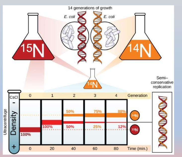
Meselson and Stahl’s experiment
The Meselson-Stahl experiment (1958) is the classic study by Matthew Meselson and Franklin Stahl that proved DNA replicates via a semiconservative mechanism
Meselson and Stahl tracked DNA generations using heavy and light nitrogen isotopes via equilibrium density-gradient centrifugation
Labeling with Heavy Nitrogen (¹⁵N): they grew Escherichia coli (E. coli) bacteria in a medium containing the heavy nitrogen isotope (¹⁵N) for many generations so all DNA incorporated heavy nitrogen
Transfer to Light Nitrogen (¹⁴N): they transferred the bacteria to a normal, lighter nitrogen medium (¹⁴N) and allowed the cells to replicate
Centrifugation and Analysis: They extracted the DNA and spun it in a cesium chloride (CsCl) density gradient to separate DNA molecules based on density
After Generation 2 (Second round of replication in ¹⁴N):
The DNA separated into two distinct bands: one intermediate-density band and one light-density band (¹⁴N)
Rule-out: this disproved the dispersive model (which would have continued to show a single blended intermediate band)
Conclusion: proved that each strand separates and acts as a template, confirming semiconservative replication

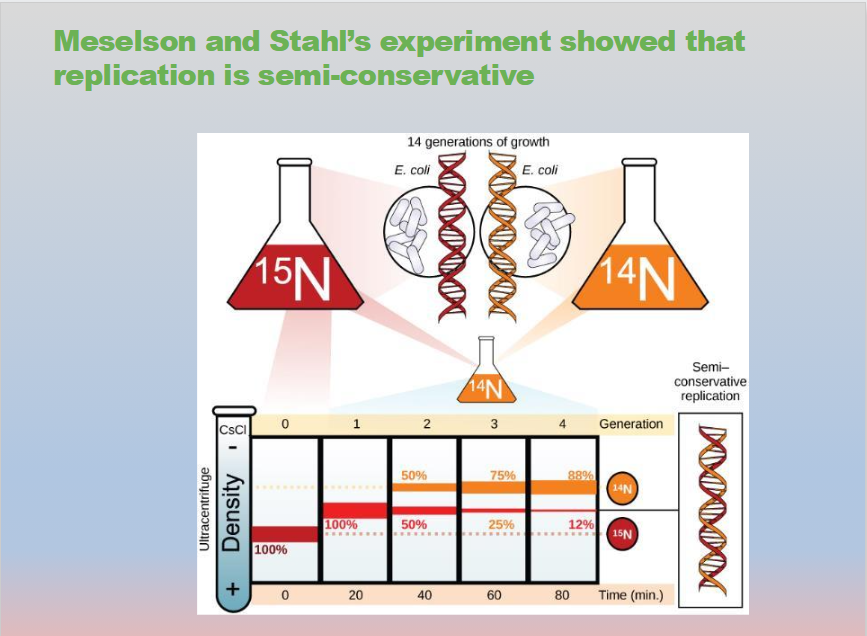
Meselson and Stahl’s experiment demonstrated that replication is semi-conservative
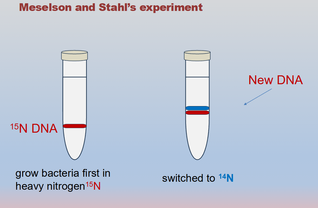
Meselson and Stahl experiment: grow E. coli first in heavy nitrogen (¹⁵N) then in ¹⁴N
DNA grown in ¹⁵N is heavier than DNA grown in ¹⁴N, and sediments to a lower level in an ultracentrifuge
DNA grown in ¹⁵N is switched to ¹⁴N, after one round of cell division the DNA sediments halfway between the ¹⁵N and 14N levels, indicating that it now contains fifty percent ¹⁴N
In subsequent divisions, DNA contains ¹⁴N only. This data supports the semi-conservative replication model

Meselson and Stahl’s experiment demonstrated that replication is semi-conservative
Meselson and Stahl experiment: grow E. coli first in heavy nitrogen (¹⁵N) then in ¹⁴N
DNA grown in ¹⁵N is heavier than DNA grown in ¹⁴N, and sediments to a lower level in an ultracentrifuge
DNA grown in ¹⁵N is switched to ¹⁴N, after one round of cell division the DNA sediments halfway between the ¹⁵N and 14N levels, indicating that it now contains fifty percent ¹⁴N
In subsequent divisions, DNA contains ¹⁴N only. This data supports the semi-conservative replication model
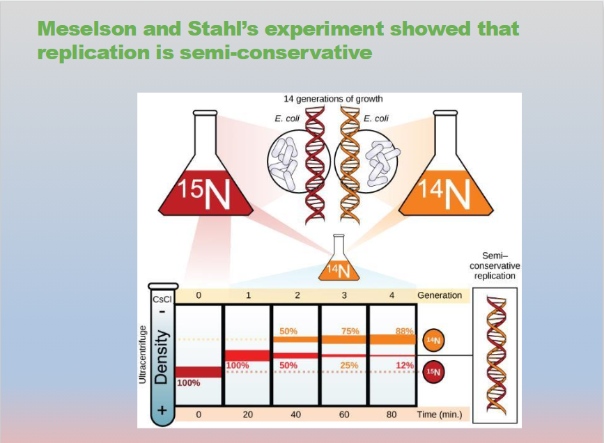
Meselson and Stahl’s experiment demonstrated that replication is semi-conservative
Meselson and Stahl experiment: grow E. coli first in heavy nitrogen (¹⁵N) then in ¹⁴N
DNA grown in ¹⁵N is heavier than DNA grown in ¹⁴N, and sediments to a lower level in an ultracentrifuge
DNA grown in ¹⁵N is switched to ¹⁴N, after one round of cell division the DNA sediments halfway between the ¹⁵N and ¹⁴N levels, indicating that it now contains fifty percent ¹⁴N
In subsequent divisions, DNA contains ¹⁴N only. This data supports the semi-conservative replication model
What did Meselson and Stahl’s experiment show?
Meselson and Stahl’s experiment showed that replication is semi-conservative
Meselson and Stahl’s experiment showed that DNA replication is semi-conservative
Key Findings
Semi-conservative mechanism: each new DNA double helix consists of one original (parental) strand and one newly synthesized strand
Disproved the conservative model: the original DNA molecule does not stay completely intact while building a separate all-new double helix
Disproved the dispersive model: DNA strands do not break apart into a patchwork or mixture of old and new segments across both strands
What did Meselson and Stahl’s experiment show?
Meselson and Stahl’s experiment showed that replication is semi-conservative
Meselson and Stahl’s experiment showed that DNA replication is semi-conservative
Key Findings
Semi-conservative mechanism: each new DNA double helix consists of one original (parental) strand and one newly synthesized strand
Disproved the conservative model: the original DNA molecule does not stay completely intact while building a separate all-new double helix
Disproved the dispersive model: DNA strands do not break apart into a patchwork or mixture of old and new segments across both strands

What did Meselson and Stahl’s experiment show?
Meselson and Stahl’s experiment showed that replication is semi-conservative
Meselson and Stahl’s experiment showed that DNA replication is semi-conservative

How the Meselson and Stahl experiment worked
Labeled with Heavy Nitrogen (¹⁵N): they grew E. coli bacteria in a medium containing heavy nitrogen so all the DNA incorporated the heavy isotope
Shifted to Light Nitrogen (¹⁴N): They moved the bacteria to a medium with normal, lighter nitrogen and let them replicate
Density Gradient Centrifugation: They spun the extracted DNA in a cesium chloride (CsCl) solution to separate molecules by weight:
After 1 generation: the DNA formed a single band of intermediate density (half ¹⁵N, half ¹⁴N), which ruled out the conservative model
After 2 generations: the DNA formed two distinct bands: one intermediate density band and one light (¹⁴N) density band, which ruled out the dispersive model
Conclusion: proved that each strand separates and acts as a template, confirming semiconservative replication

What did Meselson and Stahl’s experiment show?
The Meselson-Stahl experiment showed that DNA replicates in a semi-conservative manner, meaning each new DNA double helix consists of one old (parental) strand and one newly synthesized strand
Generation 2 (After two rounds of replication): the DNA produced two distinct bands: one intermediate density band and one light density band. This ruled out the dispersive model (which would have continued to show a single blended intermediate band)
Core Takeaway: each original DNA strand serves as a template to build a complementary new strand
Meselson and Stahl’s experiment showed that
replication is semi-conservative

Model of DNA replication in bacteria overview
The model of DNA replication in bacteria (prokaryotes) is semi-conservative, meaning each new double-stranded DNA molecule consists of one original (parental) strand and one newly synthesized strand
This was experimentally proven by the Meselson-Stahl experiment
Single Origin of Replication: Unlike linear eukaryotic chromosomes that have multiple origins, bacterial circular chromosomes have a single origin of replication where the DNA opens up to form a replication bubble and two Y-shaped replication forks
Semi-Conservative Model: each parental strand serves as a template to build a complementary daughter strand using base-pairing rules (A= T, G = C)
Directionality (5' to 3'): DNA polymerases can only add nucleotides to the free (3'-OH) end, meaning they synthesize DNA exclusively in the (5' to 3') direction

Model of DNA replication in bacteria - parental strand
A parent strand (or parental strand) in bacterial (prokaryotic) DNA replication is an original single strand of the double-stranded DNA molecule that unwinds and serves as a template to build a new complementary strand
Parent Strand (Template / Original Strand): an original single strand of the bacterial DNA double helix that separates during replication and acts as a template to guide the synthesis of a new complementary strand via base-pairing rules (A = T, G = C)
Semiconservative Model: In bacterial replication, the circular double-stranded DNA unwinds, and each of the two old parent strands acts as a template. After replication is complete, each new DNA molecule consists of one parent (old) strand and one newly synthesized daughter strand

Model of DNA replication in bacteria - parental strand
In the model of DNA replication, the original double-stranded DNA molecule that serves as the template for creating new DNA is called the parental DNA
Double stranded template: the image shows a complete, antiparallel double helix before replication begins
Complementary base pairing: the top strand (3’ → 5’) and the bottom strand (5’ → 3’) pair up perfectly with each other
The downward arrow below the sequence indicates the next step in the process, where these two original strands will seperate so that each can act as a template to synthesize new, complementary “daughter” strands

Helicase enzyme
DNA helicase is the first enzyme that loads at the origin of replication to unwind and separate the two parental DNA strands
Unzipping the Double Helix: Helicase breaks the weak hydrogen bonds between complementary nitrogenous base pairs (A-T and C-G)
Creating the Replication Fork: as it moves, it forms a Y-shaped junction known as the replication fork. Two helicases often move in opposite directions, creating a replication bubble
Seperates DNA strands
Separates the DNA strands at the origin of replication

Helicase enzyme
DNA helicase is the first enzyme that loads at the origin of replication to unwind and separate the two parental DNA strands
an ATP-powered motor enzyme that unwinds and unzips the DNA double helix by breaking the hydrogen bonds between complementary nitrogenous base pairs
Primary Function: separates double-stranded DNA into single-stranded templates
Replication Fork: creates a Y-shaped region called the replication fork (or replication bubbles) where copying can begin
Seperates DNA strands
Separates the DNA strands at the origin of replication

Topoisomerase
An enzyme that relieves twisting strain and supercoiling ahead of the replication fork during DNA replication by temporarily cutting, unwinding, and resealing one or both strands of the DNA molecule
Topoisomerase is defined as an enzyme that relaxes and relieves the torsional strain (supercoiling) in the DNA double helix ahead of the replication fork
Prevents overwinding
Topoisomerase prevents the overwinding (and underwinding) of DNA during replication and transcription
Topoisomerase prevents over-winding as DNA opens up

Topoisomerase
An enzyme that relieves twisting strain and supercoiling ahead of the replication fork during DNA replication by temporarily cutting, unwinding, and resealing one or both strands of the DNA molecule
Topoisomerase is defined as an enzyme that relaxes and relieves the torsional strain (supercoiling) in the DNA double helix ahead of the replication fork
Prevents overwinding
Topoisomerase prevents the overwinding (and underwinding) of DNA during replication and transcription
Topoisomerase prevents over-winding as DNA opens up

Topoisomerase
An enzyme that relieves twisting strain and supercoiling ahead of the replication fork during DNA replication by temporarily cutting, unwinding, and resealing one or both strands of the DNA molecule
Prevents overwinding
Topoisomerase prevents the overwinding (and underwinding) of DNA during replication and transcription
The Problem: as helicase unwinds the DNA double helix to create the replication fork, the DNA ahead of the fork gets tightly twisted and bunched up. This strain is called supercoiling or overwinding. If left unchecked, the DNA would become too knotted for replication to continue
The Solution: topoisomerase binds to the DNA ahead of the replication fork. It temporary cuts the sugar-phosphate backbone of one or both strands, allows the DNA to untwist to release the tension, and then reseals (religates) the backbone
Topoisomerase prevents over-winding as DNA opens up

DNA Polymerase
A key enzyme that synthesizes a new complementary strand of DNA by adding free nucleotides to an existing template strand during DNA replication
Direction of Synthesis: It can only add nucleotides to the 3' end of a growing strand, meaning new DNA is always synthesized in the 5' to 3' direction
Proofreading Activity: It features a proofreading function that checks newly added bases, removes mismatched nucleotides, and replaces them to prevent mutations
the primary enzyme responsible for synthesizing a new strand of DNA during DNA replication by adding complementary free nucleotides to a pre-existing template strand
Synthesizes the new strands

DNA Polymerase
A key enzyme that synthesizes a new complementary strand of DNA by adding free nucleotides to an existing template strand during DNA replication
Direction of Synthesis: It can only add nucleotides to the 3' end of a growing strand, meaning new DNA is always synthesized in the 5' to 3' direction
Proofreading Activity: It features a proofreading function that checks newly added bases, removes mismatched nucleotides, and replaces them to prevent mutations
The primary enzyme responsible for synthesizing a new strand of DNA during DNA replication by adding complementary free nucleotides to a pre-existing template strand
Synthesizes the new strands

DNA Polymerase
A key enzyme that synthesizes a new complementary strand of DNA by adding free nucleotides to an existing template strand during DNA replication
Direction of Synthesis: It can only add nucleotides to the 3' end of a growing strand, meaning new DNA is always synthesized in the 5' to 3' direction
The primary enzyme responsible for synthesizing a new strand of DNA during DNA replication by adding complementary free nucleotides to a pre-existing template strand
Synthesizes the new strands

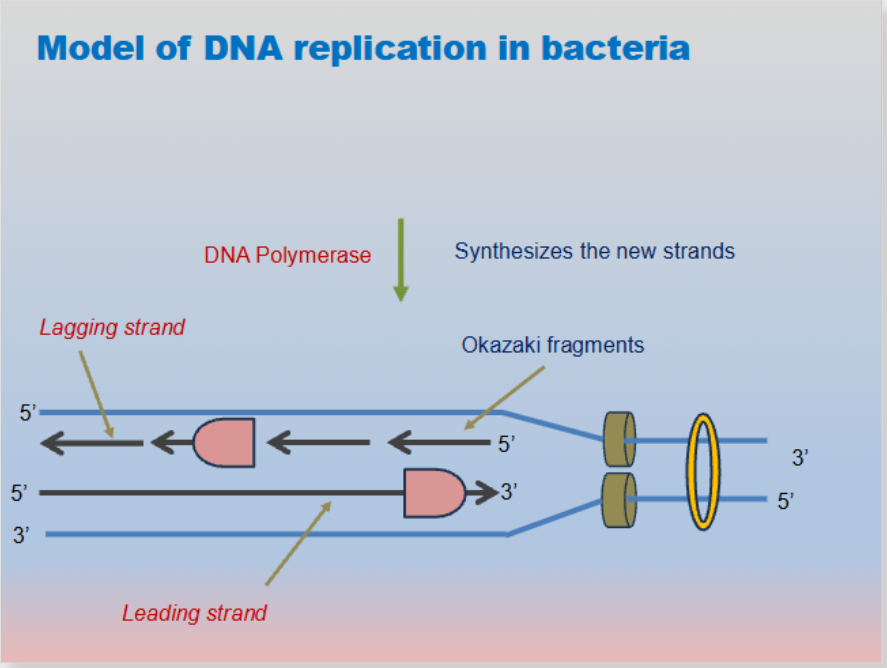
Okazaki fragments
Short, newly synthesized DNA sequences formed discontinuously on the lagging strand during DNA replication
Short sequences of DNA nucleotides synthesized discontinuously and later linked together by the enzyme DNA ligase to create the lagging strand during DNA replication
short, newly synthesized sequences of DNA formed on the lagging strand during DNA replication
Why they form: DNA strands are antiparallel (running in opposite directions), and DNA polymerase can only synthesize new DNA in the 5' to 3' direction. Because the replication fork opens up, one strand (the leading strand) is built continuously, while the other strand (the lagging strand) must be built backward in short, discontinuous segments
Okazaki fragments
Short, newly synthesized DNA sequences formed discontinuously on the lagging strand during DNA replication
Short sequences of DNA nucleotides synthesized discontinuously and later linked together by the enzyme DNA ligase to create the lagging strand during DNA replication
short, newly synthesized sequences of DNA formed on the lagging strand during DNA replication
Why they form: DNA strands are antiparallel (running in opposite directions), and DNA polymerase can only synthesize new DNA in the 5' to 3' direction. Because the replication fork opens up, one strand (the leading strand) is built continuously, while the other strand (the lagging strand) must be built backward in short, discontinuous segments

Okazaki fragments
Short, newly synthesized DNA sequences formed on the lagging strand during DNA replication
DNA polymerase can only add nucleotides to the 3' end, meaning new DNA is always built in the 5' to 3' direction
Lagging Strand: because the two template strands are antiparallel (running in opposite directions), one strand is copied continuously (the leading strand), while the other must be copied in short, backward segments away from the replication fork (the lagging strand)
Joining the Pieces: the enzyme DNA ligase seals the gaps between the fragments by creating phosphodiester bonds, turning the separate pieces into a continuous DNA strand
Short sequences of DNA nucleotides synthesized discontinuously and later linked together by the enzyme DNA ligase to create the lagging strand during DNA replication

Lagging strand
The new DNA strand synthesized discontinuously in short segments away from the replication fork during DNA replication
Direction of Synthesis: like all new DNA strands, individual pieces are still built in the 5′ to 3′ direction, even though the overall strand grows away from the opening replication fork
Okazaki Fragments: because DNA polymerase can only add nucleotides to a free 3′ end, the lagging strand is created in small, separate chunks called Okazaki fragments
the new DNA strand synthesized discontinuously in the 5'→ 3' direction away from the replication fork during DNA replication
Okazaki Fragments: the lagging strand is made in short, separate pieces of DNA called Okazaki fragments

Lagging strand
The new complementary DNA strand that is synthesized discontinuously in short pieces (called Okazaki fragments) moving away from the replication fork
DNA polymerase can only add nucleotides to an exposed 3' end, meaning new DNA is always synthesized in the 5' to 3' direction
Antiparallel Template: because the two template strands run in opposite directions (5' to 3' versus 3' to 5'), the strand opening toward the fork is continuous (leading), while the strand opening away from the fork must be built backward in pieces (lagging)
Okazaki Fragments: short, newly synthesized DNA segments formed on the lagging strand
the new daughter DNA strand that is synthesized discontinuously in the 5' to 3' direction moving away from the replication fork
Because the template strand runs in a direction that forces synthesis away from the opening fork, the lagging strand is built in short, separate segments called Okazaki fragments
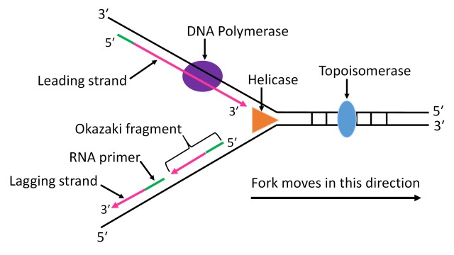
Lagging strand
The new strand of DNA synthesized discontinuously in the 5′ to 3′ direction away from the replication fork during DNA replication
Directionality Constraint: DNA polymerase can only add nucleotides to the exposed 3' end, meaning new DNA is always built in the 5'→ 3' direction. Because the two parent strands run antiparallel (5'→ 3' and 3'→ 5'), the strand opening toward the fork is continuous (leading), while the other must build backward (lagging)
Okazaki fragments: because DNA polymerase must move away from the replication fork on this template, it synthesizes the strand in short, separate pieces called Okazaki fragments
the new DNA strand synthesized discontinuously in short pieces (called Okazaki fragments) moving away from the replication fork in the 5'→ 3' direction

Leading strand
The new complementary DNA strand that is synthesized continuously in the 5' to 3' direction toward the replication fork
Direction of Synthesis: built in the 5′ → 3′ direction
Template Polarity: read along a 3′ → 5′ parental template strand
Movement of DNA Polymerase: moves toward the opening replication fork
Primers Required: needs only one RNA primer (laid down at the start)
Continuity: created in one single, uninterrupted piece without fragments
the new strand of DNA that is synthesized continuously in the 5' to 3' direction during DNA replication

Leading strand
The newly synthesized strand of DNA that is continuously produced in the 5'→ 3' direction as DNA polymerase follows the moving replication fork
the new strand of DNA that is synthesized continuously in the 5' to 3' direction toward the replication fork
Direction of Synthesis: built in the 5'→ 3' direction, meaning the new nucleotides are added to the growing 3' end
Movement of Enzyme: DNA polymerase moves toward the replication fork as it unwinds
Continuity: It is synthesized in one continuous piece, unlike the fragmented lagging strand
Primers Required: It only requires one single RNA primer at the start of replication
Template Strand: It is built using the parent (template) strand that runs in the 3'→ 5' direction
No Okazaki Fragments: unlike the opposite strand (the lagging strand), it does not break into short pieces or require DNA ligase to stitch fragments together

Leading vs. Lagging strand
The leading strand and lagging strand are the two newly synthesized DNA strands created during DNA replication. Their differences come down to one core rule: DNA polymerase can only add nucleotides to the 3′ end, meaning new DNA is always synthesized in the 5′ → 3′ direction, while the two template strands run antiparallel (in opposite 3′ → 5′ and 5′ → 3′ directions)
Leading strand
The newly synthesized strand of DNA that is continuously produced in the 5'→ 3' direction as DNA polymerase follows the moving replication fork
the new strand of DNA that is synthesized continuously in the 5' to 3' direction toward the replication fork
Direction of Synthesis: built in the 5'→ 3' direction, meaning the new nucleotides are added to the growing 3' end
Movement of Enzyme: DNA polymerase moves toward the replication fork as it unwinds
Continuity: It is synthesized in one continuous piece, unlike the fragmented lagging strand
Primers Required: It only requires one single RNA primer at the start of replication
Lagging strand
The new strand of DNA synthesized discontinuously in the 5′ to 3′ direction away from the replication fork during DNA replication
Directionality Constraint: DNA polymerase can only add nucleotides to the exposed 3' end, meaning new DNA is always built in the 5'→ 3' direction. Because the two parent strands run antiparallel (5'→ 3' and 3'→ 5'), the strand opening toward the fork is continuous (leading), while the other must build backward (lagging)
Okazaki fragments: because DNA polymerase must move away from the replication fork on this template, it synthesizes the strand in short, separate pieces called Okazaki fragments
the new DNA strand synthesized discontinuously in short pieces (called Okazaki fragments) moving away from the replication fork in the 5'→ 3' direction

DNA ligase
An enzyme that catalyzes the formation of covalent phosphodiester bonds to join DNA fragments together
A "molecular glue" enzyme that links the 3’ -hydroxyl end of one DNA strand to the 5’ -phosphate end of another, sealing the sugar-phosphate backbone
Role in DNA Replication (Lagging Strand): Joins discontinuous Okazaki fragments synthesized on the lagging strand into a single, continuous complementary DNA strand
Connects Okazaki fragments
an enzyme that facilitates the joining of DNA strands together by forming a bond between them
Seals gaps: It connects broken strands of DNA by forming chemical links (phosphodiester bonds) between the sugar-phosphate backbones.
Helps in replication: During cell division, DNA is copied. One side is built in small, separate pieces (called Okazaki fragments), and DNA ligase glues them into a single continuous strand.
seals the gaps between the Okazaki fragments

DNA ligase
An enzyme that catalyzes the formation of covalent phosphodiester bonds to join DNA fragments together
A "molecular glue" enzyme that links the 3’ -hydroxyl end of one DNA strand to the 5’ -phosphate end of another, sealing the sugar-phosphate backbone
Role in DNA Replication (Lagging Strand): Joins discontinuous Okazaki fragments synthesized on the lagging strand into a single, continuous complementary DNA strand
Connects Okazaki fragments
an enzyme that facilitates the joining of DNA strands together by forming a bond between them
Seals gaps: It connects broken strands of DNA by forming chemical links (phosphodiester bonds) between the sugar-phosphate backbones
Helps in replication: During cell division, DNA is copied. One side is built in small, separate pieces (called Okazaki fragments), and DNA ligase glues them into a single continuous strand
seals the gaps between the Okazaki fragments

An enzyme that catalyzes the formation of covalent phosphodiester bonds to join DNA fragments together
A "molecular glue" enzyme that links the 3’ -hydroxyl end of one DNA strand to the 5’ -phosphate end of another, sealing the sugar-phosphate backbone
Role in DNA Replication (Lagging Strand): Joins discontinuous Okazaki fragments synthesized on the lagging strand into a single, continuous complementary DNA strand
Connects Okazaki fragments
Helps in replication: during cell division, DNA is copied. One side is built in small, separate pieces (called Okazaki fragments), and DNA ligase glues them into a single continuous strand
Seals the gaps between the Okazaki fragments

DNA ligase
An enzyme that joins DNA strands together by catalyzing the formation of covalent phosphodiester bonds between the 3'-hydroxyl end of one DNA fragment and the 5'-phosphate end of anothe
DNA Replication: seals the gaps (nicks) in the sugar-phosphate backbone, specifically by joining Okazaki fragments on the lagging strand into a continuous DNA strand
DNA Repair: fills in and seals single-stranded breaks or gaps created when damaged sections of DNA are removed
Connects Okazaki fragments
Seals the gaps between the Okazaki fragments
Helps in replication: during cell division, DNA is copied. One side is built in small, separate pieces (called Okazaki fragments), and DNA ligase glues them into a single continuous strand
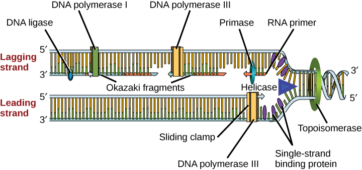
DNA replication in bacteria - new strands
The semi-conservative process where a circular chromosomal DNA molecule is copied prior to binary fission, producing two identical double helices, each containing one original template strand and one newly synthesized strand
The semiconservative process by which a circular bacterial chromosome duplicates itself before binary fission, producing two identical double-stranded DNA molecules, each containing one original template strand and one newly synthesized strand
Semiconservative Model: each new DNA molecule conserves one parental (old) strand and pairs it with one newly synthesized strand (proven by the Meselson-Stahl experiment)
Directionality: DNA polymerase can only add nucleotides to the 3′-OH end, meaning new DNA is always synthesized exclusively in the 5′ to 3′ direction (while the enzyme "reads" the template strand 3′ to 5′)
Leading Strand: synthesized continuously in the same direction that the replication fork opens
Lagging Strand: synthesized discontinuously in short segments called Okazaki fragments moving away from the replication fork
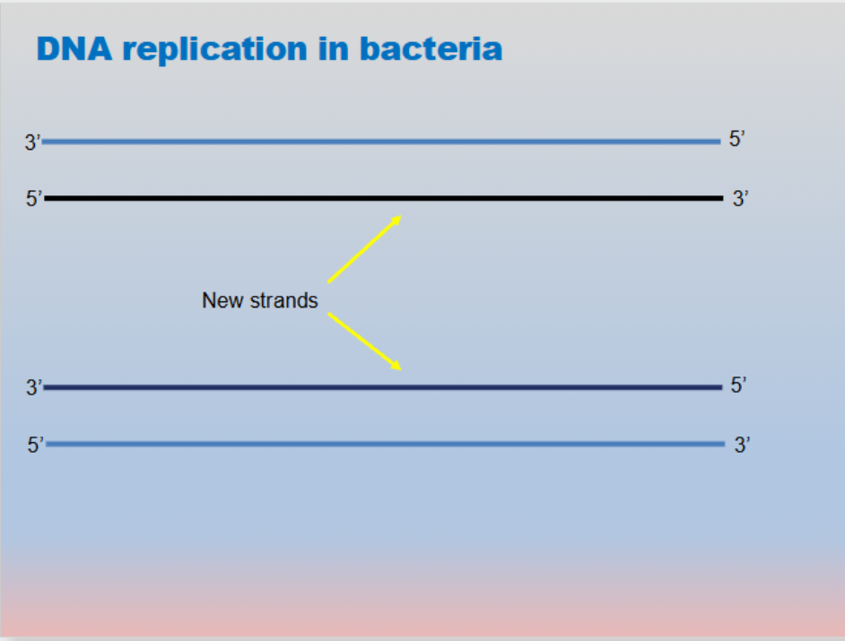
DNA replication in bacteria - new strands
The semi-conservative process where a circular chromosomal DNA molecule is copied prior to binary fission, producing two identical double helices, each containing one original template strand and one newly synthesized strand
The semiconservative process by which a circular bacterial chromosome duplicates itself before binary fission, producing two identical double-stranded DNA molecules, each containing one original template strand and one newly synthesized strand
Semiconservative Model: each new DNA molecule conserves one parental (old) strand and pairs it with one newly synthesized strand (proven by the Meselson-Stahl experiment)
Directionality: DNA polymerase can only add nucleotides to the 3′-OH end, meaning new DNA is always synthesized exclusively in the 5′ to 3′ direction (while the enzyme "reads" the template strand 3′ to 5′)
Leading Strand: synthesized continuously in the same direction that the replication fork opens
Lagging Strand: synthesized discontinuously in short segments called Okazaki fragments moving away from the replication fork

In DNA Replication, which enzyme prevents overwinding of the DNA strands?
The enzyme that prevents overwinding (supercoiling) ahead of the replication fork during DNA replication is topoisomerase (referred to as DNA gyrase in prokaryotes)
Relieves Torsion: as helicase unzips the double helix, physical tension and positive supercoiling build up ahead of the replication fork.
Cuts and Reseals: topoisomerase makes temporary cuts in one or both strands of the DNA backbone to let the helix spin, relax the twist, and then seal the strands back together
Topoisomerase prevents overwinding
Prevents over-winding as DNA opens up

In DNA Replication, which enzyme prevents overwinding of the DNA strands?
The enzyme that prevents overwinding (supercoiling) ahead of the replication fork during DNA replication is topoisomerase (referred to as DNA gyrase in prokaryotes)
Relieves Torsion: as helicase unzips the double helix, physical tension and positive supercoiling build up ahead of the replication fork.
Cuts and Reseals: topoisomerase makes temporary cuts in one or both strands of the DNA backbone to let the helix spin, relax the twist, and then seal the strands back together
Topoisomerase prevents overwinding
Prevents over-winding as DNA opens up

What is the function of the Helicase enzyme, in DNA Replication?
Helicase is an enzyme that unwinds and separates the two strands of the DNA double helix by breaking the hydrogen bonds between complementary base pairs
Unzipping the Double Helix: helicase acts like a molecular zipper, moving along the DNA molecule and breaking weak hydrogen bonds (between A-T and G-C pairs) to open up the structure
Creating the Replication Fork: as it splits the strands, it forms a Y-shaped region known as a replication fork, exposing the nucleotide template strands
Helicase is the primary enzyme that unzips and separates the DNA double helix during DNA replication by breaking the hydrogen bonds between complementary nitrogenous base pairs
Primary Function: unwinds and separates parental DNA strands
Helicase enzyme: seperates DNA strands
Separates the DNA strands at the origin of replication

What is the function of the Helicase enzyme, in DNA Replication?
Helicase is an enzyme that unwinds and separates the two strands of the DNA double helix by breaking the hydrogen bonds between complementary base pairs
Unzipping the Double Helix: helicase acts like a molecular zipper, moving along the DNA molecule and breaking weak hydrogen bonds (between A-T and G-C pairs) to open up the structure
Creating the Replication Fork: as it splits the strands, it forms a Y-shaped region known as a replication fork, exposing the nucleotide template strands
Helicase is the primary enzyme that unzips and separates the DNA double helix during DNA replication by breaking the hydrogen bonds between complementary nitrogenous base pairs
Primary Function: unwinds and separates parental DNA strands
Helicase enzyme: seperates DNA strands
Separates the DNA strands at the origin of replication

Which enzyme synthesizes the new DNA strands?
The enzyme that synthesizes new DNA strands is DNA polymerase
a key enzyme that synthesizes a new DNA strand by adding complementary DNA nucleotides to a pre-existing template strand during DNA replication
Direction of Synthesis: DNA polymerase can only add free nucleotides to the 3’ end of a growing strand, which means new DNA is always synthesized in the 5’ to 3’ direction
Template Requirement: It requires a pre-existing template strand and an RNA primer (synthesized by primase) to provide a free 3’ hydroxyl (-OH) group to start building
Proofreading: DNA polymerases (such as DNA Polymerase III and I in prokaryotes) also proofread the newly built sequence to correct errors
DNA polymerase: synthesizes the new strands

Which enzyme synthesizes the new DNA strands?
The enzyme that synthesizes new DNA strands is DNA polymerase
a key enzyme that synthesizes a new DNA strand by adding complementary DNA nucleotides to a pre-existing template strand during DNA replication
Direction of Synthesis: DNA polymerase can only add free nucleotides to the 3’ end of a growing strand, which means new DNA is always synthesized in the 5’ to 3’ direction
Template Requirement: It requires a pre-existing template strand and an RNA primer (synthesized by primase) to provide a free 3’ hydroxyl (-OH) group to start building
Proofreading: DNA polymerases (such as DNA Polymerase III and I in prokaryotes) also proofread the newly built sequence to correct errors
DNA polymerase: synthesizes the new strands

Model of DNA Replication in Prokaryotes - summarized
Helicase separates the DNA strands at the origin of replication
Topoisomerase prevents over-winding as DNA opens up
Single-strand binding proteins (ssb) bind to single-stranded DNA to prevent the helix from re-forming
DNA ligase seals the gaps between the Okazaki fragments

Model of DNA Replication in Prokaryotes - summarized
Helicase separates the DNA strands at the origin of replication
Topoisomerase prevents over-winding as DNA opens up
Single-strand binding proteins (ssb) bind to single-stranded DNA to prevent the helix from re-forming
DNA ligase seals the gaps between the Okazaki fragments

Model of DNA Replication in Prokaryotes - key components
Helicase separates the DNA strands at the origin of replication
Topoisomerase prevents over-winding as DNA opens up
Single-strand binding proteins (ssb) bind to single-stranded DNA to prevent the helix from re-forming
DNA ligase seals the gaps between the Okazaki fragments
Okazaki fragments: short, newly synthesized sequences of DNA formed in a discontinuous manner on the lagging strand during DNA replication
Lagging strand: the new DNA strand synthesized discontinuously in short pieces (called Okazaki fragments) away from the replication fork during DNA replication
Leading strand: the new complementary DNA strand that is synthesized continuously in the 5' to 3' direction toward the replication fork
Replication fork: a dynamic, Y-shaped region where a DNA double helix is unwound and separated into individual template strands so that new complementary strands can be synthesized during DNA replication; formed when the enzyme DNA helicase breaks the hydrogen bonds holding the two parental DNA strands together
DNA polymerase: an essential enzyme that synthesizes a new DNA strand by adding complementary nucleotides to a pre-existing template strand during DNA replication

Model of DNA Replication in Prokaryotes - key components
Helicase separates the DNA strands at the origin of replication
Topoisomerase prevents over-winding as DNA opens up
Single-strand binding proteins (ssb) bind to single-stranded DNA to prevent the helix from re-forming
DNA ligase seals the gaps between the Okazaki fragments
Okazaki fragments: short, newly synthesized sequences of DNA formed in a discontinuous manner on the lagging strand during DNA replication
Lagging strand: the new DNA strand synthesized discontinuously in short pieces (called Okazaki fragments) away from the replication fork during DNA replication
Leading strand: the new complementary DNA strand that is synthesized continuously in the 5' to 3' direction toward the replication fork
Replication fork: a dynamic, Y-shaped region where a DNA double helix is unwound and separated into individual template strands so that new complementary strands can be synthesized during DNA replication; formed when the enzyme DNA helicase breaks the hydrogen bonds holding the two parental DNA strands together
DNA polymerase: an essential enzyme that synthesizes a new DNA strand by adding complementary nucleotides to a pre-existing template strand during DNA replication

Single-strand binding proteins (ssbp)
Single-strand binding proteins(ssb) bind to single-stranded
DNA to prevent the helix from re-forming
A protein that binds to exposed, single-stranded DNA during DNA replication to stabilize the strands and prevent them from re-annealing (snapping back together) or forming secondary hairpin structures
Location in DNA Replication: they are found at the replication fork, right after the enzyme DNA helicase unzips the double-stranded DNA molecule
Because single-stranded DNA naturally "wants" to reform hydrogen bonds and snap back into a double helix, SSBs act as temporary "tents" holding the strands apart until replication is finished

Single-strand binding proteins (ssbp)
Single-strand binding proteins(ssb) bind to single-stranded
DNA to prevent the helix from re-forming
A protein that binds to exposed, single-stranded DNA during DNA replication to stabilize the strands and prevent them from re-annealing (snapping back together) or forming secondary hairpin structures
a group of specialized proteins that bind to exposed single-stranded DNA during DNA replication and repair to stabilize the strands and prevent them from re-annealing or folding back into a double helix
Location & Context: they work at the replication fork right after the enzyme DNA helicase unzips the parental DNA double helix
Because single-stranded DNA naturally "wants" to reform hydrogen bonds and snap back into a double helix, SSBs act as temporary "tents" holding the strands apart until replication is finished

Single-strand binding proteins (ssbp)
Single-strand binding proteins(ssb) bind to single-stranded
DNA to prevent the helix from re-forming
a group of specialized proteins that bind to exposed single-stranded DNA during DNA replication and repair to stabilize the strands and prevent them from re-annealing or folding back into a double helix
Location & Context: they work at the replication fork right after the enzyme DNA helicase unzips the parental DNA double helix
Because single-stranded DNA naturally "wants" to reform hydrogen bonds and snap back into a double helix, SSBs act as temporary "tents" holding the strands apart until replication is finished
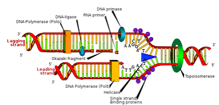
Replication fork
A dynamic, Y-shaped region where the parent DNA double helix is unwound and separated into two single strands, allowing enzymes like DNA polymerase to copy each strand
Y-Shaped Structure: Formed when the enzyme helicase breaks the hydrogen bonds between base pairs
Antiparallel Strands & Synthesis: because DNA strands are antiparallel (running in opposite 5'→ 3' and 3'→ 5' directions) and DNA polymerase can only synthesize in the 5'→ 3' direction, the two sides of the fork behave differently:
Leading Strand: synthesized continuously toward the moving replication fork
Lagging Strand: synthesized discontinuously away from the fork in short segments called Okazaki fragments (which are later joined by DNA ligase)

Replication fork
A Y-shaped region where the parent DNA double helix is unwound and separated into two single strands, allowing enzymes like DNA polymerase to copy each strand during DNA replication
A dynamic, Y-shaped region where the parent DNA double helix is unwound and separated into two single strands so that new complementary strands can be synthesized during DNA replication
Y-Shaped Structure: Formed when the enzyme helicase breaks the hydrogen bonds between base pairs
Formed at each end of a replication bubble when the enzyme DNA helicase breaks the hydrogen bonds holding the nitrogenous base pairs together
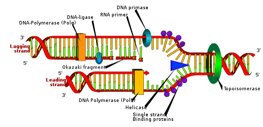
DNA polymerase proofreading activity
Mistakes during replication are detected and repaired by DNA polymerase proofreading activity
Proofreading by DNA polymerase corrects errors during replication
the built-in error-correcting process that occurs during DNA replication, where the DNA polymerase enzyme recognizes, removes, and replaces mispaired nucleotides immediately after they are added
DNA synthesis occurs 5’ → 3’, but the proofreading exonuclease removal moves backward, 3’ → 5’
Mechanism (3′ to 5′ Exonuclease Activity): If an incorrect nucleotide is detected (a mismatch like A-C or G-T), the enzyme pauses, backs up in the 3′ to 5′ direction, and cuts out the wrong nucleotide using its exonuclease domain
It happens during DNA synthesis (on the fly), not after the entire strand is finished. (For errors missed during replication, cells use a separate post-replication mechanism called mismatch repair)

DNA polymerase proofreading activity
Mistakes during replication are detected and repaired by DNA polymerase proofreading activity
Proofreading by DNA polymerase corrects errors during replication
The built-in error-correcting process that occurs during DNA replication, where the DNA polymerase enzyme recognizes, removes, and replaces mispaired nucleotides immediately after they are added
An error-checking and editing function that allows DNA polymerase to inspect newly added nucleotides against the template strand during DNA replication and immediately remove and replace mismatched bases
Mechanism (3′ to 5′ Exonuclease Activity): If an incorrect nucleotide is detected (a mismatch like A-C or G-T), the enzyme pauses, backs up in the 3′ to 5′ direction, and cuts out the wrong nucleotide using its exonuclease domain
It happens during DNA synthesis (on the fly), not after the entire strand is finished. (For errors missed during replication, cells use a separate post-replication mechanism called mismatch repair)

How does DNA polymerase proofreading work?
The built-in error-correcting process that occurs during DNA replication, where the DNA polymerase enzyme recognizes, removes, and replaces mispaired nucleotides immediately after they are added
Detection: as DNA polymerase synthesizes a new strand in the 5′ → 3′ direction, it checks the hydrogen bonding between the newly added nucleotide and the template strand. An incorrect base pairing (e.g., matching a G with an A) creates an unstable bond and alters the physical shape of the DNA double helix, causing the enzyme to pause
Excision (3’ → 5’) Exonuclease Activity): once paused, the enzyme backs up. It uses its 3’ → 5’ exonuclease domain to cleave the phosphodiester bond and remove the incorrect nucleotide from the 3’ end of the growing strand
Resynthesis: after removing the mismatch, DNA polymerase shifts back into its polymerization mode and tries again, inserting the correct complementary nucleotide and continuing elongation
DNA synthesis occurs 5’ → 3’, but the proofreading exonuclease removal moves backward, 3’ → 5’

DNA polymerase proofreading activity
Mistakes during replication are detected and repaired by DNA polymerase proofreading activity
Proofreading by DNA polymerase corrects errors during replication
the built-in error-correcting process that occurs during DNA replication, where the DNA polymerase enzyme recognizes, removes, and replaces mispaired nucleotides immediately after they are added
DNA synthesis occurs 5’ → 3’, but the proofreading exonuclease removal moves backward, 3’ → 5’
Mechanism (3′ to 5′ Exonuclease Activity): If an incorrect nucleotide is detected (a mismatch like A-C or G-T), the enzyme pauses, backs up in the 3′ to 5′ direction, and cuts out the wrong nucleotide using its exonuclease domain
It happens during DNA synthesis (on the fly), not after the entire strand is finished. (For errors missed during replication, cells use a separate post-replication mechanism called mismatch repair)

Mismatch repair
Mismatch repair (MMR) is a cellular "spell-check" mechanism that detects and corrects mispaired nucleotides (like a G paired with a T) immediately after DNA replication is complete
An incorrectly added base is detected after replication
The mismatch repair proteins detect this base and remove it from the new synthesized strand
The gap is filled with the correctly paired base
A cellular mechanism that detects and corrects mispaired nucleotides (errors) immediately after DNA replication is complete. It acts as a crucial "spell check" backup system for when DNA polymerase’s initial proofreading mechanism fails
Excision: Enzymes (endonucleases and exonucleases) cut the backbone of the faulty daughter strand and chop out a segment containing the error
Resynthesis: DNA polymerase fills the resulting gap with the correct complementary nucleotides using the original parental strand as a template
Ligation: DNA ligase seals the final nick in the sugar-phosphate backbone by forming a phosphodiester bond

Mismatch repair
Mismatch repair (MMR) is a cellular "spell-check" mechanism that detects and corrects mispaired nucleotides (like a G paired with a T) immediately after DNA replication is complete
An incorrectly added base is detected after replication
The mismatch repair proteins detect this base and remove it from the new synthesized strand
The gap is filled with the correctly paired base
A cellular mechanism that detects and corrects mispaired nucleotides (errors) immediately after DNA replication is complete. It acts as a crucial "spell check" backup system for when DNA polymerase’s initial proofreading mechanism fails

Mismatch repair
Mismatch repair (MMR) is a cellular "spell-check" mechanism that detects and corrects mispaired nucleotides (like a G paired with a T) immediately after DNA replication is complete
An incorrectly added base is detected after replication
The mismatch repair proteins detect this base and remove it from the new synthesized strand
The gap is filled with the correctly paired base
A cellular mechanism that detects and corrects mispaired nucleotides (errors) immediately after DNA replication is complete. It acts as a crucial "spell check" backup system for when DNA polymerase’s initial proofreading mechanism fails

Mismatch repair vs. Proofreading
Proofreading:
An intrinsic "spell-checking" function performed during DNA replication by the DNA polymerase enzyme itself
How it works: as DNA polymerase adds nucleotides to the growing strand, its 3' → 5' exonuclease activity immediately detects, removes, and replaces an incorrectly paired base right before continuing synthesis
Timing: active during DNA synthesis
Mismatch repair:
A secondary, post-replication quality-control system that catches and fixes errors after DNA replication has already finished.
How it works: specialized protein complexes scan the newly synthesized DNA strand, recognize structural distortions caused by mispaired bases that proofreading missed, excise the incorrect segment, and fill in the correct sequence using the parental strand as a template
Timing: occurs after DNA synthesis is completed
Nucleotide excision repair
A cellular DNA repair mechanism that removes bulky, helix-distorting DNA damage—such as UV-induced pyrimidine dimers (e.g., thymine dimers) or chemical adducts—by cutting out a short single-stranded segment around the lesion and replacing it using the undamaged strand as a template
Nucleotide excision repairs thymine dimers
When exposed to UV, thymine can form abnormal thymine dimers
In normal cells, these mutations are cut and replaced
Example: fixing thymine dimers (covalent bonds between adjacent thymines) caused by ultraviolet (UV) radiation

Nucleotide excision repair
A cellular DNA repair mechanism that removes bulky, helix-distorting DNA damage—such as UV-induced pyrimidine dimers (e.g., thymine dimers) or chemical adducts—by cutting out a short single-stranded segment around the lesion and replacing it using the undamaged strand as a template
Nucleotide excision repairs thymine dimers
When exposed to UV, thymine can form abnormal thymine dimers
In normal cells, these mutations are cut and replaced
Example: fixing thymine dimers (covalent bonds between adjacent thymines) caused by ultraviolet (UV) radiation

Nucleotide excision repair
A cellular DNA repair mechanism that removes bulky, helix-distorting DNA damage—such as UV-induced pyrimidine dimers (e.g., thymine dimers) or chemical adducts—by cutting out a short single-stranded segment around the lesion and replacing it using the undamaged strand as a template
Nucleotide excision repairs thymine dimers
When exposed to UV, thymine can form abnormal thymine dimers
In normal cells, these mutations are cut and replaced
Example: fixing thymine dimers (covalent bonds between adjacent thymines) caused by ultraviolet (UV) radiation

Telomerase enzyme
An enzyme that counteracts the shortening of linear chromosomes by elongating telomeres during DNA replication
Replication potentially shortens the ends of eukaryotic chromosomes
The ends of linear chromosomes are maintained by the action of the telomerase enzyme
prevents the progressive shortening of linear eukaryotic chromosomes during DNA replication by adding repetitive non-coding nucleotide sequences to the 3' end of the parental DNA strand
Telomerase binds to the overhanging 3' end of the parental DNA strand
It uses its internal RNA template to synthesize and add DNA repeats onto the 3' end in the 5' to 3' direction
Once the 3' overhang is sufficiently lengthened, standard DNA replication machinery (primase and DNA polymerase) can synthesize the complementary strand

Telomerase enzyme
An enzyme that counteracts the shortening of linear chromosomes by elongating telomeres during DNA replication
Replication potentially shortens the ends of eukaryotic chromosomes
The ends of linear chromosomes are maintained by the action of the telomerase enzyme
prevents the progressive shortening of linear eukaryotic chromosomes during DNA replication by adding repetitive non-coding nucleotide sequences to the 3' end of the parental DNA strand

Telomerase enzyme
An enzyme that counteracts the shortening of linear chromosomes by elongating telomeres during DNA replication
Replication potentially shortens the ends of eukaryotic chromosomes
The ends of linear chromosomes are maintained by the action of the telomerase enzyme
prevents the progressive shortening of linear eukaryotic chromosomes during DNA replication by adding repetitive non-coding nucleotide sequences to the 3' end of the parental DNA strand

Elizabeth Blackburn
Elizabeth Blackburn, 2009 Nobel Laureate, is the scientist
who discovered how telomerase works
Telomerase:
An enzyme that counteracts the shortening of linear chromosomes by elongating telomeres during DNA replication
Replication potentially shortens the ends of eukaryotic chromosomes
The ends of linear chromosomes are maintained by the action of the telomerase enzyme
prevents the progressive shortening of linear eukaryotic chromosomes during DNA replication by adding repetitive non-coding nucleotide sequences to the 3' end of the parental DNA strand
Uncorrected replication errors result in mutations
During DNA replication, DNA polymerase occasionally inserts the wrong nucleotide. While proofreading mechanisms (like DNA polymerase itself and mismatch repair enzymes) fix most of these mistakes, any errors that slip through become permanent once the cell divides. These permanent alterations are mutations
Replication is not a perfect. Errors occur and if not corrected produce mutations
Mutations are changes in the nucleotide sequence of the DNA strands
Mutations can lead to changes in the protein sequence encoded by the DNA

Uncorrected replication errors result in mutations
During DNA replication, DNA polymerase occasionally inserts the wrong nucleotide. While proofreading mechanisms (like DNA polymerase itself and mismatch repair enzymes) fix most of these mistakes, any errors that slip through become permanent once the cell divides. These permanent alterations are mutations
Replication is not a perfect. Errors occur and if not corrected produce mutations
Mutations are changes in the nucleotide sequence of the DNA strands
Mutations can lead to changes in the protein sequence encoded by the DNA

Types of mutations
Replication is not a perfect. Errors occur and if not corrected produce mutations
Mutations are changes in the nucleotide sequence of the DNA strands
Mutations can lead to changes in the protein sequence encoded by the DNA
Point Mutations (Substitutions): a single nucleotide is swapped for another. This can result in silent (no amino acid change), missense (different amino acid), or nonsense (premature stop codon) mutations
Frameshift Mutations (Insertions/Deletions): nucleotides are added or lost, shifting the triplet reading frame of the mRNA. This usually alters every amino acid downstream and drastically changes the resulting protein
Types of mutations
Point Mutations (Substitutions): a single nucleotide is swapped for another. This can result in silent (no amino acid change), missense (different amino acid), or nonsense (premature stop codon) mutations
Frameshift Mutations (Insertions/Deletions): nucleotides are added or lost, shifting the triplet reading frame of the mRNA. This usually alters every amino acid downstream and drastically changes the resulting protein
Point mutations
A change in a single nucleotide (base pair) of a DNA sequence
point mutations are typically defined specifically as a base substitution (swapping one nucleotide for another)
a genetic mutation where a single nucleotide base is substituted for a different nucleotide in a DNA sequence
Silent Mutation: the nucleotide changes, but the redundant genetic code still codes for the same amino acid (no effect on the protein)
Missense Mutation: the nucleotide change codes for a different amino acid
Nonsense Mutation: the nucleotide change turns a regular amino acid codon into a premature stop codon, cutting protein synthesis short and usually producing a nonfunctional protein

Point mutations
A change in a single nucleotide (base pair) of a DNA sequence
point mutations are typically defined specifically as a base substitution (swapping one nucleotide for another)
a genetic mutation where a single nucleotide base is substituted for a different nucleotide in a DNA sequence
a genetic mutation that occurs when a single nucleotide is substituted for a different nucleotide in a DNA sequence
3 types
Silent
Missense
Nonsense

Point mutations
A change in a single nucleotide (base pair) of a DNA sequence
point mutations are typically defined specifically as a base substitution (swapping one nucleotide for another)
a genetic mutation where a single nucleotide base is substituted for a different nucleotide in a DNA sequence
a genetic mutation that occurs when a single nucleotide is substituted for a different nucleotide in a DNA sequence
3 types
Silent
Missense
Nonsense

Point mutations - Silent
A type of point mutation (specifically a base-pair substitution) in the DNA sequence that changes a single nucleotide, but does not alter the amino acid sequence of the resulting polypeptide or protein
Mechanism: It happens when a single base changes (frequently at the 3rd "wobble" position of a codon), turning one codon into a different codon
The mutation has no effect on the amino acid because multiple distinct three-letter mRNA codons specify the exact same amino acid.
Silent mutations tend to occur at the third base of a codon
Has no effect on the protein sequence
a type of point (substitution) mutation in a gene's DNA sequence where a single nucleotide base changes, but the resulting codon still codes for the same amino acid

Silent mutations
A type of point mutation (specifically a base-pair substitution) in the DNA sequence that changes a single nucleotide, but does not alter the amino acid sequence of the resulting polypeptide or protein
Mechanism: It happens when a single base changes (frequently at the 3rd "wobble" position of a codon), turning one codon into a different codon
The mutation has no effect on the amino acid because multiple distinct three-letter mRNA codons specify the exact same amino acid.
Silent mutations tend to occur at the third base of a codon
Has no effect on the protein sequence
a type of point (substitution) mutation in a gene's DNA sequence where a single nucleotide base changes, but the resulting codon still codes for the same amino acid

Point mutations - Missense
A type of point mutation (base-pair substitution) in which a change in a single nucleotide DNA base alters an mRNA codon, resulting in the insertion of a different amino acid into the polypeptide chain during translation
It is a non-synonymous substitution because changing the nucleotide changes the encoded amino acid (unlike silent mutations, which code for the same amino acid)
Results in an amino acid substitution
a type of substitutive and nonsynonymous mutation (meaning it changes the amino acid sequence
a point mutation (a change in a single nucleotide base pair) that alters an mRNA codon so that a different amino acid is incorporated into the resulting polypeptide chain

Missense mutations
A type of point mutation (base-pair substitution) in which a change in a single nucleotide DNA base alters an mRNA codon, resulting in the insertion of a different amino acid into the polypeptide chain during translation
It is a non-synonymous substitution because changing the nucleotide changes the encoded amino acid (unlike silent mutations, which code for the same amino acid)
Results in an amino acid substitution
a type of substitutive and nonsynonymous mutation (meaning it changes the amino acid sequence
a point mutation (a change in a single nucleotide base pair) that alters an mRNA codon so that a different amino acid is incorporated into the resulting polypeptide chain

Types of missense mutations
A point mutation (a change in a single nucleotide base pair) that alters an mRNA codon so that a different amino acid is incorporated into the resulting polypeptide chain
It is a type of substitutive and nonsynonymous mutation (meaning it changes the amino acid sequence, unlike silent mutations)
Conservative Missense: the newly substituted amino acid shares similar chemical properties (e.g., polar to polar, or hydrophobic to hydrophobic) with the original amino acid. The protein's 3D shape and function are often preserved or minimally affected
Non-Conservative Missense: the newly substituted amino acid has very different chemical properties (e.g., polar to non-polar, or hydrophilic to hydrophobic). This alters protein folding, stability, or active-site function

Point mutations - Nonsense
A point mutation (a change in a single nucleotide base-pair) that converts an amino acid-coding codon into a premature stop codon (UAA, UAG, or UGA)
A point mutation (base-pair substitution) that changes an amino acid-coding codon into a premature stop codon (UAA, UAG, or UGA)
Substitutes a stop codon for an amino acid
Changes a nucleotide to a stop codon, ending translation prematurely
The ribosome reads the messenger RNA (mRNA) transcript and encounters the new stop signal too early in the coding sequence
It results in a truncated (shortened) and nonfunctional protein because translation stops before the polypeptide chain is finished

Nonsense mutation
A point mutation (a change in a single nucleotide base-pair) that converts an amino acid-coding codon into a premature stop codon (UAA, UAG, or UGA)
A point mutation (base-pair substitution) that changes an amino acid-coding codon into a premature stop codon (UAA, UAG, or UGA)
Substitutes a stop codon for an amino acid
Changes a nucleotide to a stop codon, ending translation prematurely
The ribosome reads the messenger RNA (mRNA) transcript and encounters the new stop signal too early in the coding sequence
It results in a truncated (shortened) and nonfunctional protein because translation stops before the polypeptide chain is finished

Frameshift mutations
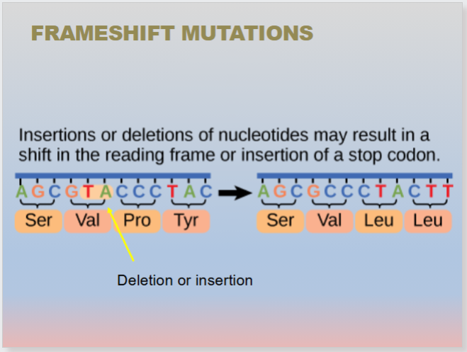
Insertions or deletions of nucleotides may result in a shift in the reading frame or insertion of a stop codon
a frameshift mutation is a change in DNA caused by inserting or deleting one or more nucleotides, which shifts the reading frame so that every codon after the mutation is misread, usually producing a nonfunctional protein.
a genetic mutation caused by the insertion or deletion of nucleotide bases in numbers not divisible by three, which shifts the triplet reading frame of the mRNA during translation
Ribosomes read mRNA transcripts in non-overlapping groups of three bases (codons). Adding or removing 1, 2, 4, or 5 bases throws off the entire downstream grouping
Downstream Effects: every amino acid encoded after the mutation site changes. It frequently introduces a premature stop codon (nonsense effect), resulting in a shortened (truncated) and usually nonfunctional protein
Frameshift mutations
Insertions or deletions of nucleotides may result in a shift in the reading frame or insertion of a stop codon
a genetic mutation caused by the insertion or deletion of nucleotide bases in a number that is not a multiple of three, which shifts the triplet reading frame of the genetic code
Ribosomes read mRNA transcripts in non-overlapping groups of three bases (codons). Adding or removing 1, 2, 4, or 5 bases throws off the entire downstream grouping
Downstream Effects: every amino acid encoded after the mutation site changes. It frequently introduces a premature stop codon (nonsense effect), resulting in a shortened (truncated) and usually nonfunctional protein
A frameshift mutation is a change in DNA caused by inserting or deleting one or more nucleotides, which shifts the reading frame so that every codon after the mutation is misread, usually producing a nonfunctional protein

Frameshift mutations
Insertions or deletions of nucleotides may result in a shift in the reading frame or insertion of a stop codon
a genetic mutation caused by the insertion or deletion of nucleotide bases in a number that is not a multiple of three, which shifts the triplet reading frame of the genetic code
a genetic mutation caused by the insertion or deletion of nucleotide bases in numbers not divisible by three, which shifts the triplet reading frame of the mRNA during translation
Downstream Effects: every amino acid encoded after the mutation site changes. It frequently introduces a premature stop codon (nonsense effect), resulting in a shortened (truncated) and usually nonfunctional protein
A frameshift mutation is a change in DNA caused by inserting or deleting one or more nucleotides, which shifts the reading frame so that every codon after the mutation is misread, usually producing a nonfunctional protein

Frameshift mutation - Insertion
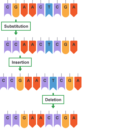
The addition of one or more nucleotide base pairs into a DNA sequence where the number of added nucleotides is not a multiple of three
Disruption of the Reading Frame: because the ribosome reads mRNA in non-overlapping groups of three nucleotides (codons), adding a number of bases not divisible by three shifts the "reading frame" of the entire message from the point of insertion onward
Downstream Amino Acid Changes: every single codon (and thus every amino acid) translated after the insertion site is altered, completely changing the downstream polypeptide sequence
the addition of one or more nucleotide base pairs into a DNA sequence, provided the number of added nucleotides is not a multiple of three; every amino acid encoded after the insertion changes
Frameshift mutation - Insertion
The addition of one or more nucleotide base pairs into a DNA sequence where the number of added nucleotides is not a multiple of three
Disruption of the Reading Frame: because the ribosome reads mRNA in non-overlapping groups of three nucleotides (codons), adding a number of bases not divisible by three shifts the "reading frame" of the entire message from the point of insertion onward
Downstream Amino Acid Changes: every single codon (and thus every amino acid) translated after the insertion site is altered, completely changing the downstream polypeptide sequence
the addition of one or more nucleotide base pairs into a DNA sequence, provided the number of added nucleotides is not a multiple of three; every amino acid encoded after the insertion changes
