Sectional Vascular Test
 Call Kai
 Learn
 Practice Test
 Spaced Repetition
 Match
 Flashcards
 Knowt Play
1/150

There's no tags or description
Looks like no tags are added yet.
Name | Mastery | Learn | Test | Matching | Spaced | Call with Kai | Chat |
|---|
No analytics yet
Send a link to your students to track their progress
151 Terms

What do the arrows represent ?
Yellow: systole (peak systole)
Orange: diastole
Compliance
blood vessels stretching with increased pressure, ability is lost in the presence of plaque build up
High resistant multiphasic/triphasic doppler velocty waveform (3 components)
1) Rapid antegrade flow reaching a peak during systole
2) Transient reversal of flow during early diastole
3) Slow antegrade flow (recoil) during late diastole
Low resistance wave form
flow profile with antegrade flow throughout the cardiac cycle (systole and diastole).
What do low resistance vascular beds need?
a constant blood supply throughout systole and diastole (high resistance vascular beds do not)
antegrade flow
flow that is going in the right direction
Extracranial
Vessels in the neck, outside the skull
Includes common carotid, external carotid (including branches), and cervical internal carotid arteries
Supplies face, neck, scalp, and flow to the Circle of Willis
Intracranial
Vessels inside the skull
Includes internal carotid (intracranial segment), vertebral-basilar system, and the Circle of Willis
Supplies brain, eyes, and meninges
Artery classification

3 Aortic Arch Branches
Supplies blood to the head, neck, and upper extremities
Brachiocephalic (Innominate) Artery → Right common carotid + right subclavian
Left Common Carotid Artery
Left Subclavian Artery
Subclavian Arteries
Supplies upper extremities, neck, and thoracic structures
Right Subclavian originates from the Brachiocephalic (Innominate)
Left Subclavian originates directly from the Aortic Arch Major branches include:
Vertebral Artery
Internal Thoracic (Internal Mammary) Artery
Thyrocervical Trunk
Costocervical Trunk
Common Carotid
Right CCA originates from the brachiocephalic trunk
Left CCA arises directly from the aortic arch
Ascends to thyroid cartilage (C2–C3) → bifurcates into ICA & ECA
Normal diameter: 0.75 – 1.25 cm
Carotid Bifurcation
Carotid bulb = dilation at bifurcation → contains baroreceptors & chemoreceptors
ECA: anterior/medial, multiple neck branches
ICA: usually posterior/lateral, no branches in neck
Common site for atherosclerotic plaque due to disturbed flow
Level of bifurcation varies → usually near thyroid cartilage (C3–C4)
Interal vs External Carotids Key Differences
collateral = backroads


Typical

Tortuous

Kinked

coiled
Vertebral Artery
First branch of subclavian artery
Enters transverse foramina at C6 → ascends to brain
Supplies ~25% of blood to brain
Segments: extracranial, intravertebral, horizontal, intracranial
left usually larger, possible trifurcation
Goals of Extracranial Duplex
Detection of nonatherosclerotic conditions
Identify patients who are at risk for stroke
Facilitate treatment
Document disease progression
Mechanisms of Cerebrovascular Symptoms
Emboli from atherosclerotic plaques
Ulcerated plaque
Reduction of flow due to high-grade stenoses (usually plaque build up)
Arterial thrombosis
Causes & Risk Factors of cerebrovascular
Atherosclerosis (≈90%)
Dissection or trauma
Fibromuscular dysplasia (FMD)
Tumors, vasculitis, compression
Primary Risk Factors of cerebrovascular
Hypertension
Diabetes mellitus
Hyperlipidemia
Smoking
Age, obesity, family history
Indications of cerebrovascular
Bruit on physical exam
Transient ischemic attack (TIA) symptoms
Amaurosis fugax (transient (temporary) monocular blindness)
Hemiparesis, syncope, vertigo
Speech issues
Stroke
Pre-op screening/clearance before
vascular/cardiac surgery
Indications for Extracranial Duplex Exam
Asymptomatic neck bruit
Hemispheric cerebral or ocular Transient Ischemic Attacks (TIAs)
History of stroke
Screening prior to surgery
Follow-up after carotid endarterectomy or stenting
General History for extracranial duples exam
Sudden onset = embolic/stroke
Transient episodes = TIA
Risk factor history
HTN
DM
Smoking
Prior vascular disease
TIA
Neurologic deficits that occur intermittently, lasting from several minutes to a few hours
Symptoms resolve within 24 hours
CVA or completed stroke
Cerebrovascular accident
FIxed or permanant neurological deficits
RIND
Reversible ischemic neurologic deficit
Neurologic deficits that last between 24 and 72 hours
Paralysis
focal weakness
Paresthesia
numbness involving some combination of face, arm, and leg on one side of body (opposite to the affected cerebral hemisphere)
Dysphasia or Aphasia
difficulty speaking or inability to speaking
Amaurosis fugax
Loss of vision in one or both eyes
What are the four symptoms of Carotid Artery lesions?
paralysis, paresthesia, dysphasia/aphasia, amaurosis fugax
What are the three symptms of vertobrobasilar insufficiency (vertebral arteries)?
dizziness, diplopia, ataxia
(less specific than carotid circulation)
diplopia
double vision
ataxia
losing coordination, balance, or speech
Arteriosclerosis
General term for hardening and loss of elasticity of arteries.
leads to restriction of blood flow to the organs and tissues.
“hard pipes”
Atherosclerosis
a specific form of arteriosclerosis causing plaque build up in the tunica intima
Plaque contains cholesterol, fat/lipids, inflammatory cells, fibrous tissue, and calcium.
Plaque can restrict blood flow and lead to stenosis
“clgged pipes”

What is the veolcity of blood at this point ?
speeds up at the narrowest part of the stenosis
What is the ICA PSV (internal coratid artery peak systole velocity) that would be concerning ?
180-230 cm/sec, causing 50-69% stenosis
Normal ICA PSV
less than 180 cm/sec (less than 50% stenosis)
greater than 230 cm/sec ICA PSV means …
greater than 70% stenosis, but less than near occlusion
Stenosis
a narrowing most often from atherosclerosis (plaque build up). Blood flows fastest
Where does stenosis commonly occur ?
in vessels going to the BRAIN (carotids), HEART (coronary arteries) and LEGS (iliac and femoral arteries).
**In abdomen we see it mesenteric arteries and renal arteries.
Carotid Exam Protocol Images
CCA (proximal, mid & distal)
CCA near flow bifurcation
ICA (ostia, proximal, mid, distal)
ECA (look for branches)
Vertebral artery
Subclavian artery
How are carotid vessels usually evaluated ?
both transverse and longitudinal planes and with B-mode imaging, color, and spectral Doppler.
Technical Standards for carotid images
Doppler angle at 60°
Record PSV & EDV at each segment
Use color Doppler to localize disturbed flow
B-Mode / Grayscale normal characteristics
Smooth vessel walls
Intimal-medial layer clearly visible and uniform (thin, gray-white line on innermost part of vessel wall)
Lumen is anechoic.
Carotid Bulb
Doppler from CCA into proximal ICA
Document flow separation in bulb
Flow reversal located along outer wall of bulb
Indicates normal flow in bulb
Velocity measurement is not needed on this signal.
Internal Carotid Artery: Scanning Technique
Must evaluate the entire length
proximal, mid, and distal segments
Measure peak systolic velocity (PSV) and end diastolic velocity (EDV) throughout
Distal segment may be difficult beyond the angle of the jaw
External Artery: Scanning Technique
ICA vs. ECA Differentiation
ECA has multiple branches within the neck.
ECA spectral waveform will oscillate with “temporal tap.”
Note:
ICA typically is larger (contains bulb).
ICA typically lies posterior to ECA.
Vertebral Artery
Place transducer at anteromedial aspect of midneck on long axis
Identify CCA and slide or angle posteriorly to identify vertebral artery between transverse processes of vertebrae
Take care to identify flow direction and waveform contour
Spectral Doppler
Normal Doppler arterial flow through the CCA.
Forward flow throughout the cardiac cycle (systole and diastole)
Systole is when the heart contracts and pumps out blood.
Diastole is when the heart relaxes and fills with blood.
Spectral Doppler ICA vs ECA
ICA is lower resistance than the ECA.
Lower resistance means the vascular bed the flow is going to needs more flow. So, your BRAIN (lower resistance) needs more flow than your FACE (higher resistance). Therefore, the ICA has a lower resistance waveform example more flow in diastole.
Triphasic Doppler velocity waveform
made up of three components which correspond to different phases of arterial flow: rapid antegrade flow reaching a peak during systole, transient reversal of flow during early diastole, and slow antegrade flow during late diastole
low resistance waveform:
flow profile with antegrade flow throughout the cardiac cycle (systole and diastole).
Triphasic Doppler velocity waveform:
made up of three components which correspond to different phases of arterial flow:
rapid antegrade flow reaching a peak during systole
transient reversal of flow during early diastole
slow antegrade flow during late diastole
Would the legs (vascular bed) be high resistance as you are sitting in class? What about when you are running a marathon?
hight when sitting class because more resistance = less flow and your legs don’t need a lot of blood flow when you’re sitting down
low when running a marathon because less resistance=more flow and your legs need blood to run
Claudication:
walking-induced muscular discomfort of thigh, hip, buttock due to ischemia
cramping pain in the leg is induced by exercise, typically caused by obstruction of the arteries.
relieved by resting 2 to 5 minutes.
Physical signs: elevation pallor and dependent rubor, ischemic ulcers, gangrene, bruits, and decreased peripheral pulses
Peripheral Arterial Disease
narrowed blood vessels, often from atherosclerotic plaque decreased blood flow to the limbs.
Risk factors of Peripheral Arterial Disease
Increasing age
Hypertension
Diabetes mellitus
Elevated cholesterol
Tobacco smoking
Documented atherosclerosis in coronary or carotid system
Family history of atherosclerosis
Symptoms of Peripheral Arterial Disease
claudication and pain at rest.
Weakened Pulses
Duplex Ultrasound
the method of choice for imaging DVT (deep vein thrombosis).
Can be used to diagnose, localize, and determine age of thrombus
Also uncover vascular and nonvascular incidental findings as
Very sonographer dependent
Duplex ultrasound is used to assess ….
Presence or absence of thrombus
Relative risk of thromboembolism
Competence of contained valves
Virchow’s triad
Virchow’s triad
Venous stasis
Hypercoagulability
Vessel wall injury
Where is thrombosis more common ?
upper extremities: due to injury to vessels wall
More frequent introduction of needles and catheters into arm veins
Signs & Symptoms of DVT (Write Out)
Extremity pain and tenderness
Swelling
Venous distention
Discoloration
Palpable cord
Pulmonary Embolism (PE)
Signs and symptoms of Pulmonary embolism (WO)
Tachypnea
Chest pain
Tachycardia
Clinical markers that can aid in diagnosis of DVT and PE include
Well’s criteria
D-dimer
Both have poor sensitivity and specificity
Superficial Phlebitis
Affects veins near skin surface (superficial)
Symptoms:
Tenderness, redness, warmth
Hardness or swelling along vein
Visible red line, itching, mild fever
Usually mild but should be evaluated
Deep Vein Phlebitis (DVT)
Involves deeper leg veins
Symptoms:
Significant pain, swelling (one leg)
Warmth, skin discoloration
Pain with walking or flexing foot
Complications if untreated:
Pulmonary embolism (PE)
Chronic venous insufficiency
Venous ulcers
Common Causes of superficial and deep phlebitis
Blood clots after surgery, injury, or IV lines
Immobility (long flights, hospital stays)
Poor circulation, smoking, or inactivity
pregnancy
Medical conditions: cancer, varicose veins
Excess weight adds vein pressure
Patient Preparation & Positioning for phlebetis patients
Bed should be tilted in reversed Trendelenburg position.
Patient lie flat on back with knee slightly bent and hip slightly externally rotated.
Allows veins to fill with blood, so easier visualize.
Legs dangled off edge of bed to visualize calf veins
Some legs need more probe pressure to compress
Deep veins
Main conduit for blood returning to the heart
Accompany (travel with) an artery of the same name
Higher risk for embolism due to squeezing action of surrounding muscles
contained within the muscular fascia
Thrombus in deep system is usually larger than in superficial system—more likely to be life-threatening pulmonary embolism (PE).
Superficial veins
saphenous trunks and tributaries.
Travel close to skin surface
lie between the dermis and muscular fascia
Smaller than deeper veins
do not have an artery
Play a major role in regulating body temperature
Less likely to cause life-threatening PE as thrombus is smaller and veins are not surrounded by muscles
Greater potential for embolus if thrombus is near junction with deep system
Communicating system:
veins connect the superficial to deep systems
Perforators cross muscular fascia; tributaries cross saphenous fascia.
Scanning technique for lower venous in transverse plane
Exam begins at groin crease
Require transducer compressions
Transverse plane, pressure is applied over the vein using transducer.
Veins under relatively low pressure, walls will collapse/close together.
Compressions performed every 2–3 cm down the entire leg.
Scanning technique for lower venous in sag plane
After compressions are completed, the vein evaluated in longitudinal /sag plane.
Compressions are NOT performed in longitudinal plane.
Color and spectral Doppler are performed in longitudinal plane.
Assess for spontaneity, phasicity with respiration, augmentation with distal compression, and cessation of flow with proximal compression
Common Femoral Vein
transverse plane: lies next to common femoral artery (CFA) just below inguinal ligament
CFV is medial to the CFA
Great Saphenous Veins
Courses medially and superficially to CFV
enters femoral vein at saphenofemoral junction
Longest vein in the body
Travels close to the skin in saphenous compartment
Becomes more anterior below the knee
Small Saphenous Vein
Superficial vein that terminates into popliteal vein (saphenopopliteal junction)
May terminate into FV or even GSV
courses along middle of posterior calf.
Receives tributaries from medial and lateral calf
Vein of Giacomini
extension of SSV above popliteal fossa
Gastrocnemius Veins
Small tributaries that join into popliteal vein
Drain gastrocnemius muscles
Paired veins with accompanying artery
When isolated painful calf muscle look here for clot.
Soleal Sinus Veins
Major storage area for blood in the calf
Common site of thrombus formation
Blood only moves out when calf muscles contract
Prone to thrombus formation due to stagnation (following surgery, long plane trip, etc.)
Communicate with PTV and peroneal veins
Thrombus can easily extend into deep system.
Doppler signals should display the following:
Spontaneous Doppler signals in all major vessels
Respiratory phasicity
Augmentation with distal compression
Cessation of flow with proximal compression or Valsalva maneuver
Flow should be unidirectional toward the heart.

1
common tibial trunk

2
great saphenous vein

3
perforating veins
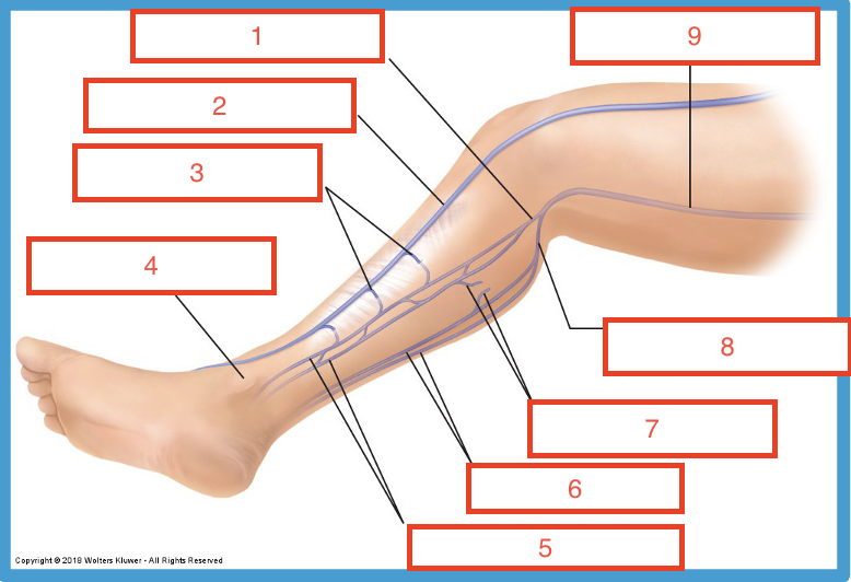
4
medial malleolus

5
posterior tibial veins

6
peroneal veins

7
soleal veins
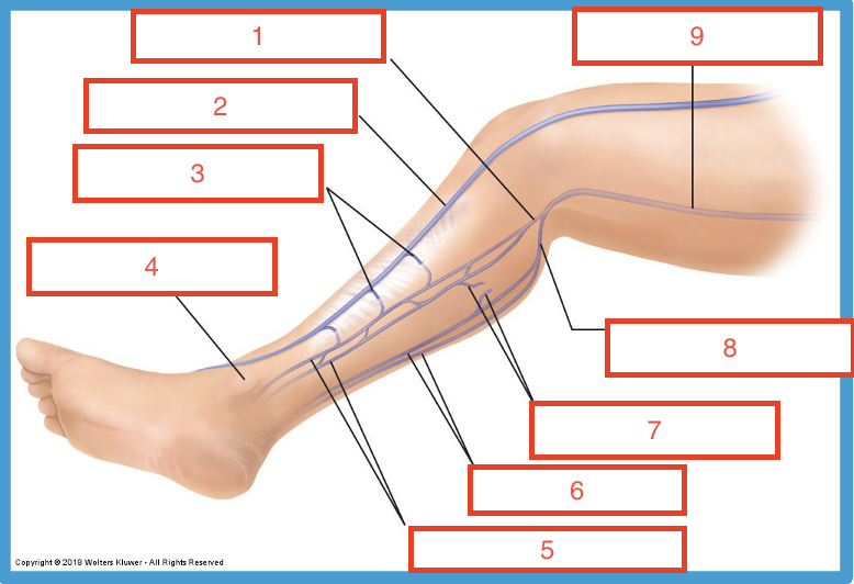
8
common peroneal trunk

9
popliteal vein

1
inferior vena cava

2
common iliac vein
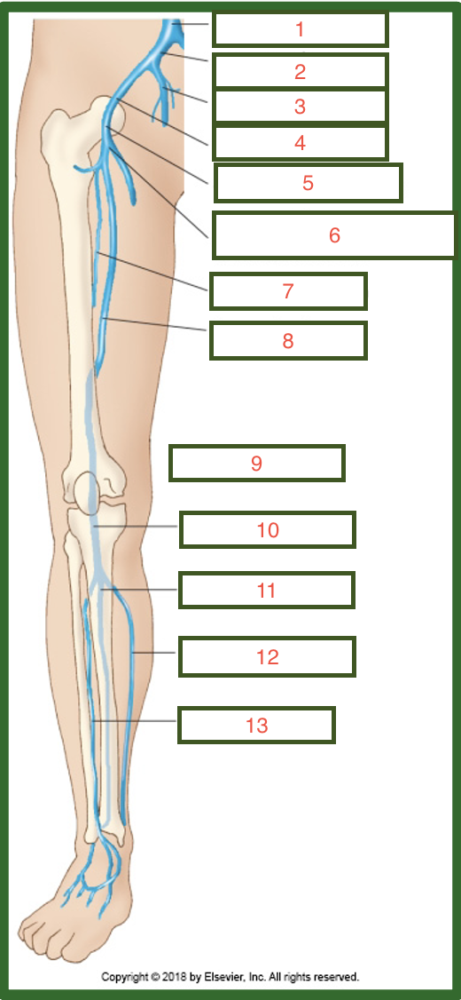
3
internal iliac vein

4
external iliac vein
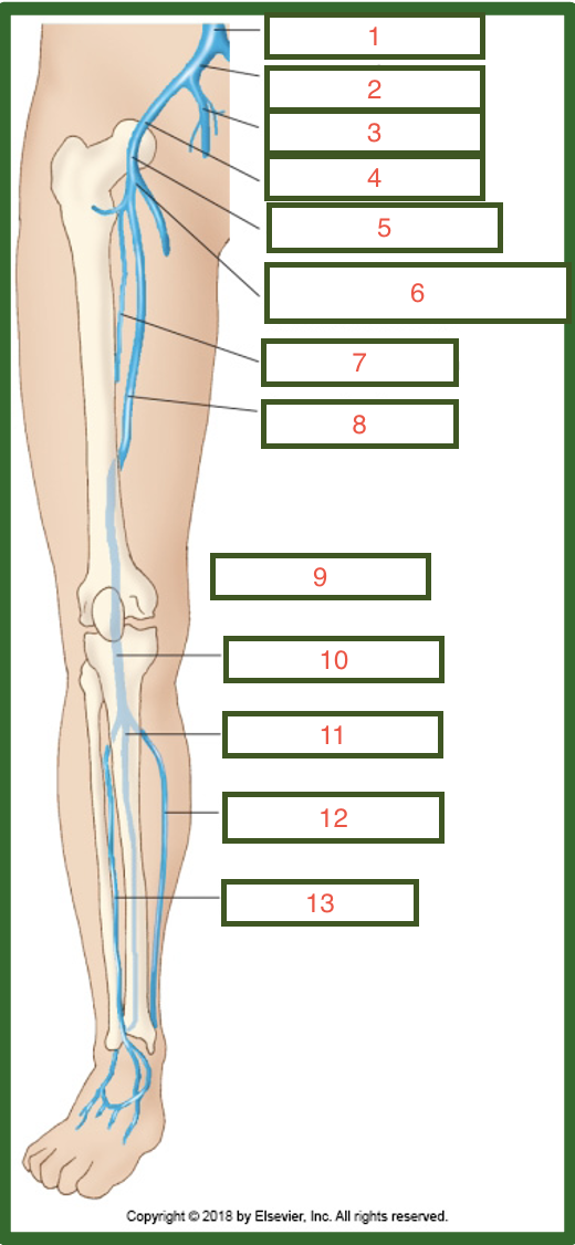
5
common femoral vein