L28 - Laboratory assessment of Bone marrow Pt.2
 Call Kai
 Learn
 Practice Test
 Spaced Repetition
 Match
 Flashcards
 Knowt Play
1/65

There's no tags or description
Looks like no tags are added yet.
66 Terms
What is the Main Goal of Bone Marrow Cell Identification?
Identify erythroid, granulocytic, and megakaryocytic lineages and their maturation stages
What Distinguishes Immature from Mature Hematopoietic Cells?
Immature:
Fine chromatin
Nucleoli
Basophilic cytoplasm
Mature:
Condensed chromatin
Differentiated cytoplasm
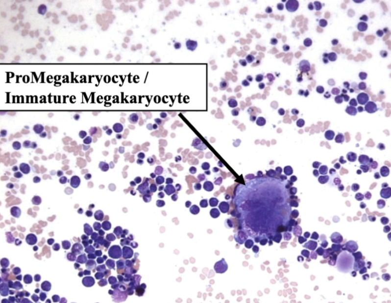
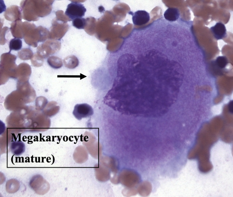
What are the Megakaryocytic Maturation Stages?
Megakaryoblast → Promegakaryocyte → Mature megakaryocyte → Platelets

How do Immature and Mature Megakaryocytes Differ?
Immature: less, deeply blue cytoplasm
Mature: abundant granular cytoplasm that releases platelets

RECOGNIZING MEGAKARYOCYTIC CELLS
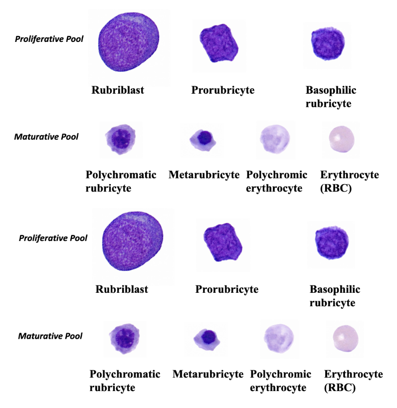
What are the Erythroid Maturation Stages?
Rubriblast → Prorubricyte → Basdophilic rubricyte → Rubricyte → Metarubricyte → Polychromatophil (Reticulocyte) → Mature RBC

How Can Proliferative Erythroid Precursors Be Recognized?
Round cells
High N:C ratio
Extremely dark-blue cytoplasm
How Can Maturative Erythroid Precursors Be Recognized?
Smaller cells
Condensed nuclei
Increasingly pink cytoplasm from hemoglobin accumulation
How do Rubriblasts and Metarubricytes Differ?
Rubriblasts: large, fine chromatin and nucleoli
Metarubricytes: small, densely condensed nuclei


RECOGNIZING ERYTHROID PRECURSOR CELLS
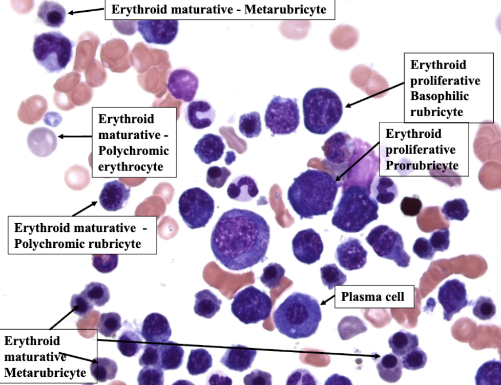
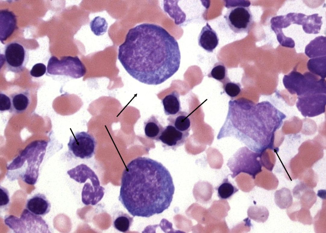

What Distinguishes Erythroid from Granulocytic Precursors?
Erythroid cells are rounder and often darker blue
Granulocytic cells are less round and may contain granules

What are the Granulocytic Maturation Stages?
Myeloblast → Promyelocyte → Myelocyte → Metamyelocyte → Band → Segmented granulocyte

Which Cells Belong to the Granulocytic Proliferative Pool?
Myeloblast → Promyelocyte → Myelocyte
These stages can divide
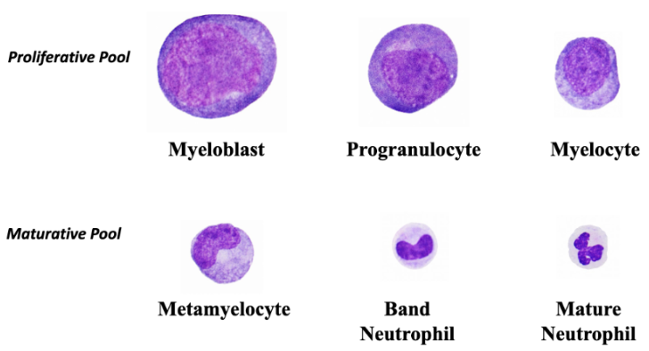
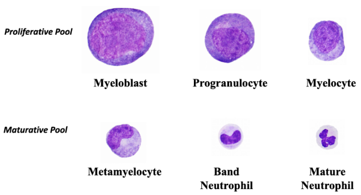
Which Cells Belong to the Granulocytic Maturative Pool?
Metamyelocyte → Band → Segmented granulocyte
They mature without dividing
Which Cells Belong to the Granulocytic Storage Pool?
Mature segmented granulocytes, including neutrophils and eosinophils
What Nuclear Feature Identifies Maturative Granulocytic Cells?
Indented, band-shaped, or segmented nuclei indicate granulocytic maturation
How Can Metamyelocytes Be Distinguished from Bands?
Metamyelocytes have indented or kidney-shaped nuclei
Bands have elongated horseshoe-shaped nuclei

How Can Eosinophil Precursors Be Recognized?
Prominent eosinophilic cytoplasmic granules
Eosinophils also have myelocyte, metamyelocyte, and band stages.


RECOGNIZING NEUTROPHIL PRECURSOR CELLS


RECOGNIZING NEUTROPHIL PRECURSOR CELLS


RECOGNIZING NEUTROPHIL PRECURSOR CELLS




What Indicates Active Proliferation in Bone Marrow?
Mitotic figures among hematopoietic precursors
Why Should Mature Cells Outnumber Immature Precursors?
Early precursors divide repeatedly, producing many more mature descendants


How Can Plasma Cells Be Distinguished from Erythroid Precursors?
Plasma cells have abundant blue cytoplasm, an eccentric nucleus, and sometimes a pale Golgi zone

What is the Normal Significance of Marrow Lymphocytes?
Small mature lymphocytes normally form a minority, usually under 5%
cats may have more
What Can Cause Increased Marrow Lymphocytes?
Immune-mediated disease, infections such as FeLV/FIV, or lymphoid neoplasia
Why do Plasma Cells Increase in Marrow with Age?
Long-lived plasma cells home to and accumulate in bone marrow


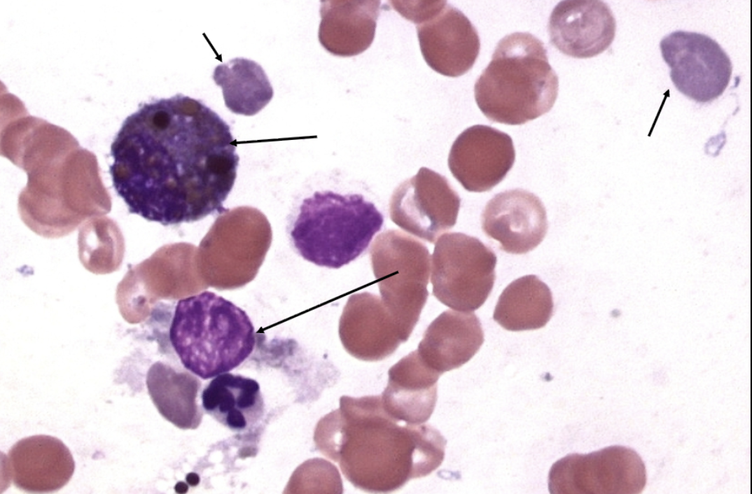

What is the Function of Hemosiderin-Containing Macrophages?
They contain stored iron derived from degraded erythrocytes
What Can Increased Marrow Macrophages Suggest?
Inflammation
Infection
Immune-mediated disease
Histiocytic disorders
What is a Skipocyte?
A damaged or lysed cell remnant, sometimes appearing as a free nucleus

How do Polychromatophils Relate to Reticulocytes?
Polychromatophils appear blue-gray on Romanowsky stains
Reticulocytes require a supravital stain

Why Must Bone Marrow Findings Be Compared with the CBC?
To determine whether marrow production and maturation adequately explain circulating cell counts
What Does a G:E Ratio of 10:1 Suggest in an Anemic Dog?
Relative erythroid deficiency
With hypocellularity and rare erythroid cells, marked erythroid hypoplasia
What is the Interpretation of the 7-Year-Old Dog Case?
A 7 yr old Dog has marked persistent, nonregenerative anemia (Hct of 12%). A bone marrow aspirate is performed.
Cytologic description: The sample contains many unit particles that are moderately hypocellular. The G:E ratio is 10:1. Only rare erythroid precursors are noted.
Hct 12%, hypocellular marrow, G:E 10:1 → marked erythroid hypoplasia to aplasia
What are the Main Differentials for Pure Red Cell Aplasia?
Immune-mediated destruction of early erythroid precursors
Selective erythroid toxicity
What Does a G:E Ratio of 1:6 Suggest in an Anemic Dog?
Marked erythroid predominance
With hypercellularity, erythroid hyperplasia.
What is the Interpretation of the Middle-Aged Dog Case?
A middle aged dog has moderate, nonregenerative anemia (Hct of 20%). A bone marrow aspirate is performed.
Cytologic description: The sample contains a few unit particles that are mildly hypercellular. The G:E ratio is 1:6. There is a moderate increase in erythroid precursors with a left shift (increase in immature erythroid cells) in the erythroid lineage.
Hct 20%, G:E 1:6, immature erythroid predominance → erythroid hyperplasia with a left shift
Why Can Erythroid Hyperplasia Occur with Nonregenerative Anemia?
Ineffective erythropoiesis, as in PIMA
Early regenerative response not yet evident in blood
What is PIMA?
Precursor immune-mediated anemia: immune injury to erythroid precursors within bone marrow
How Does PIMA Differ from Classic IMHA?
PIMA targets marrow erythroid precursors
Classic IMHA predominantly destroys circulating RBCs
Why Was Marrow Evaluated in Clinical Case #1?
5 yr old FS Terrier X
Hct = 11.7 % (2,300 retics / ul) (RI- 40-55%)
WBC = 5,780 / ul, neutrophils = 4,566/ul, lymphocytes 578/ul
Plts = 44,000 / ul (ref interval = 150 – 400k/ul)
TP = 6.2 gm/dl (RI 6-8 gm/dl)
The terrier had bicytopenia: severe nonregenerative anemia and thrombocytopenia


What Were the Key Marrow Findings in Case #1?
5 yr old FS Terrier X
Hct = 11.7 % (2,300 retics / ul) (RI- 40-55%)
WBC = 5,780 / ul, neutrophils = 4,566/ul, lymphocytes 578/ul
Plts = 44,000 / ul (ref interval = 150 – 400k/ul)
TP = 6.2 gm/dl (RI 6-8 gm/dl)
Marked hypercellularity
Megakaryocytic hyperplasia
Erythroid left shift
Erythrophagocytosis

What Does Erythrophagocytosis of Nucleated Precursors Suggest?
Destruction of developing erythroid cells within marrow, supporting PIMA
What Does Megakaryocytic Hyperplasia Suggest in Case #1?
5 yr old FS Terrier X
Hct = 11.7 % (2,300 retics / ul) (RI- 40-55%)
WBC = 5,780 / ul, neutrophils = 4,566/ul, lymphocytes 578/ul
Plts = 44,000 / ul (ref interval = 150 – 400k/ul)
TP = 6.2 gm/dl (RI 6-8 gm/dl)
Active platelet production despite thrombocytopenia, suggesting peripheral platelet loss or destruction
What Was the Diagnosis in Clinical Case #1?
5 yr old FS Terrier X
Hct = 11.7 % (2,300 retics / ul) (RI- 40-55%)
WBC = 5,780 / ul, neutrophils = 4,566/ul, lymphocytes 578/ul
Plts = 44,000 / ul (ref interval = 150 – 400k/ul)
TP = 6.2 gm/dl (RI 6-8 gm/dl)
PIMA with concurrent immune-mediated thrombocytopenia
An Evans syndrome–type presentation


Why Was Marrow Evaluated in Clinical Case #2?
6 yr old FS Std Poodle
CBC - Erythrogram WNL, mild leukopenia (4100/ul [RI 6-13K/ul] due to severe lymphopenia, 80 cells/ul, neuts = 3570/ul) and moderate thrombocytopenia. Occasional “atypical cells” in blood
Hypercalcemia - 15 mg/dl (9.7-11.5 mg/dl).
The poodle had unexplained atypical circulating cells and hypercalcemia

What Marrow Features Suggested Neoplasia in Case #2?
6 yr old FS Std Poodle
CBC - Erythrogram WNL, mild leukopenia (4100/ul [RI 6-13K/ul] due to severe lymphopenia, 80 cells/ul, neuts = 3570/ul) and moderate thrombocytopenia. Occasional “atypical cells” in blood
Hypercalcemia - 15 mg/dl (9.7-11.5 mg/dl).
Focal abnormal immature cells with high N:C ratios
Fine chromatin
Nucleoli
Frequent mitoses
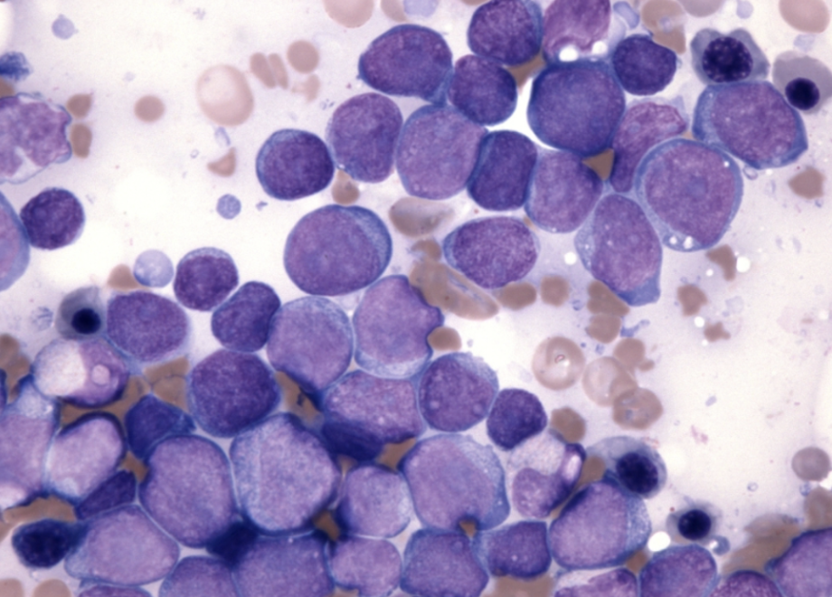

What Does Positive CD3 Staining Establish in Case #2?
T-cell lineage of the abnormal infiltrating cells

What Was the Diagnosis in Clinical Case #2?
6 yr old FS Std Poodle
CBC - Erythrogram WNL, mild leukopenia (4100/ul [RI 6-13K/ul] due to severe lymphopenia, 80 cells/ul, neuts = 3570/ul) and moderate thrombocytopenia. Occasional “atypical cells” in blood
Hypercalcemia - 15 mg/dl (9.7-11.5 mg/dl).
T-cell lymphoma with extensive bone marrow involvement (stage V)
Why Was Marrow Evaluated in Clinical Case #3?
14 yr old MC Labrador
Hct = 34.9% (46,900 retics / ul) (RI 40-55%, 7,000 – 65,000 retics / ul)
WBC = 9,430 / ul (RI 6,000 – 13,000/ul)
Plts = 815,000 / ul (ref interval = 150 – 400k/ul)
TP = 9.8 gm/dl, Alb = 2.4 gm / dl (RI=3-4.4) and Globs = 7.4 gm/dl ( 1.8-3.9 gm/dl)
The Labrador had marked hyperglobulinemia and a monoclonal gammopathy
What Does a Monoclonal Spike on Electrophoresis Suggest?
Production of a relatively uniform immunoglobulin by a clonal cell population


What Were the Key Marrow Findings in Case #3?
14 yr old MC Labrador
Hct = 34.9% (46,900 retics / ul) (RI 40-55%, 7,000 – 65,000 retics / ul)
WBC = 9,430 / ul (RI 6,000 – 13,000/ul)
Plts = 815,000 / ul (ref interval = 150 – 400k/ul)
TP = 9.8 gm/dl, Alb = 2.4 gm / dl (RI=3-4.4) and Globs = 7.4 gm/dl ( 1.8-3.9 gm/dl)
Markedly increased plasma cells, supporting plasma-cell neoplasia

What Was the Diagnosis in Clinical Case #3?
14 yr old MC Labrador
Hct = 34.9% (46,900 retics / ul) (RI 40-55%, 7,000 – 65,000 retics / ul)
WBC = 9,430 / ul (RI 6,000 – 13,000/ul)
Plts = 815,000 / ul (ref interval = 150 – 400k/ul)
TP = 9.8 gm/dl, Alb = 2.4 gm / dl (RI=3-4.4) and Globs = 7.4 gm/dl ( 1.8-3.9 gm/dl)
Multiple myeloma, supported by monoclonal gammopathy and marked marrow plasma-cell infiltration
TABLE 1. Hematopoietic Cell Lineages and Order of Development
Lineage | Order of Development | Main Recognition Feature |
Erythroid | Rubriblast → Prorubricyte → Basdophilic rubricyte → Rubricyte → Metarubricyte → Polychromatophil (Reticulocyte) → Mature RBC | Round cells; cytoplasm changes from dark blue to pink |
Granulocytic | Myeloblast → Promyelocyte → Myelocyte → Metamyelocyte → Band → Segmented granulocyte | Nuclear indentation and segmentation increase with maturity |
Megakaryocytic | Megakaryoblast → Promegakaryocyte → Mature megakaryocyte → Platelets | Exceptionally large cells; cytoplasm increases with maturation |
Key distinction: Most developing hematopoietic cells become smaller as they mature, whereas megakaryocytes become larger.
TABLE 2. Immature vs. Mature Hematopoietic Cells
Feature | Immature Cells | Mature Cells |
Cell size | Generally larger | Generally smaller |
N:C ratio | High | Lower |
Chromatin | Fine, loosely condensed | Coarse, condensed |
Nucleoli | Often visible | Usually absent |
Cytoplasm | Often deeply basophilic (blue) | More differentiated |
Erythroid cytoplasm | Dark blue | Increasingly pink with hemoglobin |
Granulocytic nucleus | Round or oval | Indented → band-shaped → segmented |
Cell division | Early stages can divide | Terminal stages do not divide |
Exam tip: If you're unsure of the exact precursor name, first identify its lineage, then determine whether it is immature or mature.
TABLE 3. Proliferative, Maturative, and Storage Pools
Lineage | Proliferative Pool | Maturative Pool | Storage / Final Stage |
Granulocytic | Myeloblast → promyelocyte → myelocyte | Metamyelocyte → band | Segmented granulocyte |
Erythroid | Rubriblast → prorubricyte → early rubricyte | Later rubricyte → metarubricyte | Polychromatophil → mature RBC |
Megakaryocytic | Megakaryoblast → promegakaryocyte | Mature megakaryocyte | Platelet release |
For the erythroid lineage, the table uses final stage rather than a true storage pool, because erythroid cells are not generally described as having the same marrow storage pool as granulocytes.
Remember: Proliferative cells divide; maturative cells undergo terminal differentiation; the granulocytic storage pool contains mature cells available for release.
TABLE 4. Other Cells Found in Normal Bone Marrow
Cell | Identification | Significance |
Lymphocyte | Small cell with dense round nucleus and scant cytoplasm | Normally a minority of marrow cells; may increase with immune stimulation or neoplasia |
Plasma cell | Eccentric nucleus, abundant deep-blue cytoplasm, sometimes a pale Golgi zone | Long-lived antibody production; may increase with age |
Macrophage | Large cell with abundant cytoplasm, sometimes containing engulfed material | Phagocytosis and recycling of cellular components |
Hemosiderin- containing macrophage | Macrophage with dark iron pigment | Indicates stored iron from RBC degradation |
Skipocyte | Damaged cell remnant or free nucleus | Artifact of cellular damage, not a separate hematopoietic lineage |
Polychromatophil | Anucleate RBC with blue-gray cytoplasm | Young RBC; corresponds to a reticulocyte on supravital staining |
Identification tip: Plasma cells can resemble proliferative erythroid cells because both have blue cytoplasm. Look for the plasma cell's eccentric nucleus and possible pale Golgi zone.
TABLE 5. Interpreting Abnormal Bone Marrow Findings
Marrow Finding | Interpretation | Possible Diagnosis |
Hypocellular marrow + markedly decreased erythroid precursors | Inadequate RBC production | Erythroid hypoplasia / pure red cell aplasia |
Hypercellular marrow + erythroid hyperplasia + left shift | Increased immature erythroid precursors, but inadequate maturation or release | Ineffective erythropoiesis, including PIMA |
Erythrophagocytosis of nucleated RBC precursors | Destruction of erythroid precursors within marrow | Supports PIMA |
Megakaryocytic hyperplasia + thrombocytopenia | Marrow is producing platelets, but peripheral counts remain low | Peripheral platelet destruction or consumption |
Atypical immature cells infiltrating marrow | Abnormal cell proliferation replacing normal hematopoiesis | Leukemia or lymphoma involvement |
Markedly increased plasma cells + monoclonal gammopathy | Clonal plasma-cell proliferation | Multiple myeloma |
Two Important Marrow Interpretation Examples
Finding | Example 1 | Example 2 |
Patient | 7-year-old dog | Middle-aged dog |
CBC | Persistent nonregenerative anemia; Hct 12% | Nonregenerative anemia; Hct 20% |
Marrow cellularity | Hypocellular | Mildly hypercellular |
G:E ratio | 10:1 | 1:6 |
Erythroid precursors | Markedly decreased | Increased, predominantly immature |
Interpretation | Marked erythroid hypoplasia to aplasia | Erythroid hyperplasia with left shift |
Main consideration | Pure red cell aplasia | Ineffective erythropoiesis, especially PIMA |
Critical exam distinction: Both dogs are nonregeneratively anemic, but their bone marrow findings are very different.
Example 1: The marrow is not producing enough erythroid cells.
Example 2: The marrow contains many erythroid precursors, but they are not successfully progressing to mature circulating RBCs.
TABLE 6. Professor's Clinical Cases — Summary
Feature | Case 1: Terrier | Case 2: Poodle | Case 3: Labrador |
Reason for marrow evaluation | Nonregenerative anemia + thrombocytopenia | Atypical circulating cells + hypercalcemia | Hyperglobulinemia + monoclonal gammopathy |
Key marrow findings | Erythroid left shift, erythrophagocytosis, megakaryocytic hyperplasia | Infiltration by abnormal immature cells | Markedly increased plasma cells |
Additional evidence | Macrophages engulf nucleated erythroid precursors | CD3-positive neoplastic cells | Monoclonal spike on electrophoresis |
Diagnosis | PIMA + immune-mediated thrombocytopenia | T-cell lymphoma with marrow involvement (stage V) | Multiple myeloma |
Key lesson | Immune destruction can occur within marrow | Neoplastic infiltration can replace normal hematopoiesis | Clonal plasma-cell proliferation can cause monoclonal gammopathy |