Histo lab 7,8: Integumentary and Endocrine System Quiz7
 Call Kai
 Learn
Learn Practice Test
 Spaced Repetition
 Match
Match Flashcards
 Knowt Play
1/131

There's no tags or description
Looks like no tags are added yet.
Name | Mastery | Learn | Test | Matching | Spaced | Call with Kai | Chat |
|---|
No analytics yet
Send a link to your students to track their progress
132 Terms
Identify the layer of the skin
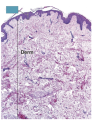
Epidermis
Identify the layer of the skin

Dermis
Identify the type of skin

Thick skin
Identify the type of skin

Thin skin

Identify the layer of the skin
Stratum Basale

Identify the specific layer

Stratum basale

Identify the specific layer
Stratum basale
Identify the specific layer

Stratum spinosum
Identify the specific layer

Stratum spinosum
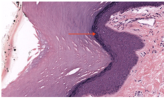
Identify the specific layer
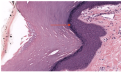
Stratum granulosum

Identify the specific layer
Stratum granulosum
Identify the specific layer (RED ARROW)

Stratum granulosum
Identify the specific layer

Stratum granulosum
Identify the specific layer
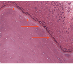
Stratum lucidum
Identify the specific layer

Stratum corneum
Identify the specific layer

Stratum corneum
Identify the specific layer
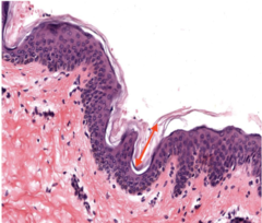
Stratum corneum

Identify the specific layer
Papillary dermis
Identify the specific layer (YELLOW ARROWS)
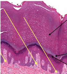
Papillary dermis
Identify the specific layer

Papillary dermis
Identify the specific layer

Reticular dermis
Identify the specific layer
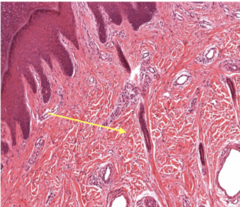
reticular dermis
Identify the specific layer

reticular dermis
Identify the specific layer

hypodermis
Identify the cells

at border of stratum basale
Keratinocytes
Identify the cells

Melanocytes
Identify the cell
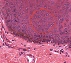
keratinocyte
Identify the cell
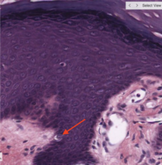
melanocyte
Identify the cells
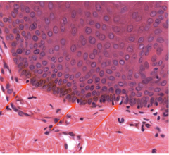
melanocytes
Identify the cell

melanocyte
Identify the structure

Hair shaft
Identify the structure

Hair follicle
Identify the structure

Dermal papilla of hair follicle
Identify the structure

Sebaceous gland
Identify the structure

Arrector pili muscle
Identify the structure

Sebaceous glands
Identify the structure

secrete sebum → hair follicle via holacrine secretion
Sebaceous glands

Identify the structure

Hair shaft

Identify the structure
hair follicle
Identify the structure

forms hair shaft
Hair bulb

Identify the structure (red box)

Hair root
Identify the structure (green line)
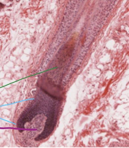
Hair root
Identify the structure (Blue line)

Hair bulb
Identify the structure (purple line)

Dermal papilla of hair follicle
Identify the structure

Dermal papilla of hair follicle
Identify the structure

Sebaceous gland
Identify the structure

Sebaceous gland
Identify the structure
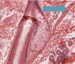
arrector pili muscle
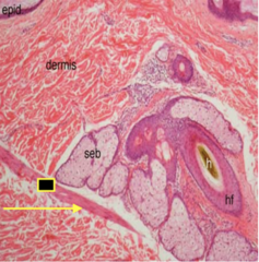
Identify the structure

Arrector pili muscle
Identify the structure

Dermal papilla of papillary dermis
Identify the structure (YELLOW)

Sebaceous glands
Identify the structure (BLACK)

Hair follicle
Identify the structure (GREEN)

Eccrine sweat gland
Eccrine glands occur over most of body and open directly onto skin surface except in lips and ___ skin and part of external genitalia
glabrous
what are the two types of sweat glands?
eccrine and apocrine sweat glands
which sweat gland type has the apical portion of the secretory cell cytoplasm shed into the lumen, whose protein/lipid-rich secretions become functional at puberty to play a role in human body odor and pheromonal signaling?
Apocrine sweat glands
which sweat gland type is responsible for the exocytosis of watery sweat with no loss of cellular cytoplasm?
**(merocrine)
eccrine sweat glands
Identify the structure (RED)
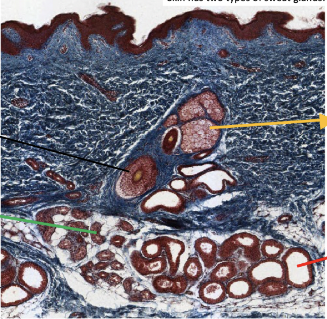
Apocrine sweat gland

Identify the structure
Apocrine sweat gland
which sweat gland type opens into hair follicle, leading to surface of skin (axilla, areola, nipple of mammary gland, skin around anus & external genitalia)?
apocrine sweat gland
Identify the structure

Nail plate

Identify the structure
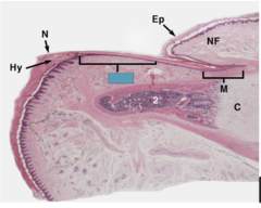
Nail bed
Identify the structure

in the dermal papilla of dermis - for light touch/textures
Meissner's Touch corpuscle

Identify the structure (2)
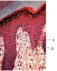
1 - stratum basale
Meissner’s Touch corpuscle
Identify the structure

Pacinian corpuscle
which corpuscle is found in the reticular dermis and hypodermis and is for coarse touch, preassure, & vibration?
pacinian corpuscle

Identify the structure

Pacinian corpuscle

Identify the structure

Pacinian corpuscle

Identify the organ

Pituitary gland

Identify the organ

~unmyelinated
posterior pituitary gland

Identify the organ

Anterior pituitary gland

Identify the cells

Acidophils

Identify the cells

Basophils
Identify the region of the organ

Adenohypophysis

Identify the cells

chromophobes
Identify the region of the organ

~responsible for synthesis of own hormones
adenohypophysis
Identify the cell
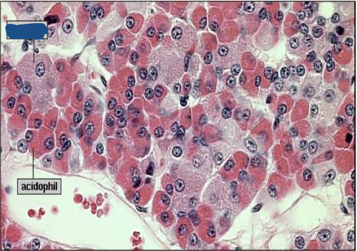
Basophil
Identify the cell

Acidophil

Identify the region of the organ

Neurohypophysis
the posterior pituitary is also known as what? why?
neurohypophysis; it’s pars nervosa
the neurohypophysis has axons from the _______ that release _____ into the pars nervosa
hypothalamus, hormones
neurohypophysis’ neurosecretory cells secrete what?
ADH and oxytocin
what type of glial cells surround the axons in the neurohypophysis?
pituicytes
what occurs in the ventral portion of the neurohypophysis where it’s capillary rich?
most hormone release
Identify the organ

Pituitary gland
Identify the region of the organ (1)

adenohypophysis
Identify the region of the organ (2)

bottom image: notice colloid-filled cyst on (#2; top image) of the anterior lobe
pars intermedia
Identify the region of the organ (3)
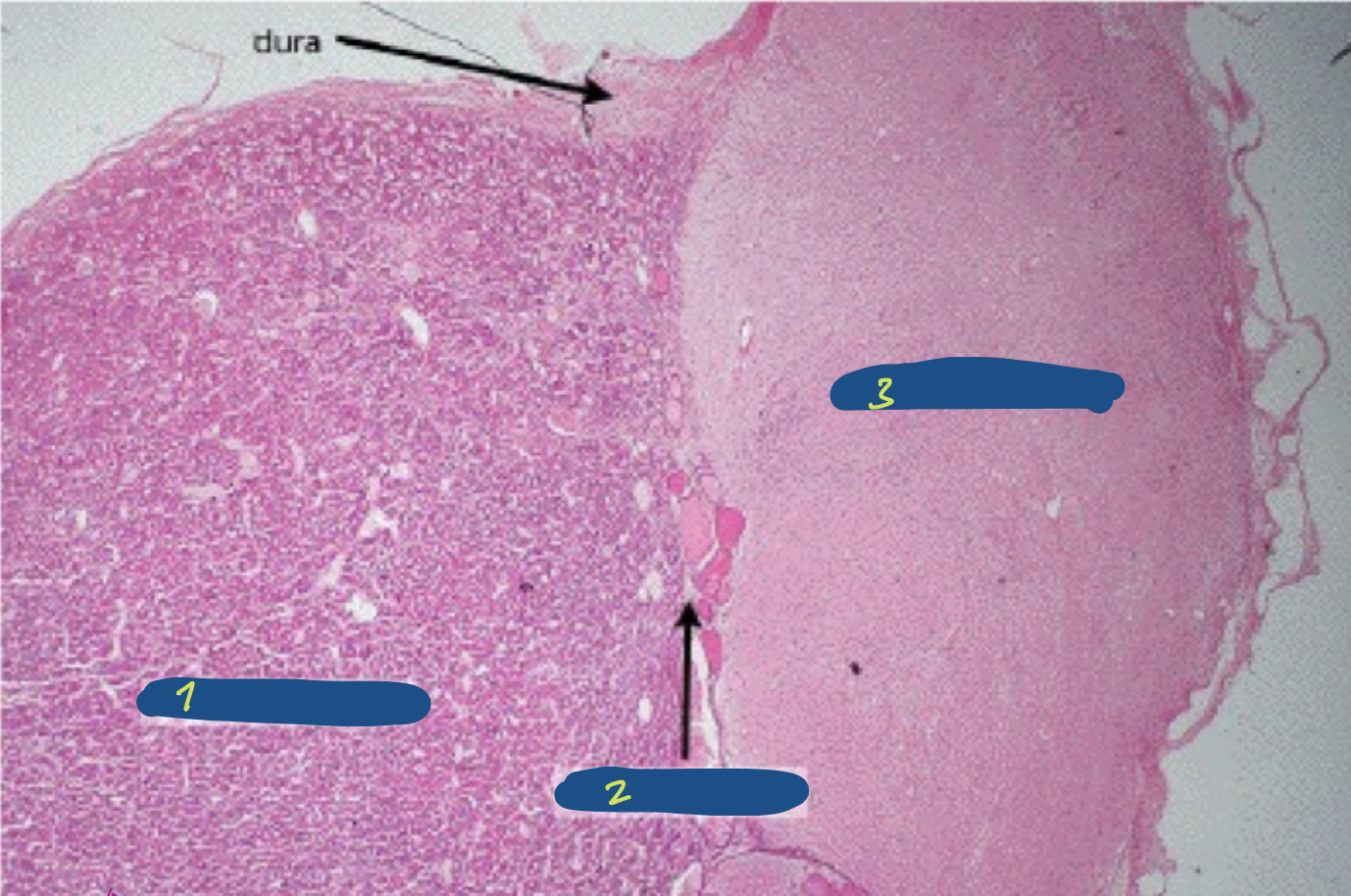
(pars nervosa)
Neurohypophysis
Identify the cells

Pituicytes

Identify the organ
Pineal gland
Identify the cells (1)

(synthesize & secrete melatonin)
pinealocytes
Identify the structure (2)
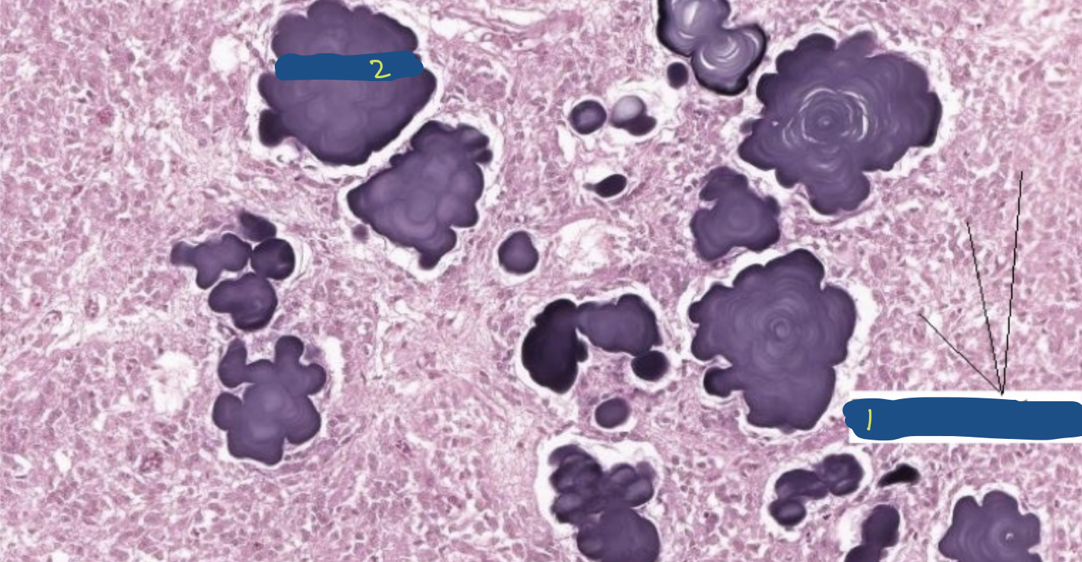
[calcium salt]; accumulates with age
brain sand
brain sand AKA
Copora arenacea
Identify the organ

Pineal gland
Identify the organ

Thyroid gland
the thyroid gland is a bi-lobed endocrine gland surrounded by CT with two types of secretory cells called ______ &______ cells.
Which makes up most of the thyroid cells ? —(hollow balls)
what do they contain? (acidophilic gelatinous)
follicular, parafollicular; follicular cells; colloid
Identify the organ (red square)

Thyroid gland
Identify the structure

thyroid gland
thyroid follicle
Identify the substance

Colloid
Identify the cells

follicular cells