6. Musculoskeletal System: Lower Extremity
 Call Kai
 Learn
 Practice Test
 Spaced Repetition
 Match
 Flashcards
 Knowt Play
1/243

There's no tags or description
Looks like no tags are added yet.
244 Terms
lower extremity bones
pelvic girdle
2 pelvic bones
sacrum + coccyx (axial skeleton)
thigh
femur
lower leg
tibia
fibula
ankle + foot
tarsals
metatarsals
phalanges

pelvic bone
3 bones fused together to lateral acetabulum + obturator foramen
ilium
ischium
pubis

ilium
part of pelvic bone
has
anterior → iliac fossa
posterior → gluteal surface
anterior superior iliac spine (ASIS)
anterior inferior iliac spine (AIIS)

ischium
part of pelvic bone
features ischium tuberosity

pubis
part of pelvic bone

pelvic girdle joints
sacroiliac joint
plane joint
fibrous joint components
sacrococcygeal joint
plane joint
pubic symphysis
symphysis
pelvic joint
ball-and-socket joint

hip joint
articulation b/w femoral head + acetabulum of pelvic bone
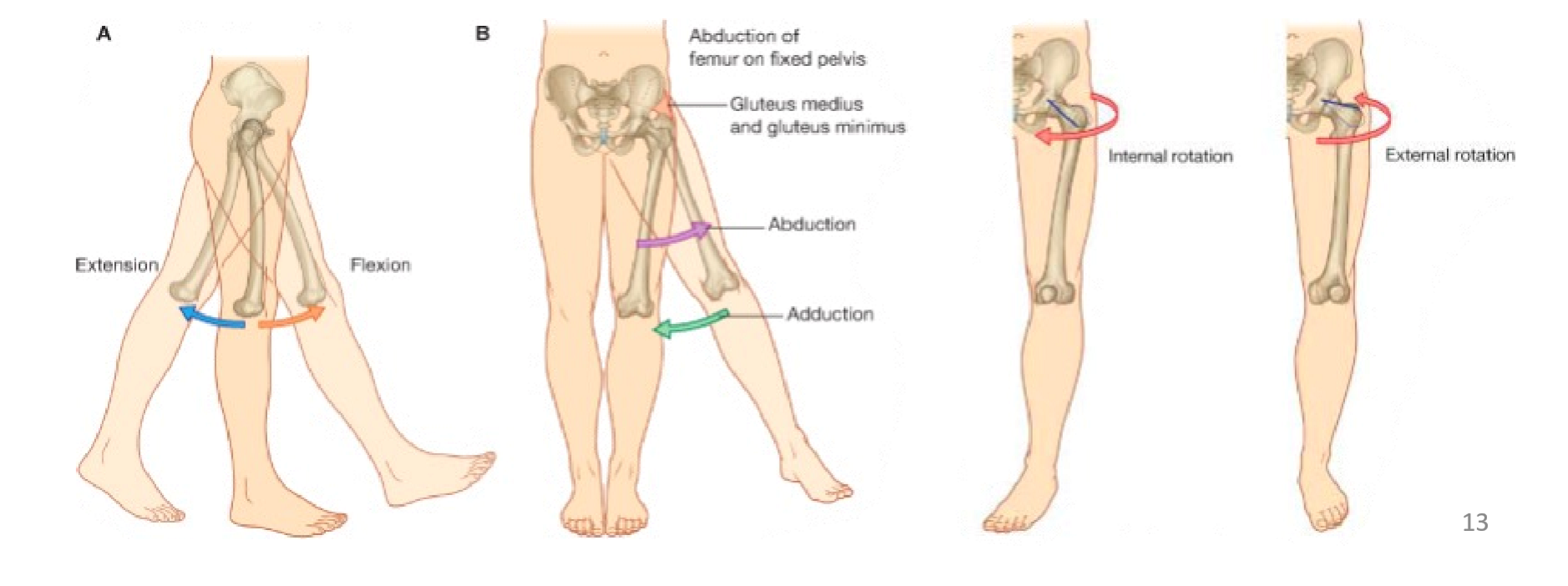
triaxial ball-and-socket joint
movements
flexion + extension
abduction + adduction
internal medial + external lateral rotation
hip joint muscles
anterior
iliopsoas = iliacus + psoas major
flexion
posterior
gluteus maximus
extension
gluteus medius
abduction
gluteus minimus
abduction
deep lateral rotators
piriformis
obturators internus + externus
superior + inferior gemelli
quadratus femoris
lateral rotation at hip joint

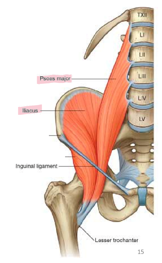
iliopsoas
acts on hip joint → flexion at hip joint
formed by iliacus + psoas major
origin
sides of lumbar vertebral bodies + iliac fossa
passes deep to inguinal ligament
insertion
lesser trochanter of femur
gluteus maximus
acts on hip joint → extension at hip joint
origin
ilium
sacrum
insertion
gluteal tuberosity of femur
iliotibial tract

gluteus medius and minimus
acts on hip joint → abduction at hip joint
origin
posterior ilium (gluteal surface)
insertion
greater trochanter of femur

deep lateral rotators
act on hip joint → lateral rotation at hip joint
muscles
piriformis
obturators internus + externus
superior + inferior gemelli
quadratus femoris
origin
anterior sacrum
posterior ischium
obturator foramen
runs posterior to hip joint
insertion
greater trochanter of femur

proximal lower limb arteries
aorta
common iliac artery
internal iliac artery
pelvic organs
gluteal muscles
external iliac artery
lower limb
femoral artery
profunda femoris artery
popliteal artery

proximal lower limb veins
deep veins
share names w/ associate arteries
superficial veins
dorsal venous arch
great saphenous vein → medial
drains into femoral vein
small saphenous vein → lateral
drains into popliteal vein
blood from lower limb veins drain to external iliac vein → drain into inferior vena cava

lower limb innervation
lumbar plexus = L1-L4
major nerves
femoral nerve
anterior thigh compartment
obturator nerve
medial thigh compartment
connects to sacral plexus via lumbosacral trunk = L4-L5
lumbosacral plexus = lumbosacral trunk + S1-S4
major nerve
sciatic nerve + branches
posterior thigh compartment
compartments of lower leg + foot

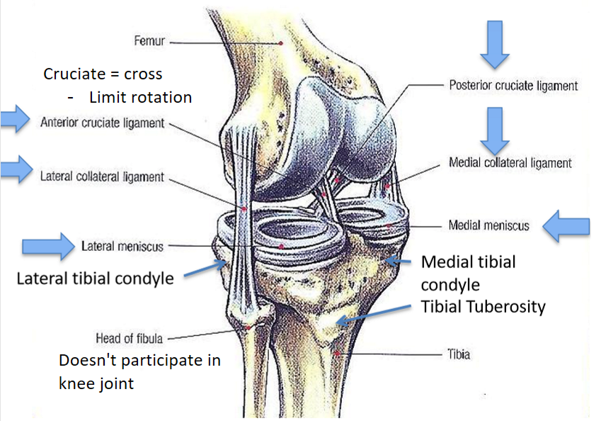
knee joint
biaxial modified hinge joint
consists of
femoral condyles articulate w/ tibial plateau
medial + lateral collateral ligaments
anterior + posterior cruciate ligaments (ACL + PCL)
medial + lateral menisci (fibrocartilage)
optimize joint congruency
shock absorption
sits on tibial plateau + acts as cushion
movements
flexion + extension
slight medial + lateral rotation
locking + unlocking of knee
knee locking
knee moves into extension
slight medial rotation of femur
tightening of collateral ligaments
full extension = reinforces extended state + prevents buckling

knee unlocking
flex knee from extended position
popliteus contracts
slight lateral rotation of femur on tibia
knee ligaments loosened

popliteus
deep muscle posterior to joint
attachments to distal femur + proximal tibia
action
unlocks knee
contraction → slight rotation of femur on tibia
loosens knee ligaments

cruciate ligaments
attach to sides of intercondylar fossa + tibial plateau
action
limit rotational movement at knee joint
2 structures
anterior cruciate ligament (ACL)
posterior cruciate ligament (PCL)

anterior cruciate ligament (ACL)
attach to sides of intercondylar fossa + tibial plateau
action
limit rotational movement at knee joint
restraint against anterior translation of tibia compared to femur
prevents tibia from moving too far anteriorly

posterior cruciate ligament (PCL)
attach to sides of intercondylar fossa + tibial plateau
action
limit rotational movement at knee joint
restraint against posterior translation of tibia compared to femur
prevents tibia from moving too far posteriorly

anterior thigh compartment
muscles
sartorius
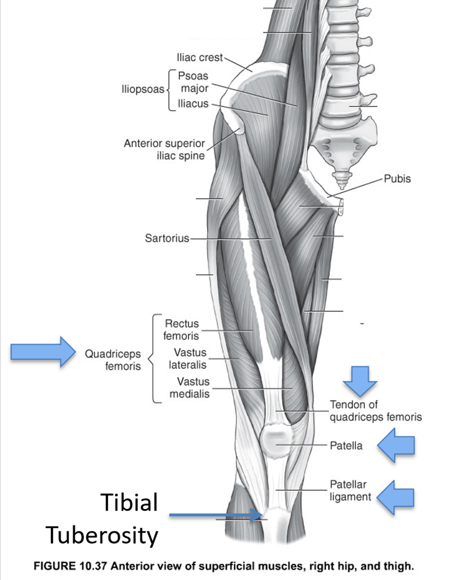
quadriceps femoris
rectus femoris
vastus lateralis, intermedius, medialis
action
sartorius → flexion at hip and knee
quadriceps femoris → extension at knee joint
innervation
femoral nerve

quadriceps femoris
part of anterior thigh compartment → flexion at knee joint
4 muscles
rectus femoris
vastus lateralis
vastus intermedius
vastus medialis
origin
RF → anterior superior iliac spine
others → proximal anterior femur
insertion
tibial tuberosity via patella + patellar ligament
innervation
femoral nerve
sartorius
part of anterior thigh compartment → flexion at hip and knee
origin
anterior superior iliac spine (ASIS)
insertion
medial proximal tibia
innervation
femoral nerve

hamstrings
part of posterior thigh compartment → flexion at knee joint
3 muscles
biceps femoris (BF)
long + short heads
semitendinosus (ST)
semimembranosus (SM)
origin
ischial tuberosity
insertion
BF → lateral tibial condyle/head of fibula
ST + SM → medial tibial condyle
innervation
sciatic nerve

adductors
part of medial thigh compartment → adduction at hip joint
4 muscles
pectineus
adductor longus
adductor brevis
adductor magnus
origin
pubis
insertion
linea aspera of femur
adductor magnus → adductor tubercle
innervation
obturator nerve

adductor hiatus
hole in adductor magnus for passage of popliteal artery + vein to posterior knee

gracilis
part of medial thigh compartment → adduction at hip joint
part of adductors
origin
pubis
insertion
medial proximal tibia
innervation
obturator nerve

sciatic nerve
innervates
posterior thigh compartment
muscles of lower leg + foot
exits pelvis via greater sciatic foramen
descends b/w medial + posterior compartments
divides into tibial + common peroneal (fibular) branches at popliteal fossa

femoral artery
external iliac artery passes deep to inguinal ligament + enters thigh → becomes _____ artery
major branch = profunda femoris artery
runs deep + parallel
descends b/w anterior + medial compartments of thigh
passes through adductor hiatus to enter posterior knee → becomes popliteal artery

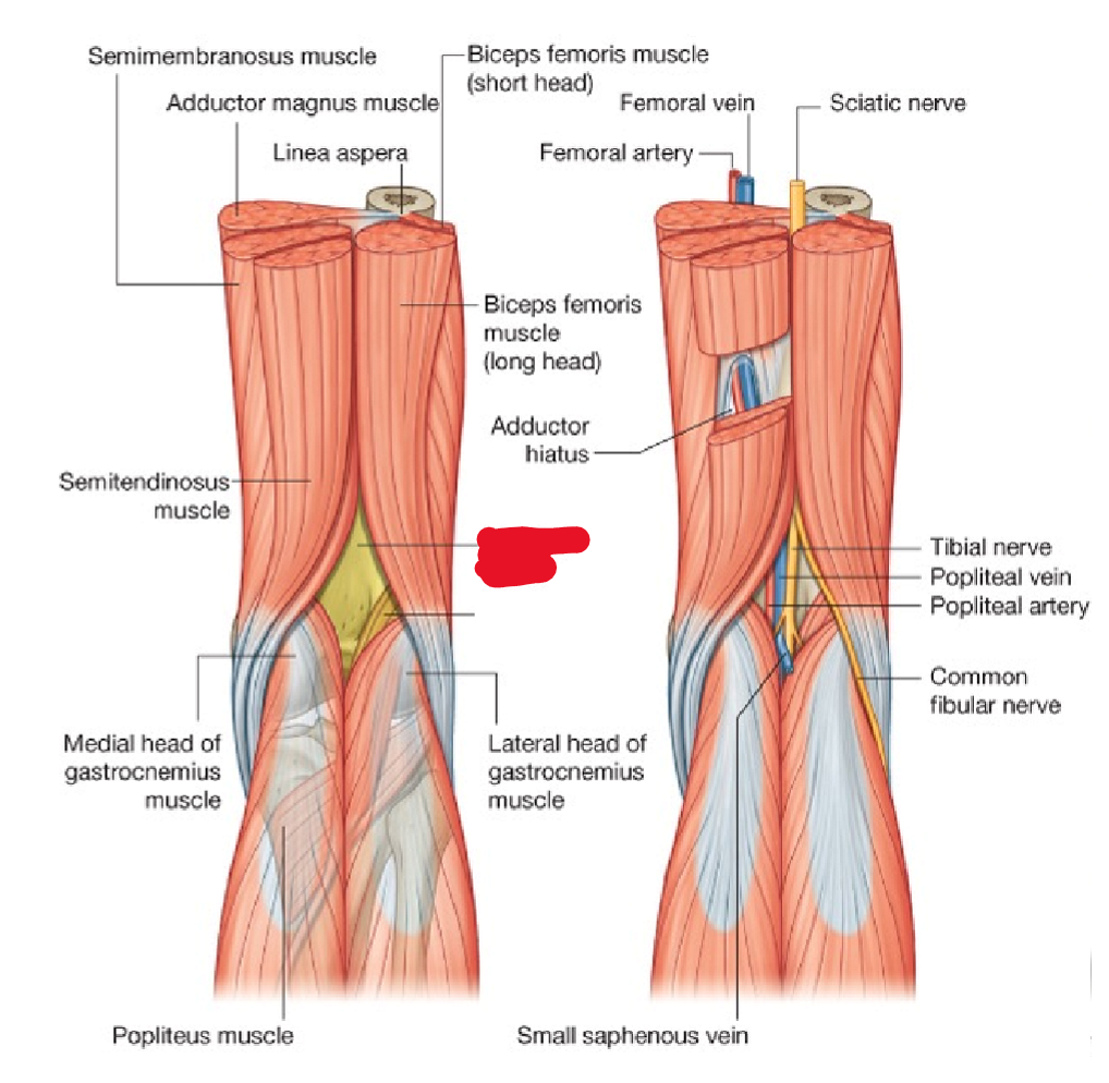
popliteal fossa
superficial landmark behind knee
boundaries
superior border
biceps femoris
semitendonosus
semimembranosus
inferior border
gastrocnemius
contents
popliteal artery + vein
tibial + common peroneal (fibular) nerves → derived from sciatic nerve
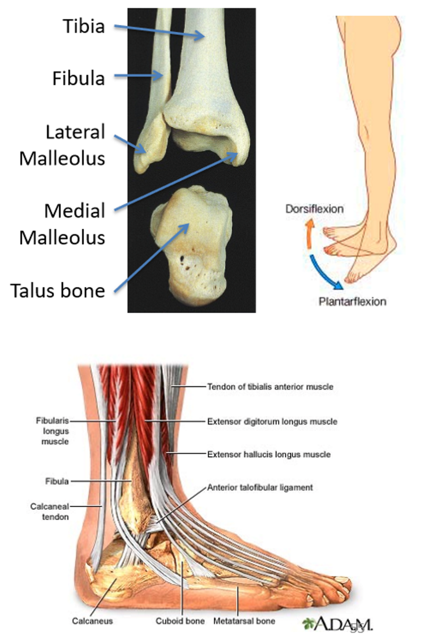
ankle joint
articulation b/w distal tibia + fibula, some talus bone
uniaxial hinge joint
supported by collateral ligaments, tendons running distally into foot
movements
dorsiflexion
plantarflexion
anterior lower leg compartment
muscles
tibialis anterior (TA)
right against shin bone
extensor digitorum longus (EDL)
extensor hallucis longus (EHL) → big toe
origin
anterior tibia
fibula
interosseous membrane
insertion
distal to ankle joint
EDL + EHL tendons out to respective digits
action
dorsiflexion at ankle joint
EDL + EHL → extension of digits
innervation
deep peroneal nerve

superficial posterior lower leg compartment
muscles
gastrocnemius
soleus
action
plantar flexion at ankle joint
innervation
tibial nerve

gastrocnemius
part of superficial posterior lower leg compartment → plantar flexion at ankle joint
origin
medial + lateral femoral condyles
insertion
calcaneus via calcaneal (Achilles) tendon
innervation
tibial nerve

soleus
part of superficial posterior lower leg compartment → plantar flexion at ankle joint
origin
posterior proximal tibia + fibula
insertion
calcaneus via calcaneal (Achilles) tendon
innervation
tibial nerve

medial thigh compartment
muscles
adductors
pectineus
adductor longus, brevis, magnus
gracilis
action
adduction at hip joint
innervation
obturator nerve

posterior thigh compartment
muscles
hamstrings
biceps femoris
semitendinosus
semimembranosus
action
flexion at knee joint
innervation
sciatic nerve

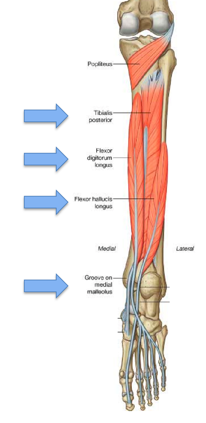
deep posterior lower leg compartment
muscles
tibialis posterior (TP)
flexor digitorum longus (FDL)
flexor hallucis longus (FHL)
origin
posterior tibia + fibula
interosseous membrane
tendons pass posterior to medial malleolus
insertion
distal to ankle joint
FDL + FHL → respective digits
action
plantar flexion at ankle joint
FDL + FHL only → flexion of digits
innervation
tibial nerve
lateral lower leg compartment
muscles
fibularis (peroneus) longus
fibularis (peroneus) brevis
origin
along length of fibula
pass posterior to lateral malleolus
insertion
longus → onto 1st metatarsal
brevis → onto 5th metatarsal
action
plantar flexion at ankle joint
eversion of foot → movement at subtalar joint
support arches of foot
innervation
superficial peroneal nerve

sciatic nerve branches
2 branches
tibial nerve
superficial + deep layers of posterior compartment of lower leg
common peroneal (fibular) nerve
superficial peroneal (fibular) nerve
lateral compartment of lower leg
deep peroneal (fibular) nerve
anterior compartment of lower leg

popliteal artery
2 branches
anterior tibial artery
moves anteriorly through proximal hiatus in interosseous membrane
supplies anterior compartment of lower leg
posterior tibial artery
moves distally b/w superficial + deep layers of posterior compartment of lower leg
supplies posterior compartment of lower leg

piriformis
deep lateral rotator muscle → lateral rotation at hip

obturators internus and externus
deep lateral rotator muscle → lateral rotation at hip

superior and inferior gemelli
deep lateral rotator muscle → lateral rotation at hip

quadratus femoris
deep lateral rotator muscle → lateral rotation at hip

important bony landmarks

rectus femoris
anterior compartment of thigh → extension at knee joint
part of quadriceps femoris
origin
anterior superior iliac spine (ASIS)
insertion
tibial tuberosity via patella + patellar ligament
innervation
femoral nerve

vastus lateralis
anterior compartment of thigh → extension at knee joint
part of quadriceps femoris
origin
proximal anterior femur
insertion
tibial tuberosity via patella + patellar ligament
innervation
femoral nerve

vastus intermedius
anterior compartment of thigh → extension at knee joint
part of quadriceps femoris
origin
proximal anterior femur
insertion
tibial tuberosity via patella + patellar ligament
innervation
femoral nerve

vastus medialis
anterior compartment of thigh → extension at knee joint
part of quadriceps femoris
origin
proximal anterior femur
insertion
tibial tuberosity via patella + patellar ligament
innervation
femoral nerve

biceps femoris
posterior compartment of thigh → flexion at knee joint
part of hamstrings
origin
ischial tuberosity
insertion
lateral tibial condyle/head of fibula
innervation
sciatic nerve

semimembranosus
posterior compartment of thigh → flexion at knee joint
part of hamstrings
origin
ischial tuberosity
insertion
medial tibial condyle
innervation
sciatic nerve
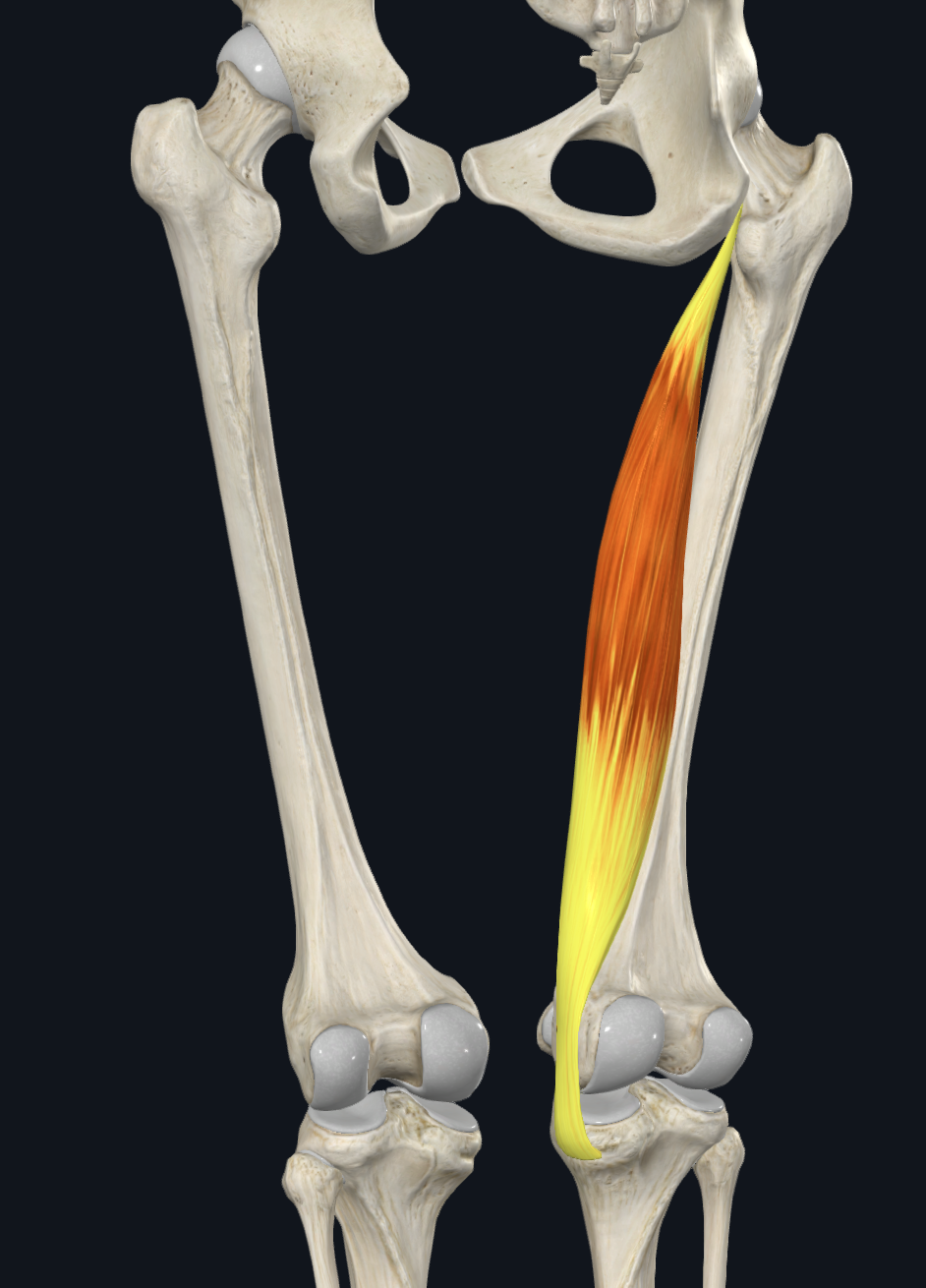
semitendinosus
posterior compartment of thigh → flexion at knee joint
part of hamstrings
origin
ischial tuberosity
insertion
medial tibial condyle
innervation
sciatic nerve

pectineus
medial compartment of thigh → adduction at hip joint
part of adductors
origin
pubis
insertion
linea aspera of femur
innervation
obturator nerve

adductor longus
medial compartment of thigh → adduction at hip joint
part of adductors
origin
pubis
insertion
linea aspera of femur
innervation
obturator nerve

adductor brevis
medial compartment of thigh → adduction at hip joint
part of adductors
origin
pubis
insertion
linea aspera of femur
innervation
obturator nerve

adductor magnus
medial compartment of thigh → adduction at hip joint
part of adductors
origin
pubis
insertion
adductor tubercle
innervation
obturator nerve
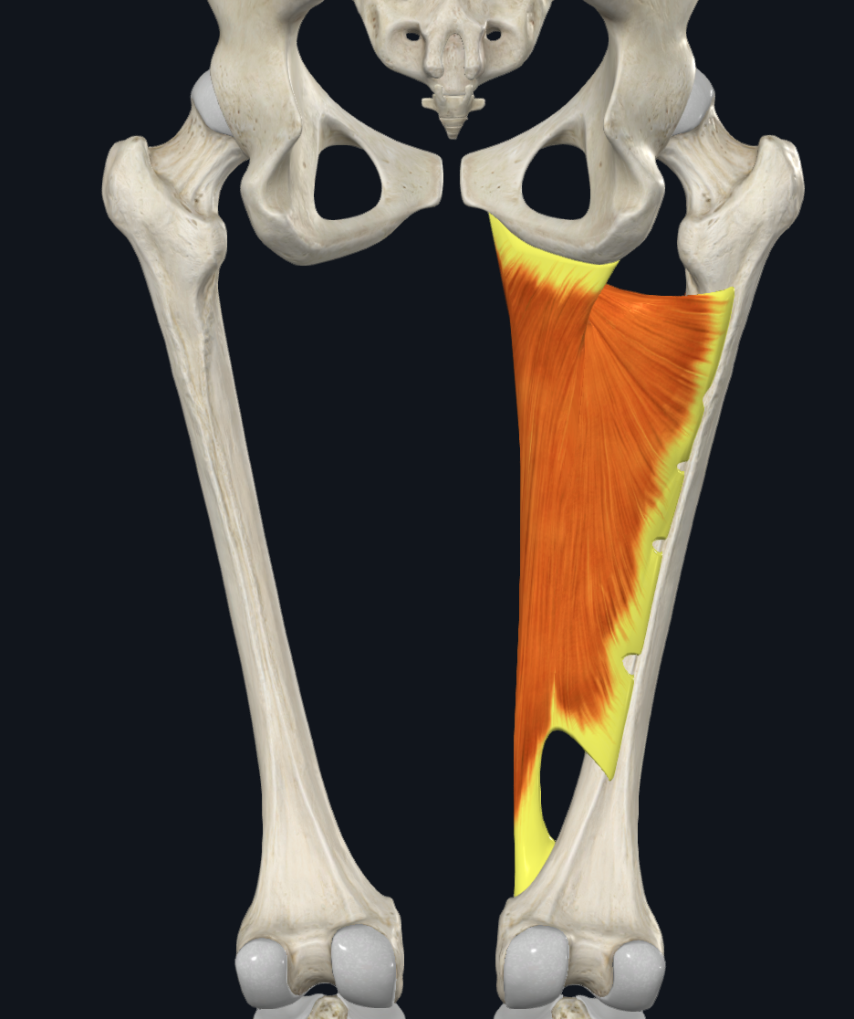
tibialis anterior
anterior compartment of lower leg → dorsiflexion at ankle joint
origin
anterior tibia (lateral condyle)
interosseous membrane
insertion
distal to ankle joint
innervation
deep peroneal nerve

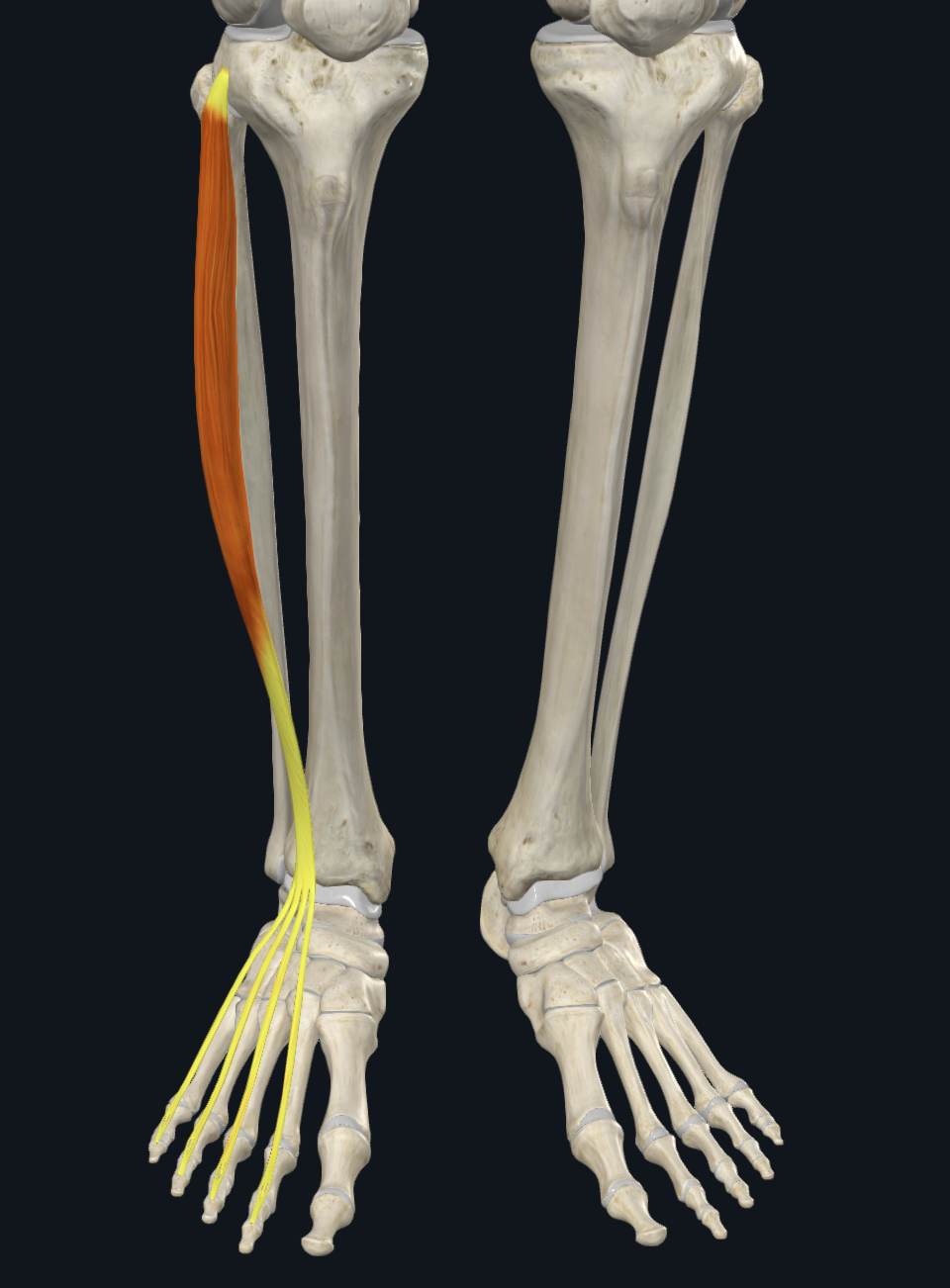
extensor digitorum longus (EDL)
anterior compartment of lower leg → dorsiflexion at ankle joint + extension of digits
origin
anterior tibia (lateral condyle)
fibula
interosseous membrane
insertion
tendons out to respective digits (2-5)
innervation
deep peroneal nerve
extensor hallucis longus (EHL)
anterior compartment of lower leg → dorsiflexion at ankle joint + extension of digits
origin
fibula
interosseous membrane
insertion
tendons out to respective digit (1)
innervation
deep peroneal nerve

tibialis posterior
posterior compartment of lower leg → plantarflexion at ankle joint
origin
posterior tibia + fibula
interosseous membrane
insertion
distal to ankle joint
innervation
tibial nerve

flexor digitorum longus
posterior compartment of lower leg → plantarflexion at ankle joint + flexion of digits
origin
posterior tibia + fibula
insertion
tendons to respective digits (2-5)
innervation
tibial nerve

flexor hallucis longus
posterior compartment of lower leg → plantarflexion at ankle joint + flexion of digits
origin
posterior tibia + fibula
insertion
tendons to respective digit (1)
innervation
tibial nerve
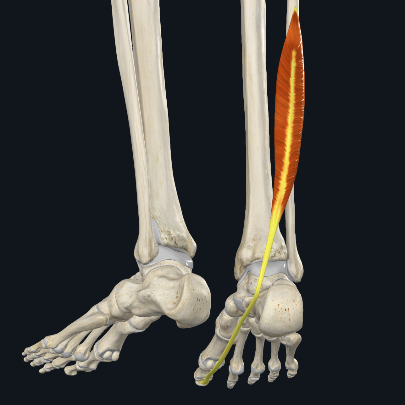
fibularis longus (peroneus longus)
lateral compartment of lower leg → plantarflexion at ankle joint + eversion of foot (at subtalar joint)
origin
along fibula
insertion
onto 1st metatarsal
innervation
superficial peroneal nerve

fibularis brevis (peroneus brevis)
lateral compartment of lower leg → plantarflexion at ankle joint + eversion of foot (at subtalar joint)
origin
along fibula
insertion
onto 5th metatarsal
innervation
superficial peroneal nerve

ilium
(1)

sacrum
(2)
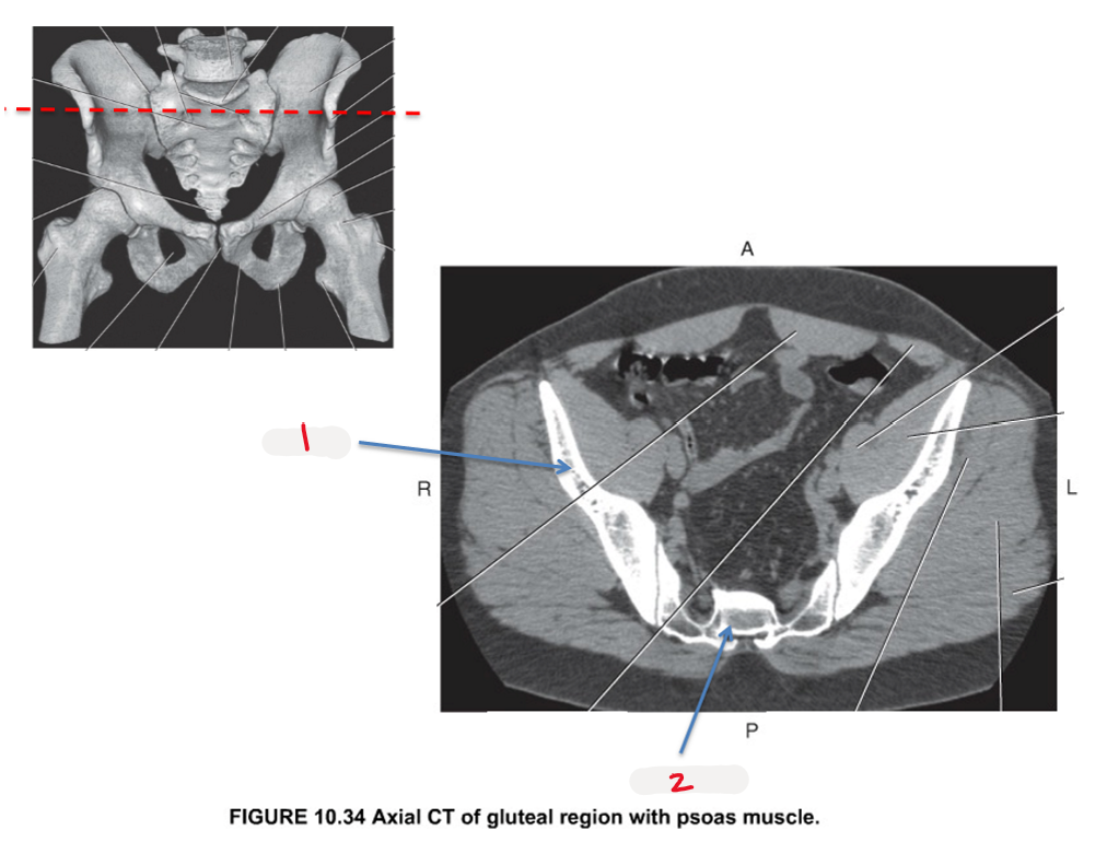
ilium
(1)

sacrum
(2)

pubis
(1)

femoral head
(2)

femoral neck
(3)

greater trochanter
(4)

ischium
(5)

femur
(1)

lesser trochanter
(2)

inferior ramus of pubis
(3)

greater trochanter
(1)

acetabulum
(2)

ilium
(3)

femoral head
(4)

acetabulum
(1)

femoral head
(2)

ilium
(3)

ischial tuberosity
(4)

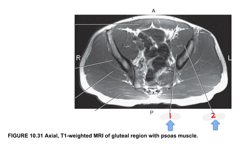
psoas
(1)

iliacus
(2)
iliopsoas
(1)

femoral head
(2)
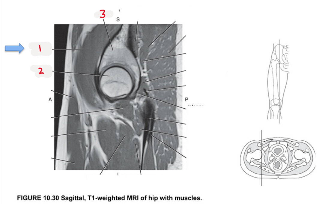
ilium
(3)

gluteus minimus
(1)

gluteus maximus
(2)

gluteus medius
(3)

iliopsoas
(4)

gluteus medius
(1)
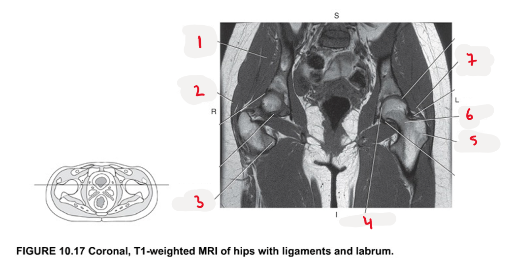
gluteus maximus
(2)

lesser trochanter
(3)

acetabulum
(4)
