Biochem/MolE Exam 1
 Call Kai
 Learn
Learn Practice Test
 Spaced Repetition
 Match
Match Flashcards
 Knowt Play
1/157

Earn XP
Description and Tags
Go over slides, HWs, reflections, one note
Name | Mastery | Learn | Test | Matching | Spaced | Call with Kai | Chat |
|---|
No analytics yet
Send a link to your students to track their progress
158 Terms
What are some reasons why it is hard to make an effective vaccine?
Pathogens rapidly mutate
Can evade immunity through humanized sugar coating called glycocaclyx
Logistical issues complicating vaccination
Antibodies generated by vaccine have weak binding to pathogen
Definition of K_eq = Ka vs Kd
Which binding is stronger, Kd = 10, or kd = 1000?
Ka = [RL] / ([R]•[L])
Kd is the inverse
Kd = 10 is stronger
What is a “good” target Kd value for an antibody to a pathogen?
10^-9
How many amino acids occur naturally in proteins? How many in the human body specificially?
22-24 total, 21 only in humans
21 is selenocysteine, 22-24 is pyrrolysine and its derivatives
Secondary protein structure
Hydrogen bonding. Arrows on the protein structure are beta sheets, twisting parts are alpha helices. Remember that there are also turns and loops connecting them; not just alpha and beta for secondary structure
Which is stronger, primary or secondary structure?
Primary b/c covalent bondign (200-600 kJ/mol) vs secondary h-bonds (<7 kJ/mol)
Conversion of kcal to kJ
1 kcal = 4.18 kJ (kJ is smaller unit)
Give one example of a pathology related to secondary protein structure
Alzheimer’s - Diseased brain equilibriates to amyloid fibril B-sheets instead of correct Amyloid precursor protein tetramers, causing a chain reaction
What are protien motifs?
Bridge secondary and tertiary structure. There are dozens of motifs that include, turns, loops, zinc finger, helix-loop domains, forkhead, etc. Used for binding to DNA for example.
All parts of tertiary structure
alpha helices, B-pleated sheetss, turns & loops, instrinically disordered domains
Held together by ionic bonds (charged molecules), hydorgen bonds, van der waals forces (hydrophilic/phobic)
Intrisically disordered domains
Regions of proteins that are very fluid and have the ability to bind through multiple partner proteins through induced fit. Very fe proteins are entirely intrisically disordered.
These structure tend to be invisible via X-ray
More likely to have serine than tryptophan
Why are IDDs more likely to haveserine than tryptophan?
Nonpolar/hydrophobic amino acids tend to clump together to bury the nonpolar side chains within their center in a globular structure. Tryptophan is hydrophobic whereas serine is hydrophilic and can stay solubilized
Give an example of a highly intrinsically disordered protein
p-53 (allows it to bind to hundreds of partners)
Why are weak bonds important in proteins?
Allows for flexibility and function. If everything were rigid, proteins couldn’t function. It is better to regulate these proteins with modifications than rigidly structure them
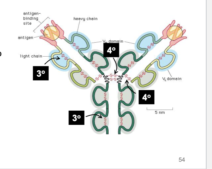
How do antibodies use Disulfide bonds?
They bridge parts of the antibody chain to create a looped structure that allows for better binding
How strong is a disulfide bond?
60-65 kcal/mol
How does a perm work?
keratin in hair is chemically treated with ammonium thioglycolate to break disulfide bridges and then reformed with thioglycolate in a difference structure.
Affinity vs avidity
Affinity measures binding strength of single bond, aviditiy measures strength of all interactions between two binding partners.
Likely exam question is saying avidity instead of affinity
For multivalent binding, what happens from Kd1 to Kd2?
Kd2 is lower because binding usually becomes more favorable (although there are some cases of negative multivalent interactions)
∆G formulas
∆Gº = ∆H - T∆S
∆Gº = RT ln(Kd)
Must add a negative sign if using K_eq/K_ac instead of Kd
R = 8.314 × 10^-3 kJ/mol*K
T is 298 K @ STP
∆G vs ∆Gº
∆Gº is for 1.0 M, 25ºC, STP
∆G is “real world conditions”
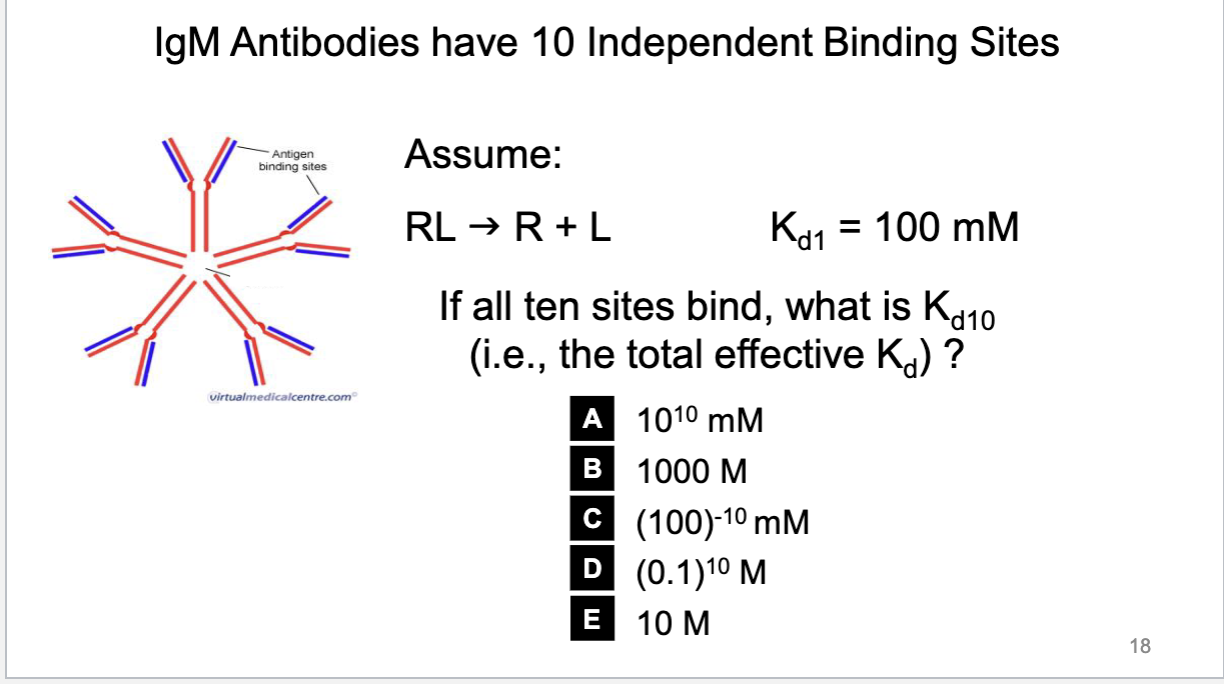
How to find the ratios of Kd’s for multivalent binding of an antibody
Use addivity: ∆G2º = ∆G1º + ∆G1º = 2∆G1º. Note that you CANNOT sum of Kd values from Kd1 to find Kd2
What is the relationship for independent binding between Kd values for multivalent binding?
Kd# = (Kd1)^#
Note that units on Kd always remain in M

Types of Antibodies
IgG (bivalent)
IgA (tetravalent)
IgM (decavalent)
IgD
IgE


Hemoglobin
Tetramer, binds to 4 O2
pO2 in lungs only gets to 100 mHg. Need to be able to carry a lot of oxygen. Ligand binding brings 40-70x more O2 carries than can be dissolved in blood
Myoglobin
very similar 3D structure to hemoglobin subunit but significantly different AA sequence (24/141 AAs match)
Still binds to O2, univalent binding
Hill Equation
Theta is fractional occupancy. When theta = 0, no binding sites occupied; 1 means all sites occupied
When theta = 1/2, [L] = Kd, receptors half saturated
![<p>Theta is fractional occupancy. When theta = 0, no binding sites occupied; 1 means all sites occupied</p><p>When theta = 1/2, [L] = Kd, receptors half saturated</p><p></p>](https://assets.knowt.com/user-attachments/522569e4-0380-4c7f-b380-cb32b9952ec3.png)
Hill Log Plots
Slopes are hill coefficients (n) - how you can tell if an enzyme has multivalent binding
If slope = 1, independent. >1 cooperative, <1 negatively cooperative

Allosteric definition
A change to protein structure and function by binding of an effector to a different site on the protein
Myoglobin vs Hemoglobin
Univalent binding of O2 to myoglobin is much stronger than hemoglobin binding. Gets more saturataed quickly and “outcompetes”. Myoglobin only in muscles though and hemoglobin on in vascular tissue.
Log vs sigmoidal curve

What factors shift the Hb curve right?
Increasing Co2
Increasing temperature
Exercising
BPG increase
When is myoglobin oxygen released
Depends onlocal PO2 in. muscle, Hemoglobin has to drop a lot of oxygen for myoglobin to start releasing
Carbon Monoxide poisoning
CO binds to hemoglobin 200-300x stronger than O2/Co2, causing poisoning
What is the ceiling for Kd1 values in nature?
Around 1 M and significantly below. Only exception is water’s concentration being 55.5 M.
By this logis, theta can never be 0 (only when Kd is infinity or [L] = 0) or 1 (vice versa logic)
What is the ∆G for ATP? Respiration?
-7.3 kcal/mol
686 kcal/mol
K_eq formula for equilibrium

∆G determines…
Favorability and amount of completion reaction will go to. Rate is determining by kinetics, Ea
Why do graphs of energy levels in glycolysis differ so much?
Depends on ∆G conditions and whether it is at ∆G or not. Glycoylsis seems energetically unfavorable (+∆G) at ideal conditions, but under biological reality, it is favorable
Enzyme Mechanisms
Binds to two substrates and encourages proper orientation (basically all enzymes do this)
Rearranges electrons in substrate to create partial charges (like neuraminidase, where a cell surface sugar is cleaved off - mechanism for virusees unbinding from cell membrane)
Enzyme bends substrates
Combination of these
Tamiflu
Competitive inhibitor
Can be taken prohpylactically but is too expensive, has side effects, and could lead to viral resistance Works by blocking sialidase activity to stop viruses from detaching from cells to spread
Michaelis Menten Equation
Enzyme kinetics. An enzyme only follows MM kinetics if it is bivalent (single binding), or multivaent with Independent binding (non-cooperative)

MM Curve
Note Km = x value of 0.5V_max

Lineweaver-Burke Plot
Linearization of MM Equation.
Note axises and intercepts are reciprocals, slope is the ratio.

How does the line change on a LB Plot with Enzyme inhibition?
Competitive?
Non-Competitive?
Uncompetitive?
Competitive keeps y-intercept, increases slope (Km increases)
Non-competitve increases slope and y intercept but keeps horizontal intercept (V_max decreases)
uncompetitive inhibition is a shift to the left (both intercepts change, both v_max and K_m decrease b/c ES complex is inhibited - binding is too tight).

Reversible vs Irreversible Inhibitors
Most enzyme inhibitors are reversible but but some (like sarin gas that permanent binds to AChE which degrades ACh)are not.
What does the graph of an allosteric enzyme look on an MM curve?
Sigmoidal graph. Can actually have worse affinity when inactive and then shoots up and has greater affinity once first substrate binds.

How do activators/inhibitors affect MM curve?
Activators (allosteric) shift to the left, inhibitors to the right.
Pyruvate kinase is an example of what?
An allosteric enzyme. Its substrates are pyruvate and ATP. Activated by Fructose-bis-1,6-phosphate (FBP) and inhibited by AAs noncompetitively.
Glycolysis inputs and outputs
1 glucose in
2 pyruvates, 2 ATP, 2 NADH out
Pyruvate decarboxylation input and ouptu
2 pyruvates in
2 Acetyl CoA, 2 CO2, 2 NADH out
Citric Acid cycle inputs and outputs
2 Acetyl CoA in
4 CO2, 2 ATP, 6 NADH, 2 FADH2 out
Oxidative Phosphrylation ins and outs
10 NADH, 2 FADH2 in, 30-34 ATP out
Total ATP made by respiration
30-38 ATP (varies because of transporters used, where electron carries enter ETC,
How much ATP do cells contains?
Only a few minutes worth
How much ATP do we make a day?
Our body weight
How much ATP do we have at any given point?
80-100g
How many times is our ATP replenished in a day?
500-1500x
Glycolysis Steps
Glucose phosphorylated twice in 3 steps
Broken into two 3-C molecules in step 4 (becomes 2 G3P)
Each G3P changes CHO to CHOH and is then phosphrylated (Step 5)
NADH is taken (Step 6)
ATP is made using the phosphate that was attached (7)
Steps 8-9 involves changes in conformation
Step 10 extracts one more ATP before giving pyruvate
Enzymes in glycolysis
Hexokinase/glucokinase give glucose 1st ATP
Phosphofructose kinase (PFK-1) gives the second (makes Fru-1, 6P2, Irrversible step_
pyruvate kinase extracts the last ATP in step 10.
Ways to lower blood sugar
Main ways are insulin (affects pancreatic B cells to stop breaking down glycogen),
somatosin (affects pancrease delta cells)
How does insulin work?
Insulin activates insulin receptor to turn on Glucose Transporters (GLUTs) to take glucose out of the blood and into cells via diffusion
Ways to increase blood sugar
glucagon (pancreas alpha cells)
epinhephrine (adrenal)
cortisol (promots glucoseneogenesis)
ACTH (pituitary, releases cortisol and fatty acid decomp)
Growth hormone
Thryoxine (enhances sugar absorption in intestines)
Hexokinase vs
Both phosphylate glucose to keep in the cells as G6P.
Hexokinase has very low km and almost always work at V_max
Present in most body cells
Liver cells produce glucokinase, which prefentially phosphrylates glucose but repsonds to [Glc]. Liver does not express HK, allowing it to break down glycogen and release glucose when levels are low
PFK-1
Takes F6P and products F1,6 Bisphosphate using ATP.
AMP positive regulates, Fru-2, 6P2 also positively looops back (also promoted via insulin)
ATP negatively regulates even though its a substrate
Citrate also negatively regulates (from downstread CA cycle)
What happens to pyruvate?
Anaerobically is converted to lacate + NAD+ or ETOH+Co2+NAd+ (yest)
Aerobic respiration in mitochondria
How does pyruvate get into the mitochondrial matrix
Diffuses across permeable outer membrane. Transported acros inner matrix via PDHC
What are pathways revolve around Acetyl CoA?
AA breakdown, lipid breakdown, pyruvate
Acetyl CoA can be made into cholesterol, and then of course Citric Acid Cycle
PDHC
pyruvate dehydrogenase complex
Multienzyme complex. Catalyzes irreversible step. Activated by dephophorylation (remember kinases and phosphatases)
Pyruvate loses Co2 and electrons (oxidized) by unit E1 and then NAD+ is reduced in E3. Then E2 helps to attach remaining 2-C molecule to Coenzyme A (via thiol linking)
What is PDHC regulated by?
Activated by pyruvate, AMP/ADP, NAD+, CoA
Inhibited by ATP, NADH, citrate
Citric Acid Cycle
C2 goes in (Acetyl CoA)
Oxaloacetate attached to form citric acid
Rearranges into isocitrate
Loses CO2 and NADH beocming alpha-ketoglutarate
Loses Co2 and NADH to become succinyl-CoA
Loses GTP to become succinate
Loses FADH2 to become fumarate
gains H2O to become malate
Loses NADH (to regenerate C4 oxaloacetate)
Enzymatic oxidation captures about 93% of glucoses energy
Oxidative Phosphrylation
Uses electron carriers in ETC to fuel pmf to catalyze ATP formation
10 protons per ETC run.
ETC components in order
NADH-Q Reductase (pumps 4H)→ CoQ (ubiquinone)→ Cytochrome reductase (pumps 4)→ Cyt C → cytochrome oxidase (pumps 2H and give e- to Oxygen) then to ATP synthase
Note this is a stepdown process to pump protons
Where does FADH2 fit in?
FADH2 uses succinate-Q reductase to enter at Co-Q spot, thus only transferring 6 protons.
How efficient is oxidative phosphorylation?
41% Because protons are disorderly, can wander off, picked up by a base, energy lost as heat (beneficial).
How many ATP do NADH and FADH2 produce?
2.5 for NADH, 1.5 for FADH2
How does NADH from cytosol get in the ETC?
Uses malate-asprtate shuttle to create mito-NADH to enter at Complex I, or glycerol-3-phosphate shuttle (NOT G3P) to form FADH2 inside an enzyme acaledd mitochondrial G3P dehydrogenase to deliver to CoQ.
Which side of the mitochondrial inner matrix is positive/negative?
IMS is positive, matrix is negative
How many protons does ATP synthase need to make 1 ATP?
4
PMF
proton motive force generated by protons being pumped out by ETC. Stores potential energy in concentration gradient and voltage.
How does cyanide work?
Binds to iron in cytochrome oxidase, halting ETC
What is DNP?
Uncoupler that shuttles protons across IMM to disrupt ATP synthase, losing energy as heat and can lead to death
Hwo efficient is the complete conversion of glucose to ATP?
32%
No better than a gasoline-powered car
ROS
Reactive oxygen species - 0.2% of oxygen is converted to ROS in ox-phos.
When oxygen gets one electron instead of two, forming radicals or strongly charged compounds. Superoxide anions and hydroxide radicals are dangerous.
Could have been developed on purpose, as ROS is a signaling
How are ROS “defused?
Super enzymes like superoxide dismutases and catalase. Some of the highest catalystic efficencies (K_cat/K_M)of any enzymes.
Note that k_cat is the max number of substrate molecuels converted to product by a single enzyme active sit per unit of time when fully saturated (
How far can you run using only he glucose in your blood?
¼ of a mile
Glycogen
70 kg adult muscles have 400g glycogen, up to 700g in trained athlete
Average person can run 12.8 miles by burning glcyogen, athlete: 22.4
IMCLs
intramyocellular lipids. Lipids in muscle (distribution differs in athletes). Provide energy source via small vesicles around muscle fibers
Fat breakdown
Fatty acids broken down into acetyl-CoA via beta-oxidation.
This involves use of ATP, reduction of electron carriees, cleaving of carbons to form Acetyl-CoA.
Amount of energy produced depends on length of fatty acid chain
Fatty acids activated in cytoplasm into fatty acyl-CoA then carnitine shuttle brings into mitochondrial matrix
Carnitine shuttle
brings fatty acyl-CoA across both membranes with transferases and a translocase channel
Energy calculations of fatty acid oxidation
-1 ATP, +1 FADH2, +1 NADH, then (FADH, 3 NADH, GTP from Krebs)
Note that the -1 ATMP counts as “two ATPs” because it is converted to AMP and requires two phosphates to regenerate
Only for first two carbons of FA chain.
Every two carbons in a fatty acid produce an acetyl coA and go into CA cycle
How many ATPs made from palmitic acid C16?
106 ATP: -2ATP, 7 cycles of Beta oxidation, 8 cycles of Krebs, 28+80 -2
On a per mass basis, the energy content of a fatty acid is approximately ___ of glucose? On a per carbon basis?
265%.
150%
Energy distribution in molecules for the average person
Carbs (primarily glycogen) - 1%
proteins - 21%
fat - 78%
Protein oxidiation
Broken down into AAs int o Acetyl-COA.
Ammonia is removed from AAs and goes into urea cycle or to form new nucleotides/AAs
Why is there a range of 30-38 ATPs from glucose?
30-32 is produced from one molecule becauseof the uneven yields. 38 is assuming perfectly ideal conditions where NADH corresponds to 3 ATP and FADh2 to 2
The plasma membrane makes up what proportion of all cellular membranes?
0.5%
What class of molecules diffuse across membranes?
Small, hydrophobic molecules.
Note that molecules like water do move by facilitated diffusion (aquaporins)
Fick’s Law


Note D is in cm²/s
What is the ∆G for diffusion?
Always <0