Principles of Molecular and Cellular Biology Unit 2
 Call Kai
 Learn
Learn Practice Test
 Spaced Repetition
 Match
Match Flashcards
 Knowt Play
1/109

There's no tags or description
Looks like no tags are added yet.
Name | Mastery | Learn | Test | Matching | Spaced | Call with Kai | Chat |
|---|
No analytics yet
Send a link to your students to track their progress
110 Terms
How can the cell regulate transcription?
Gene-adjacent regions of DNA (transcriptional control regions), proteins that bind to DNA to alter transcription, manipulation of chromatin structure (access to genes)
RNA polymerases in eukaryotes
Pol I: rRNAs, nucleolus
Pol II: mRNAs, nucleus
Pol III: tRNAs, nucleus

RNA polymerase II structure
RPB1 and 2 (RNA Pol B1 and RNA Pol B2, similar to betas of prokaryotic RNA), prokaryotes have Greek letters while eukaryotes have numbers/English letters

Pol I,II,III compare/contrast
Pol I and III are most similar,
Pol II has unique alpha subunits and a unique CTD (C-terminal domain) on RPB1, all subunits are required for transcription and all require more factors to recognize promoters (General Transcription Factors)
RPB1 Clamp Domain
Moves to hold DNA template and transcript (creating channel), RPB1 is clamp domain while RPB2 is wall domain
DSIF (elongation factor) supports by stabilizing the closed clamp

C-Terminal Domain of RPB1
Heptapeptide repeats (YSPTSPS), yeast have 26 repeats while vertebrates have 52 repeats
Essential for viability:
Modification of RPB1 CTD
CTD is initially unmodified and after transcription initiation, CTD is phosphorylated (mainly Serines 2 and 5): phospho-CTD supports other binding regulatory proteins (mRNA processing, chromatin remodeling)
Transcriptional control region
A DNA sequence that a protein binds to, DNA-protein interaction influences transcription
Focused/regulated promoters
TATA boxes, initiator sequences, BREs (TFIIB recognition element), DPE (downstream promoter element)
Found in genes with varied expression levels, help position RNA polymerase on start site, binding sites for general transcription factors (GTFs), found between -40 and +40
Focused vs. Dispersed promoters
While for focused/regulated promoters all mRNAs use the same start site, for dispersed/housekeeping promoters each mRNA uses a different start site
Dispersed/housekeeping promoters
CpG Islands (less flexible and hard to bend around nucleosomes, makes them free and easy to access for transcriptional proteins)
Each mRNA uses a different start site, found in genes with constant expression levels, multiple TSS (transcription start sites), do not directly position RNA pol
Location of focused/regulated promoters
BRE: -37 to -32
TATA box: -31 to -26
Inr: -2 to +4
DPE: +28 to +32
CpG islands and methylation
CpG islands allow for non-specific binding of GTFs, good for constant low-level expression (housekeeping), recruit RNA pol II but in a non-specific direction (transcripts can be sense or anti-sense)
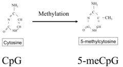
CpG repeats are normally pretty low in mammalian DNA due to methylation (5-methyl C), 5-methyl C can spontaneously deaminate to T (Cme G>>T G) or U (CG>UG>CG), methylation of CpGs is a signal for trasncriptional repression
Transcription sequence
Recruitment>pre-initiation complex>initiation>pausing>elongation>termination
General Transcription Factors (GTFs)
Proteins or multi-subunit protein complexes (TFIIA,B,D,E,F,H), functions are positioning RNA Pol II at TSS and aid in binding to promoter core elements, help separate DNA strands to allow template strand to enter Pol active site, GTFs are required for all genes transcribed by RNA Pol II and are highly conserved across all eukaryotes

TFIIA
3 subunits, stabilize TBP-TATA
TFIIB
1 subunit, stabilize TFIID-TATA, start site selection
TFIID
Subunits: TBP + ~13 TAFs, core promoter recognition
TFIIE
2 subunits, TFIIH recruitment
TFIIF
2 subunits, chaperone Pol II to promoter
TFIIH
10 subunits, CTD kinase, helicase, DNA repair
Sequential assembly of the pre-initiation complex, TATA promoter example
1. TATA promoters recognized by the TFIID (TBP binds first)
2. TFIIA binds DNA and TBP to stabilize
3. TFIIB binds DNA and TBP to stabilize
3. TFIIB binds DNA and TBP, later separates strands
4. TFIIE binds complex and forms binding site for TFIIH
5. Pre-initiation complex is formed
Transcription initiation from the PIC
1. TFIIH helicase subunit (XPB), ATP hydrolysis
2. TFIIB and ol II melt DNA, opening bubble (open complex)
3. Pol II initiates transcription
4. TFIIH kinase domain phosphorylated RPB1 CTD (Serine of repeats), an enzyme complex binds phospho-CTD and caps mRNA
5. GTFs dissociate from the promoter
TBP-associated factor (TAF) writers and readers
Writers: some TAFs have enzymatic activity (HATs, kinases)
Readers: some TAFs have PTM binding domains (bromodomains reading acetylated lysines and PHD finger domains with methylated lysines)
CTD code
No phosphorylation: RNA pol II assembled into PIC
Ser-5 phosphorylation: transcriptional initiation and pausing (phosphorylated by TFIIH, recruits capping complex, recruits elongation inhibition factors NELF/DSIF)
Ser-2/5 phosphorylation: processive elongation (phosphorylated by P-TEFb, recruits splicing complex)
RNA Pol II pausing and phosphorylation
In PIC, TFIIF and TFIIE cover binding sites for NELF/DSIF:
1. Once initiation starts GTFs separate opening up binding site
2. NELF/DSIF bind within 100 bases, pausing transcription
3. P-TEFb relieves pausing by phosphorylating NELF and DSIF and Serine 2 of CTD repeats
P-TEFb - BRD4 connection
P-TEFb is recruited to euchromatin by BRD4, BRD4 has bromodomain which finds acetylated histones
SET1 and SET2 HMTs
SET1 and SET2 are recruited by Ser-2/5 phosphorylated CTD, they methylate H3K4 and H3K36 which are markers for actively transcribed chromatin
Pol I and Pol III
Each pol has its own GTFs, do not require ATP hydrolysis to separate DNA template strands, initiation rates are linked to growth/proliferation
Types of transcriptional control regions/elements
Core promoters: bound by GTFs
Proximal promoters: bound by activator proteins, short sequences within 200 bp of TSS
Enhancers: bound by activator proteins, longer sequences, 50kb or more upstream/downstream (enhancers in yeast are called upstream activating sequences)
Repressors: bound by repressor proteins
Unique transcription nature of eukaryotes
Transcriptional control regions are often very far from genes (upstream or downstream), transcriptional control regions can be within genes, and genes often have many transcriptional control regions
Core promoters
Specific sequences, great for tight regulation for high expression when needed
Examples: TATA Box (bound by TBP of TFIID), Inr and DPE (bound by TAFs of TFIID), and BRE (bound by TFIIB)
Inr and DPE often occur together and more frequently, BRE and TATA often occur together and less frequently
Differences/smiliarities of proximal promoters and enhancers
Differences: enhancers are further away from TSS, proximal promoters are specific to promoter and enhancers have limited specificity, enhancer moving won't affect ability but proximal promoter won't work if moved >50bp
SImilarities: Both have 6-10 bp elements where TFs bind, many TFs can bind and work at both, orientation is often unimportant
Transcription factor basic anatomy
Modular domain proteins: DNA binding protein, protein binding domain (activator or repressor), flexible unstructured region that allows for flexibility of distance between transcriptional control elements
Activation domains/repression domains: bind co-activator or co-repressor
Many TFs exist as heterodimers, which can create variety
Cooperative binding of multiple TFs
More TF binding interactions>>increased transcription, multiple promoter/enhancer binding proteins often stabilize each other
Large complexes at a regulatory region=enhancesome
Super-enhancers and transcriptional condensates
Multiple close-spaced enhancers (within 10 kb), highly acetylated histones, TF flexible regions (IDRs) interact with each other and co-activators, hold together in a liquid-like state and increases availability of transcriptional machinery
Insulators
Specific long range DNA elements, bind specific proteins, proteins create DNA loops to sterically block promoter interactions (heterochromatin barrier and enhancer blocker)
Heterochromatin barrier
Blocks heterochromatinization by removing nucleosomes, recruiting acetyltransferases, etc.
Enhancer blocker
Blocks enhancer activation of a protein
CTCF/Cohesin example: CTCF binds DNA and cohesin
if cohesin binds the enhancer: promotes transcriptional activation
If cohesin doesn't bind the enhancer: acting as an insulator, enhancer binding factors can't access promoter
Chromatin remodeling
Regulators: HATs (histone acetyltransferase), HDACs (histone deacetylase), HMTs/KMTs (histone methyltransferase), HDMs/KDMs (histone demethylase), DNMTs (DNA methyltransferase), chromatin remodelers (SWI/SNF)
*Methylation is not always repressive

Activator/repressor chromatin modification
Co-activators/co-repressors can be HDACs/HATs
All TFs need is access to the DNA somewhere to exert their influence on surrounding nucleosomes, acetylation tends to promote euchromatin and also serves as a signal for bromodomains in TFIID, and BRD4
Chromatin repositioning (SWI/SNF family)
Can slide DNA along the nucleosome, can eject nucleosomes, and can be used to promote open or closed chromatin
Conserved ATPase and helicase domain, ATPase domain contains bromodomain (finds acetylated regions in DNA), actin-related domain is related to nucleoskeleton
Mediator Complex
Large mega complex of proteins, forms a molecular bridge between TFs and Pol II, participates in PIC assembly (different subunits bind different TFs), a single activator can have multiple touch points and mediator enables multiple regulatory regions to influence PIC at the same time

Regulatory elements
DHS: DNA hypersensitivity (due to open chromatin)
Methyl: presence of CpG methylation (marker of closed chromatin)
Med: mediator binding
CTCF: factor which binds insulator
Epigenetic "writers"
Histone acetyltransferases (HATs), histone methyltransferases (HMTs/KMTs), DNA methyltransferases (DNMTs)
Epigenetic "erasers"
Histone deacetylases (HDACs), histone demethylases (HDMs/KDMs), DNA demethylases
Epigenetic "readers"
Bromodomains (finds acetylated lysines), chromodomains/PHD finger domains (find methylated lysines), DNA methyl binding proteins
Epigenetic markers preservation in replicating cells
Self-reinforcing systems, after replication epigenetic markers are divided across daughter cells nucleosomes, often PTM readers will PTM adjacent tails to fill back in
H3K9 Methylation review (heterochromatinization)
Methylation is a stable mark and is epigenetically inherited
HP1 binds to H3K9me3 (chromodomain)>>HP1s bind to each other (chromoshadow domains), aggregation of nucleosomes>>HP1 recruits H3K9-HMT (spreading of methylation)
DNA methylation (methylcytosine)
Cytosine in CpG is methylated by DNA methyltransferase (DNMT), 80% of CpGs are methylated in mammals (CpG islands are typically not methylated), non-CpG island CpGs are less common than odds would predict
DNA methylation=repression
DNA methylation proteins
Writers: DNMT1/UHRF1-maintenance methylation, DNMT3a/DNMT3b/DNMT3L-de novo methylation
Erasers: TET proteins
Reading: Methyl-binding proteins (MBPs), UHRF1/2
De novo DNA methylation (DNMT3)
Early embryo (pre-implantation): most CpGs are unmethylated
H3K4 near active genes gets methylated
during transcription (SET1 and SET2, due to
Serine 2/5-Phospho-CTD): Methylated H3K4 prevents binding of DNMT3L>>DNMT3a/b can't bind or methylate CpGs
Non-transcribed regions do not get H3K4 methylated: Exposed H3K4 can be bound by DNMT3L>>DNMT3a/b can methylate CpGs

Maintenance of DNA methylation during replication (DNMT1)
During DNA replication, methylated CpGs are converted to "hemi-methylated" CpGs, These sites are recognized by UHRF1, DNMT1 restores methylation symmetry
DNA methylation erasers (TET proteins)
TET proteins oxidize 5-methyl C to 5-hydroxymethyl C, these products can be removed by base excision repair which resets them to unmodified Cs
Oxidized methyl Cs inhibit maintenance methylation: inhibits UHRF binding, impairing DNMT1 activity, over time CpGs will lose DNA methylation through replication cycles
TET proteins in early embryogenesis
Post-fertilization: TET3 oxidizes most methyl-CpGs
Implantation: TET1 and 2 are turned on, DNMT3 jump onto non-transcribed areas, together they reset DNA methylation
Silencing/methylating actively transcribed DNA (G9a)
Actively transcribed area>>G9a is recruited (SET domain-HMT)>>G9a methylates H3K9>>G9a recruits DNMT3a/b (ANK domain), DNA is methylated
3 Mechanisms for DNA methylation-mediated repression
1. MBD (methyl CpG-binding proteins): reocgnize methylated DNA, recruit HDAC and H3K9-HMT
2. DNMTs: recruit HDACs and H3K9-HMT
3. Blocking binding: methylCs can sometimes block binding of TFs; alternatively, blocking repressors/insulators can promote expression
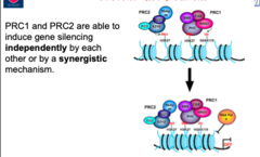
Polycomb group (PcG) protein complexes (for silencing genes)
PRC2 complex: writer, methylates H3K27, has a SET (HMT) domain
PRC1 complex: reader, finds and acts on methyls
PRC1
Chromodomain that finds and binds H3K27me3, PRC1 mediates chromatin compaction and catalyzes ubiquitination of H2AK118/119 (blocks nucleosome removal during transcription, causes stalling of RNA Pol II which blocks transcription)
X chromosome inactivation (targeting PRC2 to genes)
1) Xist gene on the chosen X chromosome activates and produces a long non-coding RNA (lncRNA)
2) This Xist RNA sticks directly to the same X chromosome that made it, coating it completely
3) Recruit the PRC2 protein complex to the chromosome and binds to Polycomb Response Elements (PRE)
4) Methylates H3K27 and recruits PRC1
Trithorax Group (TrxG): Mediators of Activation
Some TrxG proteins are HMTs (with SET domains): methylate H3K36 and H3K4
Trx C-ter (MLL C-ter): interacts with H3K4me (recruits CREB-binding protein, facilitates acetylation of H3K27 and promotes decondensation)
BRD4 (Fsh in drosophila): bromodomain binds acetylated lysines, Recruits P-TEFb, P-TEFb phosphorylates CTD of Pol II at Ser 2
Maintaining silencing following replication
New nucleosomes need to be methylated: PRC2 may reset H3K27me3, PRC1 binds H2K27me3-may reset H2A ubiquitination
Maintaining active state following replication
Trx C-ter may reset H3K4me, Ash1 may reset H3K36me, Trx N-ter may recruit CBP, CBP may reset H3K27ac
Connection between PcG silencing and TrxG activation
Both states can influence the other with constant cross-talk, once a state is established it's stable but can be switched with the right machinery
RNA instability
RNA is unstable because 2' hydroxyl can act as nucleophile-breaks the strand itself, RNA is degraded at alkaline pH and it is susceptible to enzymatic digestion, mRNAs never occur as free molecules (protected by proteins and ribonucleoprotein complexes)
Eukaryotic pre-mRNA processing
Localized in nucleus
1. 5' capping
2. 3' cleavage and polyadenylation
3. RNA splicing
Co-transcriptional RNA processing
5' capping happens as transcript emerges (protects)
5' cap and 3' polyA tail distinguishes mRNA from other RNAs, the CTD of RNA Pol II coordinates
RNA Pol II CTD for RNA processing
Phosphorylated CTD recruits/binds processing proteins, the cap (7-methyl-G) is placed within 25 bp of transcription as transcript exits Pol II (protects from exonucleases)
3' cleavage and polyadenylation
PolyA protein complex: binds to polyA signal (AAUAAA), cleaves mRNA, adds 200-250A PolyA tail
hnRNPs
heterogeneous ribonucleoprotein particles, mRNA chaperones, regulators throughout RNA processing: prevent RNA self-base pairing, support both movement in the cell and protein complex recruitment, involved in many steps in transcription

hnRNP RRM Domain
RNA Recognition Motif: region for RNA binding, beta sheet with positive charges on one side (interact with negative charge phosphates on RNA backbone)
Critical conserved splicing sequences (in introns)
5' GU
Branch point A
Polypyrimidine tract (10-12 Cs/Us)
3' AG
Each of these conserved sequences is a binding site for a protein that regulates splicing
Splicing reaction
Due to two sequential transesterification reactions (switching one OH for another in ester bond)
Reaction 1: Branch point A hydroxyl attacks 3' O of exon 1, exon 1 ends in free hydroxyl
Reaction 2: exon 1 hydroxyl attacks 5' PO4 of exon 2, intron is released as a lariat

snRNPs (splicing protein complexes)
snRNA + proteins, each complex is named for the snRNA inside (U1,U2,U4,U5,U6), snRNA does base pairing with mRNA while proteins do the rest of the work

Key sequence binding elements in snRNPs
U1: snRNA binds across the exon1/intron boundary, recognizes the conserved GU
U2: snRNA binds across the branch point A, it is not base paired so it sticks out (U2AF: U2 associated factor that binds to the pyrimidine tract and 3' splice site)
Spliceosome complex assembly
Complex assembly:
Step 1: U1 binds the 5' splice site, splicing factor 1 binds branch point A, U2AF binds polypyrimidine tract and 3' splice site
Step 2: U2 binds branch point A, displaces SF1, U2AF stabilizing binding of U2
Step 3: U4/5/6 come in together, U6 and U2 base pair to hold splicesome together, eventually U1 and U4 release and U2/5/6 do the rest of the work because of a base-pairing rearrangement between snRNPs

Splicing Regulatory (SR) Proteins
Mostly non-snRNP proteins, often have RRMs for RNA binding and RS domains for protein-protein interactions
Bind regions of the pre-mRNA: exon splicing enhancers (ESEs) help with exon recognition and can be activated through phosphorylation
SRs also bind with eachother, results in "cross-exon recognition complex"
Exon splicing enhancers (ESEs)
6-8 nucleotide sequences in exons, SR binding "labels" the exon for the spliceosome machinery, helps with exon recognition
Example for alternative splicing
Female vs male embryos in sex determination of drosophila: female embryos have complexes that bind ESEs and ensure exon 4 is recognized as exon while exon 4 is not recognized in male embryos
Backsplicing circular RNAs
If snRNPs aren't present at exon boundary, it will skip exon when splicesome assembles
Machinery can work backwards to splice out an exon by itself, exon gets circularized by the splicing machinery
ESSs and ISSs (exon and intron silencers)
Silencers that prevent the splicing of the region, exon exclusion
Concentration of cytoplasmic mRNA is determined by
rate of synthesis and rate of degradation
unstable mRNAS vs stable
stable mRNA: encode proteins required in large quantities (ribosomal proteins)
unstable mRNA: encode proteins needed in short bursts (cytokines)
Mechanisms of mRNA degradation
1. deadenylation-dependent pathway
2. deadenylation-independent pathway
3. endonuclease-mediated RNA decay
Deadenylation-dependent mRNA decay
1. Deadenylase complex shortens polyA tail to
Deadenylation-independent mRNA decay
5' cap is removed by the DCP1/DCP2 decapping complex, uncapped mRNA is degraded 5'>3’ by XRN1
Endonucleolytic pathway
No capping/deadenylation (ex. RNAi pathway)
Internal cleavage via endonuclease, mRNA fragments can be digested by exonucleases
RNA editing
Chaning the sequence of RNAs, occurs in nucleus during transcription
C to U: early stop codon
A to I: inosine is read by translation machinery as G, generates alternative splice sites
apoB pre-mRNA (RNA editing)
Unedited: ApoB in liver cell and expresses apoB-100, NTD associated with lipids and CTD binds to LDL receptor on cell (lipids taken into cell)
Edited: C>A edit makes early stop codon, expresses apoB-48 (intestinal cell), NTD associated with lipids and does not bind to intestinal cells (lipids aren't taken in)
Processing of rRNA
Occurs in nucleolus
rRNAs from Pol I: primary RNA is large 45 precursor that contains 28S, 18S, 5S RNAs with spacers
rRNAs from Pol III: 5S processed separately

Processing of tRNA
Synthesized by Pol III, tRNAs are the most modified RNAs (between 10-20% of tRNA bases are modified), processing includes cleavage and base modification while some tRNAs are splices

Modification of RNA nucleotides
Over 100 distinct chemical modifications (cap, tail, internal regions), non-coding RNAs are more heavily modified (m6A most abundant in mRNA)
Positional conservation of mRNA modifications
Ex: m1A is primarily found near translation start codons/first splice sites while m6A is found at many sites in mRNA near stop codon

How modifications can influence mRNA function
1. Accelerated mRNA processing and translational initiation for faster expression
2. Accelerates decay when you want expression to be quick and then off
m6A and differentiation
When embryonic cells differentiate, they need to dramatically change their gene expression profile
Ex. METTL3 mutation: METTL3 normally adds m6As to target mRNAs for quick degradation but in METTL3 mutants, transcripts stick around longer and differentiation is slowed down
Key experiments that led to RNAi discovery
RNA carrying the code for a muscle protein is injected (in worm), ssRNA has no effect, but when dsRNA is injected, the worms starts twitching in a similar way to worms carrying a defective gene for the muscle protein
Discovery of RNAi: in situ hydridization
dsRNA was injected into the gonads of adult C. elegans, embryos from these worms are subjected to in situ hybridization (stains RNA in cells/tissues), the gene that was silenced in adult gonads was also silenced in their progeny
RNAi components and processes
Production and processing of regulatory RNAs
Small interfering RNAs (siRNA)
MicroRNAs (miRNA)
Processing proteins: Drosha (nucleus), Dicer (cytoplasm)
Dicer
RNA endonuclease makes short dsRNAs (used by both siRNA and miRNA)
Key domains: PAZ domain, dsRNA-binding domain, RNAse III catalytic domains (cut dsRNAs from within, endonucleolytic)
Processing and packaging RNA: siRNA
1. Processing of dsRNA: dicer cuts them into 22nt dsRNAs
2. Packaging of dsRNA into RISC