Lecture 4 GEOMETRICAL FACTORS
 Call Kai
 Learn
Learn Practice Test
 Spaced Repetition
 Match
Match Flashcards
 Knowt Play
1/18

There's no tags or description
Looks like no tags are added yet.
Name | Mastery | Learn | Test | Matching | Spaced | Call with Kai | Chat |
|---|
No analytics yet
Send a link to your students to track their progress
19 Terms
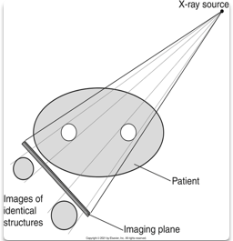
Photons travel in straight, divergent lines
Since photons move in straight but different directions (divergence)
magnification and distortion are seen on CT and portal images
Divergence of an x-ray beam
Because the beam widens as it gets the farther from the source:
More tissue is exposed as the beam exits the patient than at the anterior skin surface
This also accounts for magnification
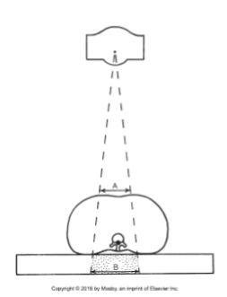
Magnification
When an image appears larger than it actually is
All images on a CT, portal image or LCD
monitor are larger or smaller than the
structures actual size
Magnification may depend on:
Geometrical arrangement of the target
Patient size
The medium on which the image is displayed
Sometimes determining the size of internal anatomy is not
possible.
To determine the size of an object inside a patient, there are 2
ways to measure the magnification factor.
Magnification Factor
is directly proportional to the distance of the object from the target and is dependent on the distance of the object from the image receptor
2 formulas to measure Magnification Factor

To determine the size of an object inside a patient, magnification can be calculated by using the ratio of TID (target to image distance) and TOD (target to object distance)

Magnification- Measuring Devices In Radiographic Imaging
Devices that are used for comparing objects of a predetermined size with objects of an undetermined size
Fiducial plate / graticle
Rulers / Calipers
Rings
Coins
Digital Scaling Software
Distortion
a change in the size, shape or appearance of the structures being examined
Magnification
is a type of size distortion
Size distortion

Shape distortion
is the misrepresentation of the actual shape of the structure that is examined
This happens when the object plane or part being imaged is not parallel with the image plane
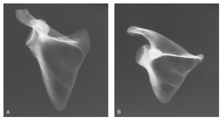
Accessing Radiographic Distortion (2 types)
Radiographic misrepresentation of either the size or shape of the anatomic part is called distortion.
2 types
Size
Shape
When angling the beam, shape distortion will occur on the image,
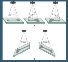
If the 2 planes are parallel:
The only size distortion that occurs is Magnification
OID
OID has a direct relationship with size distortion
As OID increases = size distortion/magnification increases
As OID decreases = size distortion/magnification decreases

Close the Object is to the film (OID), the less distortion


When looking at the obturator foramina, why do they do look of unequal size?
Answer: the patient is rotated (patient also has a Ewing's sarcoma of the pelvis)
Gantry angle examples

3 rule for accurate image formation to reduce distortion:
Decrease OID = decrease in distortion and magnification
Have central ray perpendicular to film
Have plane of object parallel to film