A&P Skeletal System
 Call Kai
 Learn
 Practice Test
 Spaced Repetition
 Match
 Flashcards
 Knowt Play
1/76

There's no tags or description
Looks like no tags are added yet.
77 Terms
Functions of the skeletal system
support: bearing the weight of the body
protection: encasing essential organs
movement: joints provide movement for bones
storage: storage of minerals to be released into the bloodstream; storage of fat in yellow bone marrow
manufacturing: production of red and white blood cells from red bone marrow (hematopoiesis)
Bone classification
Solid matrix of living cells and fibers surrounded by calcium deposits. Classified by shape.
Diaphysis
Shaft, long part of long bones
Epiphysis
Ends of long bones and contains spongy bone
Epiphyseal plate (growth plate)
Thin layer of internal cartilage between the diaphysis and epiphysis. Area of growth
Articular cartilage
External layer of cartilage covering the epiphyses which provide smooth movement of joints and cushion from shock
Medullary cavity
Hollow cavity found in the diaphysis of the long bone. Filled with bone marrow
Red bone marrow
Fills the medullary cavity in young people
Yellow bone marrow
Fatty bone marrow that replaces red bone marrow in the medullary cavity when people age. Stores fat for later usage
Hematopoiesis
Production of blood cells within bone marrow
Periosteum
Tough connective tissue that makes up the outer layer of bone and is the location of muscle attachment and bone repair

Compact bone
Underneath the periosteum; osteons are arranged in concentric circles (lamellae) which surround a central (haversian) canal
Haversian canal
Canal in osteons in compact bone that contains blood vessels and nerves
Volkmans (perforating) canals
Connects the central canals and run perpendicularly

Spongy bone
A lattice of trabeculae that are found along the lines of stress to resist force of compression. No osteons or a haversian canal. Between the trabeculae are spaces filled with bone marrow
Osteon
Unit of compact bone; long cylinders surrounding haversian canals

Osteocytes
Mature bone cells that make up majority of the bone structure and turn into calcified matrix

Osteoblasts
Cells that form (build) new bone, secrete calcified ground substance, becomes spongy bone. (Line haversian canals and the surfaces of compact and spongy bone)

Osteoclasts
Bone destroying cells, cells will remodel/repair, grow bone. Secrete acid to enlarge the medullary cavity as the bone grows so that bone marrow is available for all cells. (Line haversian canals and the surfaces of compact and spongy bone)

Canaliculi
Connect all bone cells (osteocytes), allowing them to receive nutrients and remove wastes

Ossification
The process of incorporating calcium and minerals into cartilage to become bone
(Embryo skeleton made up of cartilage, osteoblasts begin to secrete mineral deposits that replace cartilage in the third month of development, osteoblasts mature into osteocytes)

Chondrocytes
Cartilage cells in the epiphyseal plate that divide and deteriorate as the matrix around them calcifies, becoming osteoblasts
Osteoid (organic)
Makes up 35% of the bone, ground substance and collagen fibers (flexibility), protects bones from being broken
Mineral salts (inorganic)
Makes up 65% of the bone, mineral crystalline salt made up of hydroxyapatite, provides bone strength/hardness

Bone remodeling
Calcium levels in the blood is maintained by bone being created or dissolved and this process is triggered by calcitonin and parathyroid hormone
Calcitonin
Deposits extra calcium from blood into bones
Parathyroid hormone
Stimulates osteoclasts to break down bone which adds calcium to blood

Bone healing
Hematoma will form, blood enters the wound, cells begin to die, phagocytes will ingest dead bone cells and debris
Callus will form, blood vessels grow, cartilage forms to hold bone together
Callus ossifies, formation of spongy bone replacing cartilage
Compact bone forms, osteoclasts will increase size of medullary cavity, spongy bone replaced with compact bone

Axial skeleton
Central axis of the body: skull, ribs, sternum, and vertebrae (80 bones)

Appendicular skeleton
Pectoral and pelvic girdles, bones of the arms, legs, pelvis, and shoulders (126 bones)

Label 1
Frontal

Label 2
Sphenoid
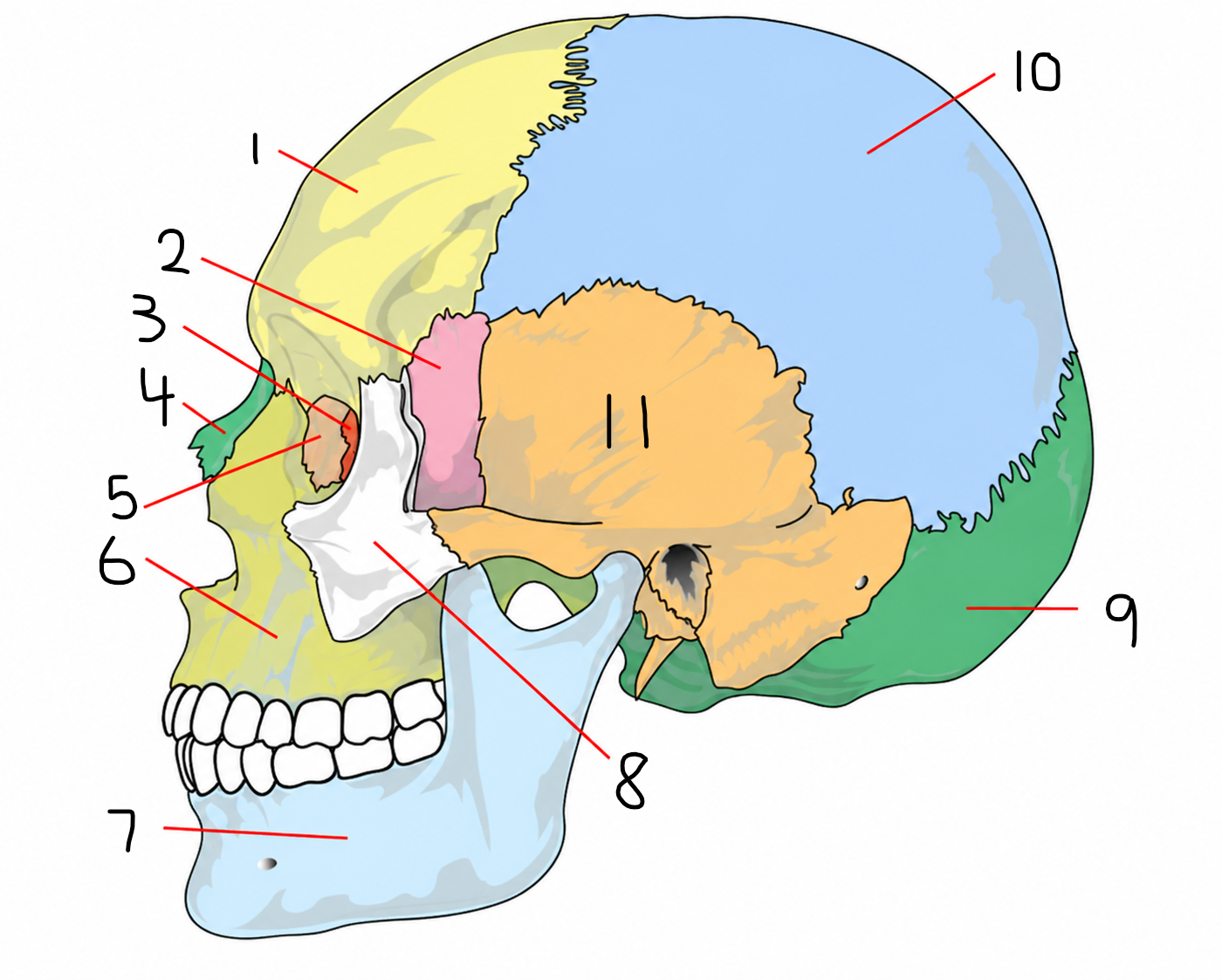
Label 3
Ethmoid

Label 4
Nasal

Label 5
Lacrimal
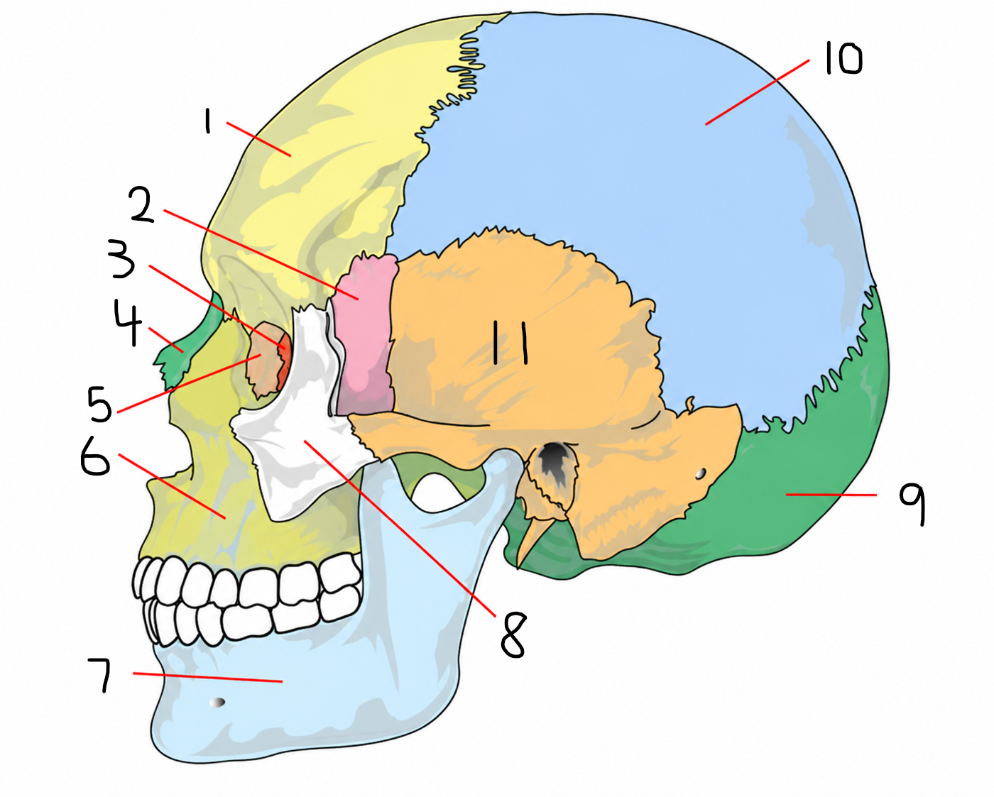
Label 6
Maxilla

Label 7
Mandible

Label 8
Zygomatic
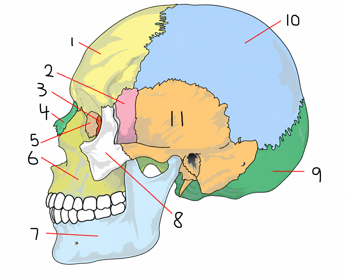
Label 9
Occipital

Label 10
Parietal

Label 11
Temporal
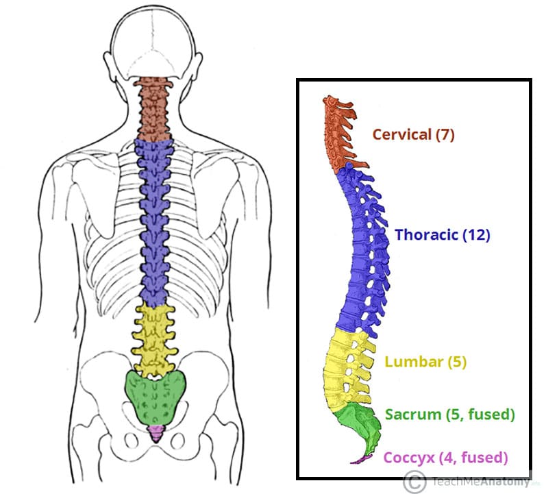
Vertebral coloumn
Extends from skull to pelvis; provides support and protects the spinal cord running through it. Consists of 33 vertebrae at birth, but the 5 sacral vertebrae and 4 vertebrae of the coccyx fuse in adolescence.
Intervertebral discs
Separates the vertebrae, provides cushioning, and absorbs shock
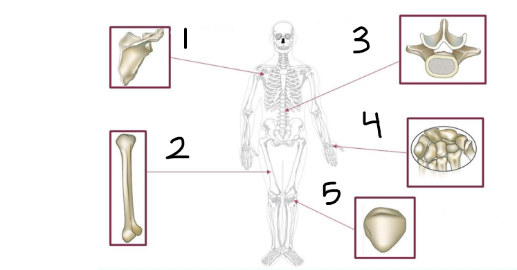
Classify bone shape and label 1
Flat bone, scapula

Classify bone shape and label 2
Long bone, femur
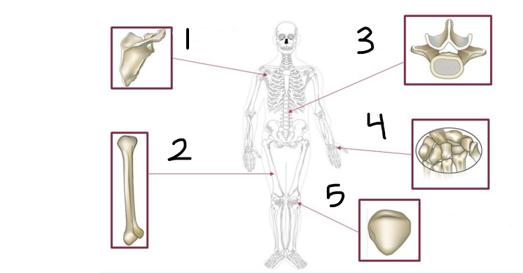
Classify bone shape and label 3
Irregular, lumbar vertebrae
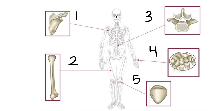
Classify bone shape and label 4
Short, carpals (and tarsals)

Classify bone shape and label 5
Sesamoid, kneecap/patella

Fibrous joints
Immovable/slightly movable, held together with fibrous connective tissue (ex. sutures of skull, attachment of teeth)

Cartilaginous joints
Slightly movable, held together with fibrocartilage (ex. intervertebral disks, meniscus knee, ribs)

Synovial joints
Freely movable, contains synovial fluid for frictionless movement, articular cartilage
Synovial joint
A joint capsule filled with synovial fluid surrounds the end of the bones and a synovial membrane and articular cartilage line the joint cavity
How do ligaments and tendons differ?
Ligaments connect bone to bone while tendons connect muscle to bone

Classify joint and label 1
Pivot joint, c1 and c2 (protraction vs. retraction)
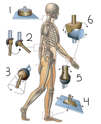
Classify joint and label 2
Hinge joint, elbow (extension vs. flexion)
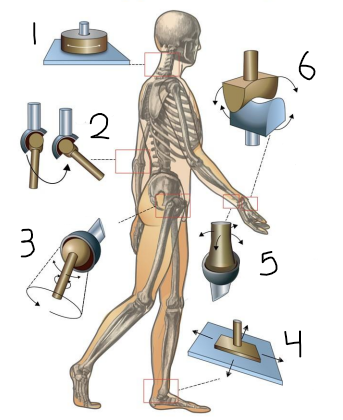
Classify joint and label 3
Ball and socket, femur/pelvic girdle (abduction vs. adduction)

Classify joint and label 4
Gliding joint, between tarsals
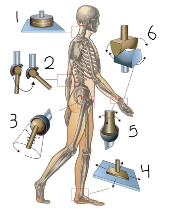
Classify joint and label 5
Condylar joint, radius/carpal (pronate vs. supinate )

Classify joint and label 6
Saddle joint, thumb (grab motion)
Flexion vs. extension
Bending a limb/decreasing the angle of a joint vs. straightening a limb/increasing angle of a joint
Abduction vs. adduction
Movement away from the midline of the body vs. movement toward the midline of the body
Pronation vs. supination
Rotation of arm/leg so the palm is down or sole out vs. rotation arm/leg so palm is up or sole in
Eversion vs. inversion
Movement of sole away from midline of the body vs. movement of sole towards the midline of the body
Lateral rotation vs. medial rotation
Rotation away from the midline vs. rotation towards the midline
Protraction vs. retraction
Movement forward vs. movement backward
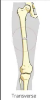
Transverse
Fracture line is perpendicular to the shaft of the bone (straight line)

Oblique
Fracture line is on an angle through the bone

Comminuted
Bone is broken into three or more pieces
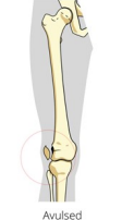
Alvused
A tendon is ligament pulls a fragment of bone away

Spiral
The bone is broken in a spiral pattern due to twisting or rotational force

Greenstick
The bone is broken, but not broken all the way through

Label anatomy of synovial joint 1
Fibrous layer

Label anatomy of synovial joint 2
Periosteum

Label anatomy of synovial joint 3
Synovial membrane
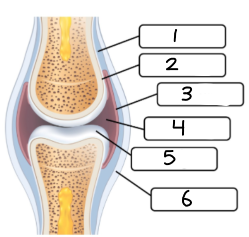
Label anatomy of synovial joint 4
Joint cavity

Label anatomy of synovial joint 5
Articular cartilage

Label anatomy of synovial joint 6
Ligament