Physics exam 3
 Call Kai
 Learn
 Practice Test
 Spaced Repetition
 Match
 Flashcards
 Knowt Play
1/78

There's no tags or description
Looks like no tags are added yet.
79 Terms
CXR
Chest x ray, evaluates the lungs, heart, pleura and tube placement
CT
Computed tomography, uses x ray to get cross sectional images of lung lesions, diseases
MRI
Magnetic Resonance imaging, magnetic fields to see soft tissue, vascular and mediastinal issues
Ultrasound
Sound waves to see pleural effusions, pneumothorax and to guide procedures
V/Q
Ventilation/ perfusion scan compares air reaching the lungs with blood flow to the lungs
What can a V/Q scan indicate?
Pulmonary embolism: sudden blockage of an artery in the lung
PET
Positron emission tomography, shows metabolic activity combined with CT to see cancers
How to read a CXR
Is the trachea midline or shifted due to fluid or air buildup, are the lungs clear, collapsed, opaque , dark, is the heart enlarged, is the diaphragm flatted from excess air ?
What are the components of the chest tube collection system
(1) Suction control chamber , (2) Water seal chamber, (3) Collection chamber,
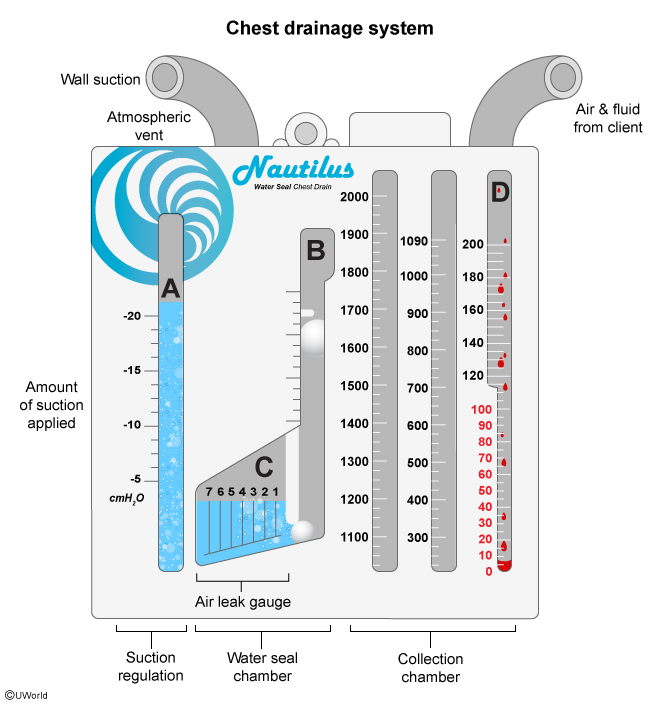
What does the collection chamber do?
collects fluid and blood from the pleural space
What does the water seal chamber do?
one way valve that allows air to escape but prevents liquid from entering
What does the suction control chamber do?
regulates suction, wet suction systems will have a gentle bubble, if its a dry system a bubbling will mean that there is a leak
Where does the chest tube go for a pneumothorax?
The apex of the lung since air is in the upper space of the pleural cavity
Where does the chest tube go for a pleural effusion?
posterior or lateral since the fluid collects in dependent regions, they sit lower and are affected by gravity
What is a pleural effusion?
abnormal build up of fluid in the pleural cavity
What is a hemothorax?
blood pooled in the pleural space
Where does the chest tube go for a hemothorax?
dependent pleural region
What is empyema?
Collection of PUS in the pleural space
Where does the chest tube go for a empyema?
Must be determined by the x-ray but it is placed wherever the fluid is built up
What do you do if the chest tube collection system is nonstop bubbling in the water seal chamber?
This indicates that there is a leak present, check all tubing and move patient if needed
What are indications for a chest tube insertion?
Pneumothorax to release air, tension pneumothorax to decompress, hemothorax to remove blood, pleural effusion to remove pleural fluid, empyema to remove pus, thoracic surgery to remove any fluid or air
What are common contradictions for chest tubes?
Infections, emergency situations, distorted anatomy, pleural adhesion that causes the pleural walls to stick, nearby organs are at risk and an unstable patient
What is a Spontaneous pneumothorax?
Occurs without an external injury or procedure, a collapsed lung from blebs with no prior disease history
What is a Primary spontaneous pneumothorax?
No history of disease pneumo/ collapsed lung
What is a Secoundary spontaneous pneumothorax?
Occurs in someone with an underlying lung disease, still spontaneous and more life threatening than a primary
What is a Traumatic pneumothorax?
Occurs from blunt or penetrating chest injuries such as car crashed, falls, stabbings
What is a Iatrogenic pneumothorax?
Accidental pneumothorax caused by medical intervention such as biopsies or a thoracentesis to remove fluid from around the lungs
What is a Open pneumothorax?
Sucking chest wound, opening in the chest wall that lets air enter and exit the pleural space, can be caused by trauma or accidental medical intervention
What is a Tension pneumothorax?
When air gradually enters the pleural cavity and has no where to escape to, results from a one way valve into the pleural space
What is atmospheric pressure?
760 mmHg
What is intrapulmonary pressure?
The pressure inside of the alveoli, it drops -1 during inspiration but is usually at 0 or atmospheric pressure
What is Intrapleural/ Intrathoracic pressure?
the pressure within the pleural cavity, inspiration is -8 and expiration is -4
What is the inspiration intrapleural pressure?
-8
What is the expiration intrapleural pressure?
-4
What is the trans pulmonary pressure
the pressure difference between the alveoli and the pressure in the pleural cavity
What is the order of pleural layers, from outer to inner
Partietal pleura
Pleural cavity
Visceral Pleura
What is a tracheostomy?
A stoma in the neck passed the vocal cords to maintain airway access, indicated by prolonged intubation, upper airway obstruction and difficulty clearing secretions
What equipment should a trach patient always have?
Trach tube, inner cannula, obturator for insertion, trache collar and ties, cuff and pilot balloon , suction yonker and sterile catheter , O2 delivery device, humification, spare trach tube the same size and one size small
When should the trach cuff be inflated/ Deflated?
The cuff should be deflated for patients who can clear secretions, aren’t on mechanical vent and those who are learning to speak again
Inflated for patients on mechanical ventilation, post operative and high aspiration risks
What is an ETT tube
Endotracheal tube that is inserted through the mouth or nose to secure an airway through the trachea indicated by respiratory failure, inability to protect their airway, severe hypoxemia, inadequate ventilation, controlled ventilation and surgery
What equipment is needed for ETT?
ETT the correct size, laryngoscope, AMBU bag and O2 delivery device, suction, syringe to inflate/ deflate the cuff, ETT face holder
When should the cuff be inflated on an ETT tube?
Once the tube is inserted into the vocal cords
When should the cuff be deflated on an ETT tube?
When intubating or extubating a patient
What pressure should the ETT cuff be at?
25-30
When do you know that the cuff is deflated on an ETT tube?
The patient will make a gurgling sound
Where should the ETT be place?
Through the vocal cords 3-5 cm above the carina

What is a combitube?
Dual lumen rescue airway that is inserted blindly indicated by an emergency airway management when intubation isn’t possible.
Combitube equipment?
Two lumens, two cuffs, ventilation ports and a syringe for the cuffs

What is a LMA?
Laryngeal Mask Airway, a supraglottic airway device positioned above the vocal cords indicated by an emergency airway rescue situation when intubation isn’t possible or during a surgery
LMA equipment
Correct LMA size tube, lube, AMBU bag, O2 delivery device, suction yonker
Where is a LMA positioned?
Over the laryngeal opening confirmed by chest rises and SPO2%, only used in emergencies because it doesn’t offer protection from aspiration like the ETT does
What is a Laryngoscope?
A tool used to visualize the vocal cords and larynx during intubation, includes the handle , blade and light source/ camera
What are the different types of laryngoscopes?
Macintosh- curved blade in the vallecula
Miller- straight blade to life the epiglottis
What is the adult amount of pressure for suction?
-100 t0 -150 mmHg
What is the child amount of pressure for suction?
-80 to -120 mmHg
What is the infant amount of pressure for suction?
-60 to -100 mmHg
What is the neonate amount of pressure for suction?
-60 to 080 mmHg
What is excessive pressure and suction bad?
It can cause trauma, hypoxia and atelectasis/ lung collapse from barotrauma
What is the Adult catheter size?
10 to 16 french but it depends on the ETT size and the quality of the secretions
What is the Child Cather size?
6 to 10 french depending on the airway size
What is the infant/ neonate Cather size?
5 to 8 french
Boyles law and how it applies to chest tubes
When volume increasing, pressure decreases; when volume decreases, pressure increases
during inspiration the chest expands increasing the volume and lowering the pressure
During expiration the chest deflates decreasing the volume and increasing the pressure
chest tube systems are a lower pressure so the trapped air and liquid gravitate towards the chest tube exit instead of staying in the pleural cavity
When does the mediastinum shift towards the problem?
When the affected side is collapsed from atelectasis
When does the mediastinum shift away from the problem?
When the affected side is filled of air or fluid —> tension pneumothorax, pleural effusion , emphysema and hyperinflation
What does air look like on a CXR?
Black, radiolucent, doesn’t absorb well
What does Fat look like on a CXR?
Dark gray, absorbs a small amount
What does Water/ Soft tissue look like on a CXR?
Medium gray
What does bone look like on a CXR?
Radiopaque, absorbs large amounts of the x ray
what is indicated by gray to white on a CXR?
Pneumonia, edema, masses, pleural fluid
what is indicated by abnormally dark on a CXR?
Extra pleural AIR, pneumothorax with no lung markings, flattened diaphragm
Which film makes the heart look enlarged?
AP, anteroposterior
Where does the beam go in for AP and PA
AP- beam goes in the front, anterior
PA- beam goes in the back, posterior
What does Pneumonia look like on a CXR?
Patchy dense opacities
What does Pulmonary edema look like on a CXR?
bilateral hazy congestitises
What does Atelectasis look like on a CXR?
Opacity with signs of volume loss and lung collapse
What does Pneumothorax look like on a CXR?
Dark pleural space with absent lung markings
What does Emphysema look like on a CXR?
Hyperinflation with flattened diaphragm from excess air
What does it mean to have the ability to protect your airway?
The body is able to keep saliva and food out of the trachea, able to swallow, cough, gag, and prevent aspiration
What is decannulation
Process of removing the tracheostomy tube from the neck after being weened off