PSYC 301 midt 2
 Call Kai
 Learn
 Practice Test
 Spaced Repetition
 Match
 Flashcards
 Knowt Play
1/55

There's no tags or description
Looks like no tags are added yet.
56 Terms
Circle of Willis: ACA? AComA? MCA? PComA? PCA? ICA? BA?

MCA

feeds basal ganglia
MCA Superior division: parietal frontal
MCA Inferior division: temporal lobe
ACA

medial frontal
sweeps back into parietal
What areas of cortex do ACA, MCA, PCA apply to?

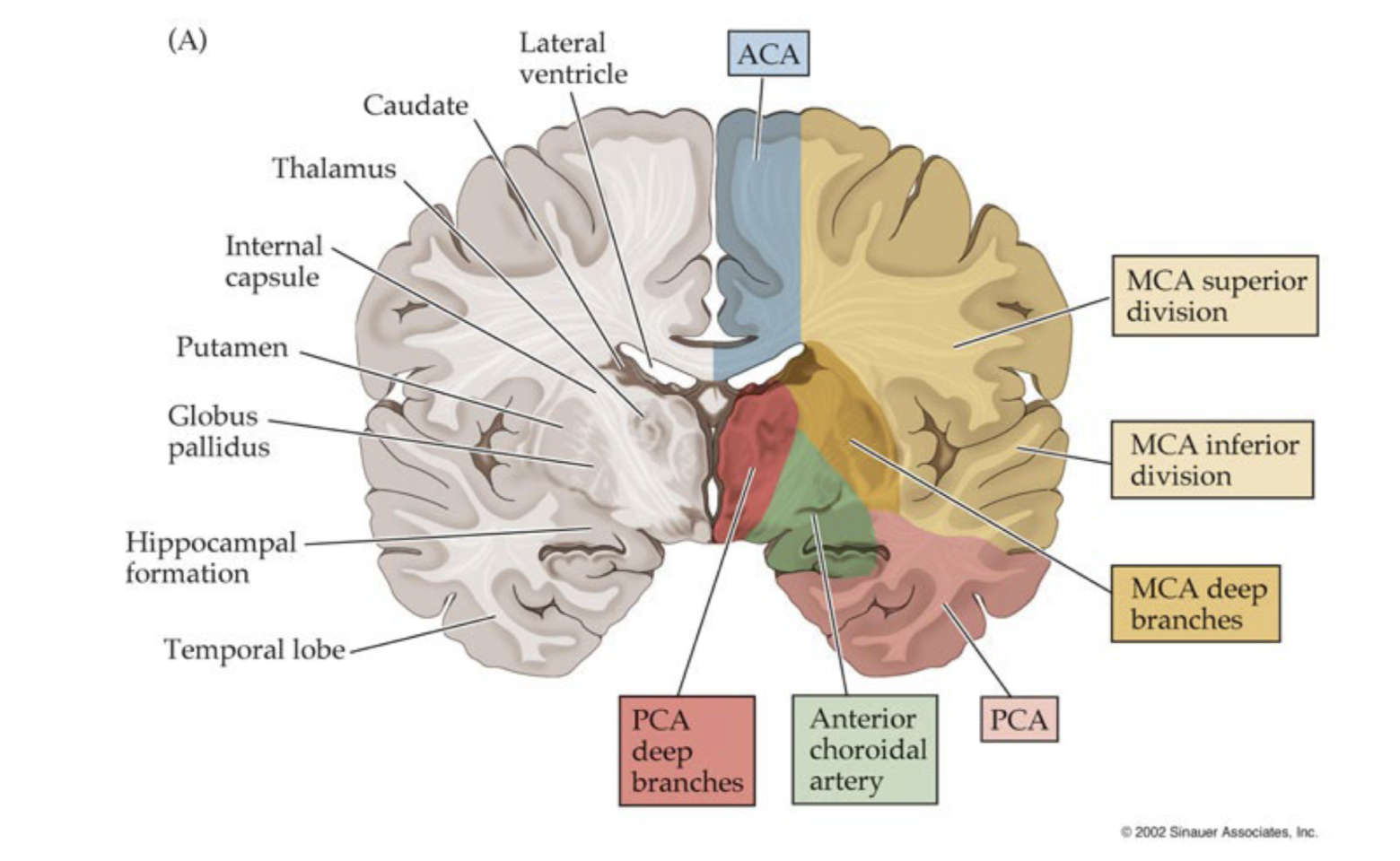

MCA: lateral, prefrontal, parietal, temporal
ACA: medial, prefrontal, parietal
PCA: occipital
Types of stroke: Ischemic? Hemorrhagic?
I. Ischemic: a clot of plaque buildup that floats and blocks blood flow to an area of the brain, and then artery narrows
II. Hemorrhagic: rupture/bleed that weakens vessel
hematoma, on the outside

Aneurysm
a kind of bleed:
the ballooning out and weakening of blood vessels (small and not likely to rupture)
runs in the family
most common to take place in orbitofrontal
confabulation, personality changes
can clip or coil the balloon
Epidemiology for Stroke
major cause of disability and death
likelihood increases w age
people who have risk factors (hypertension, diabetes, smoking, sodium intake) can modify these things
(hemorrhage) anticoagulant meds—that help for clots
(ischemic) atrial fibrillation—heart
Pathophysiology of Ischemic Stroke; Penumbra? Ischemic Core?
Ischemic core: brain tissue that’s dead
Penumbra: brain tissue at risk of dying

Pathophysiology of Hemorrhagic Stroke; Midline Shift? Herniation?
increased intracranial pressure
brain tissue getting pushed elsewhere
Midline Shift: brain matter swells and decentres middle structure of the brain
Herniation: brain matter shifts into places it shouldn’t
Clinical Presentation: Acute Stroke
focal, unilateral deficits
(hemorrhagic): bad headaches, vomiting, severe hypertension, coma
Prognosis for Acute Stroke
prog is better for ischemic (hemorrhagic more likely for death)
most recovery happens in first 3 months
predicted by age, stroke size, severity of initial deficits
Treatment for Acute Stroke? For ischemic, hematoma?
ischemic: thrombolytics—tPA, thrombectomy
hematomas: anticoag reversal, IV antihypertensive drugs, surgical evac—open up skull
secondary prevention (meds and lifestyle management of risk factors for second stroke)
early mobalization after 24 hours
multirehab (PT, OT, SLP)
What Consists of Brain Aging? (4) How Does the Brain Adapt with Age?
volume shrinks 5% per decade after 40, then accelerates after 70
declined serotonin and dopamine levels
decreased cerebral blood flow and white matter lesions
mild Alzheimer’s pathology (not disease)
adaptive
ongoing neuroplasticity
downregulates neural metabolism; doing the least it could do
increases clearance of cellular debris and protein buildup
Least vs. Greatest Cognitive Functions Declining with Age?
decline: episodic memory, learning speed, exec function
increased: wisdom/verbal knowledge

Mild Cognitive Impairment
mild cog difficulties (more than expected) but no dysfunction
Dementia
impairment of multiple cog functions + dysfunction
slow and lowk decline
onset in middle-to-late adulthood
List the Strongest Modifiable Risk Factors for Dementia
hearing loss
depression
TBI
less education
hypertension
obesity
smoking
social isolation
Diagnosis of Dementia? Blood Biomarkers?
history of progressive devline
interview people around them to survey activities of daily living
blood biomarkers
what allele of APOE gene; apoE4/4 has 3-4x risk for AD
only available for Alzheimer’s pathology (no other dementia)
high positive rate in people without cognitive impairment and then don’t develop dementia
confounding med conditions
confirmed to work on white people (probably diet/genes)
Dementia vs. Delirium vs. Depression

Clinical Presentation for Alzheimer’s? (Early, Middle, Late)

Alzheimer’s Pathophysiology? What Happens in the Brain?
ventricles get larger, brain tissue around them breaking
healthy aging but exaggerated
accumulation of amyloid-b (or plaque) between neurons

Treatment for Alzheimers? (NOT Reversible)
acetylcholinesterase inhibitors
glutaminergic antagonists (slows the rate of decline memory func)
anti-amyloid antibody (lecanemab)
Frontotemporal Dementia (FTD)? Behavioural Variant (bvFTD)?
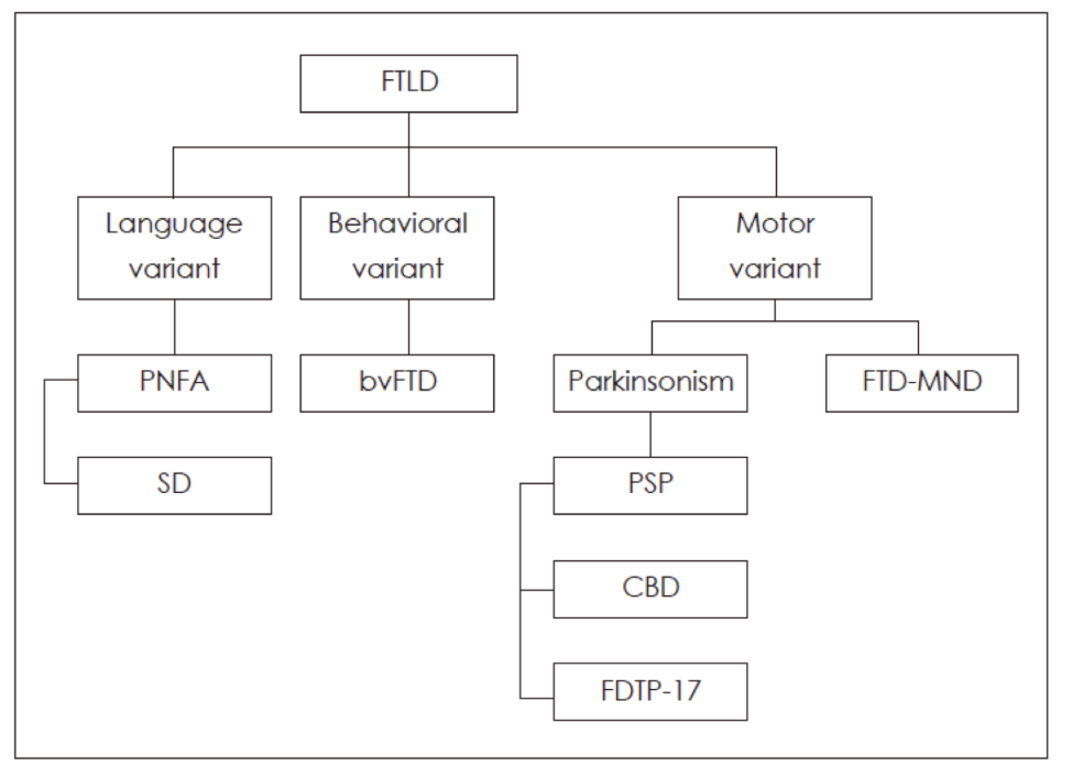
most common to have early onset, 50s
behavioural
inhibition
apathy
social inappropriateness
exec dysfunction
anosognosia (unaware of own deficit)
language
pnon-fluent aphasia ; brocas
semantic dementia (losing concepts, can’t name object knowledge) ; ant temporal
Neuropathology FTD?
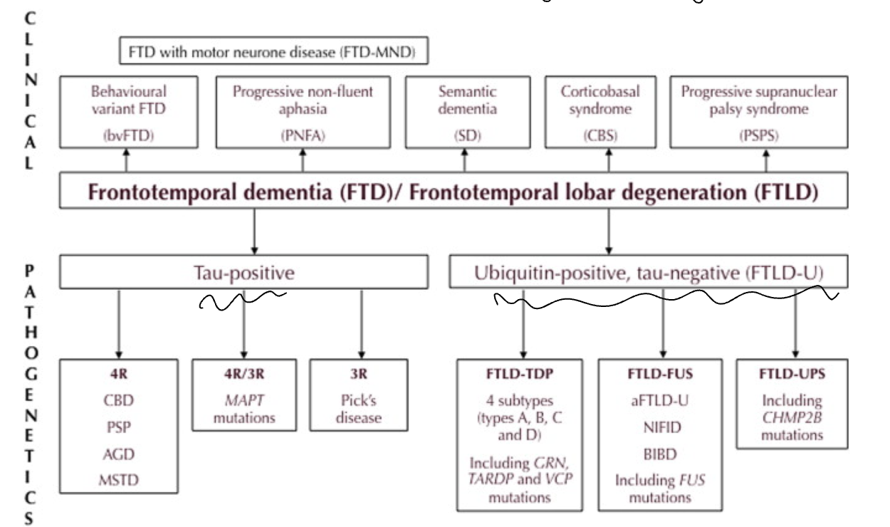
Treatment for FTD?
symptom management only—for behaviours
diff pharmacological agents needed for each underlying pathology
Clinical Presentation for Lewy Body Dementia?
fluctuations in attention and alertness
visuospatial difficulties; can’t copy or interpret objects
hallucinations
parkinsonism
REM sleep behaviour disorder (they act out their dreams since that inhibitor is lost)
Neuropathology for Lewy Body Dementia?
protein clumping
aggregation of a-synuclein
Treatment for Lewy Body Dementia? Why Not Antipsychotics?
acetylcholinesterase inhibitors
L-Dopa (helps parkinsonism)
sensitivity b/c personality problem
Vascular Dementia?
ischemic vascular disease +- clinically symptomatic strokes
subcortical dementia profile (cog slowing, exec dysfunc, language and visuospatial preserved)
Epidemiology for Parkinsons?
onset between 45-70
male>female
white>black>asian
familial risk
Clinical Presentation for Parkinsons? (5)
Bradykinesia (slowness—in movement and thought)
rigidity
upper extremity resting tremor
postural instability with shuffling gate (compensating for imbalanced torso)
expressionless face
Neuropathology for Parkinsons?
substantia nigra degeneration
neuronal loss, and remaining cells have Lewy bodies

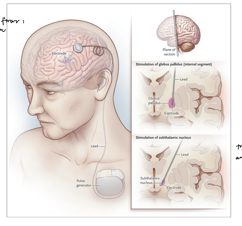
Treatment for Parkinsons?
L-dopa (building block to make dopamine)
dopamine agonists—to mimic dopamine
deep brain stimulation (lead electrode trying to stimulate action potential)
Epidemiology for Huntington’s?
hereditary (autosomal dominant)
child of a person who has it has 50% chance
Clinical Presentation for Huntington’s?
chorea (dance-like fidgeting)
dystonia (bursts of muscle tone, toes curling for ex)
personality change from exec func
subcortical dementia
Neuropathology for Huntingtons?
increased number of trinucleotide CAG on chromosome = cell toxicity
bilateral caudate degen
frontal-subcortical circuits
Treatment for Huntington’s?
none
Functional Neurological Disorder (FND)? Voluntary or Involuntary?
sensory, motor, cognitive, or speech problems not fully explained by known neurological disease
What are the Clinical Phenotypes for FND?
func weakness: face is not affected
non-epileptic seizures: crying (ep are disoriented), stop/start (ep one and done)
func cog disorder: vivid memory of forgetting
Pathophysiology for FND?
ex: trauma/childabuse + injury/strain/new sensation

Diagnosis for FND?
internal inconsistency: girl being able to walk into the office but can’t lift her leg while she’s on the table
incompatibility: no lesion in the brain can output that
distractibility: reduction/disappearance in the symptom when their mind gets pulled off it
EEG for seizures
EMG for tremor
Treatment for FND?
educate
therapy
physiotherapy
Epidemiology for TBI?
males get injured almost twice as much
adolescents and young adults by sports and rec
fall-related old people
Pathophysiology for TBI?
non-specific repolarization: extra firing
hours to days
ions imbalanced
energy crisis where one pumps and cells work hard to release it; trying to reach homeostasis

Structural Neuropath in TBI? Prim-Acute? Secondary-Acute? Post?
Primary-Acute
diffuse axonal injury (DAI)
bleeds
focal contusions (bruising)
Secondary-Acute, the killing one
edema (swelling)
ischemic-hypoxic injury (further damage from blood not flowing)
seconds to weeks
Post-Acute
risk factor for alzheimers
neuroinflammation
impaired clearance and cerebrovasc autoregulation
months to years after
Diffuse Axonal Injury (DAI), Diffuse or Focal?
stretching and tearing of axons
diffuse
can have TBI but not DAI

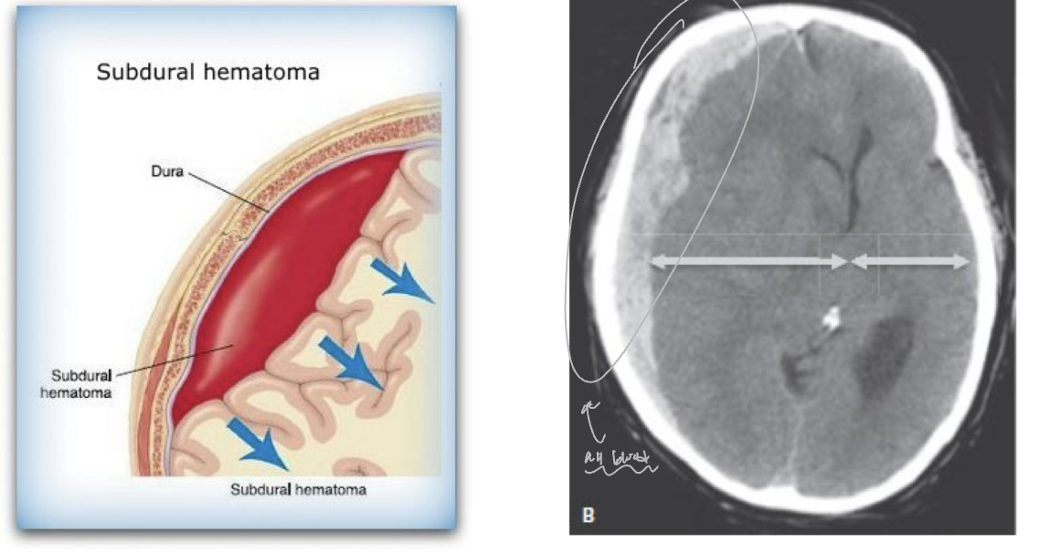
Subdural Hematoma
too much blood where not supposed to be ; pressure and pushing of brain tissue
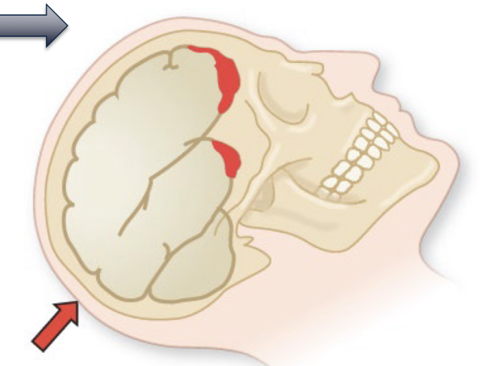
Contusions
bruising, brain bumping with skull
TBI; Why is it always Frontal-Temporal
Contracoup
Bony inner skull
What are the Causes for Anoxia
blood not flowing
suffocation
carbon monoxide poisoning (blood still flows but blood is not oxygenated; cm binds to hemoglobin where oxygen would normally go)
respiratory distress (ex, COVID)
respiratory supression (ex, fent)
Pathophysiology for Anoxia? Brain Regions Most Sensitive to it?
lack of oxygen supply to brain
insuff cerebral flow
no oxygen saturated in blood
motor
basal ganglia
hippocampi
cerebellum
watershed areas of cortex
Clinical Presentation for Anoxia?
severe prolonged —> coma or brain death (beyond 15min)
if oxygen is resumed within 5-15min
amnesia
exec dysfunc
ataxia (lack of vol coordination)
proximal limb weakness (person-in-the-barrel)
visual agnosia and cortical blindness
Prognosis for Anoxia?
go to hopital, depth of coma? okay bet
if outside hotpal, 10% survive, 5% make full neurorecov
if inside hoptal, 50% survival
Primary vs. Metastatic Brain Tumour? Types of Primary?
metastatic is more common, that tumour originates in another part of the body (cancer) and those malfunctioning cells go to the brain
primary:
meningiomas: from meninges (outside of brain)
gliomas: from glial cells
pituitary tumours: pressure onto optic chiasm; hormonal changes and headaches, loss of peripheral vision
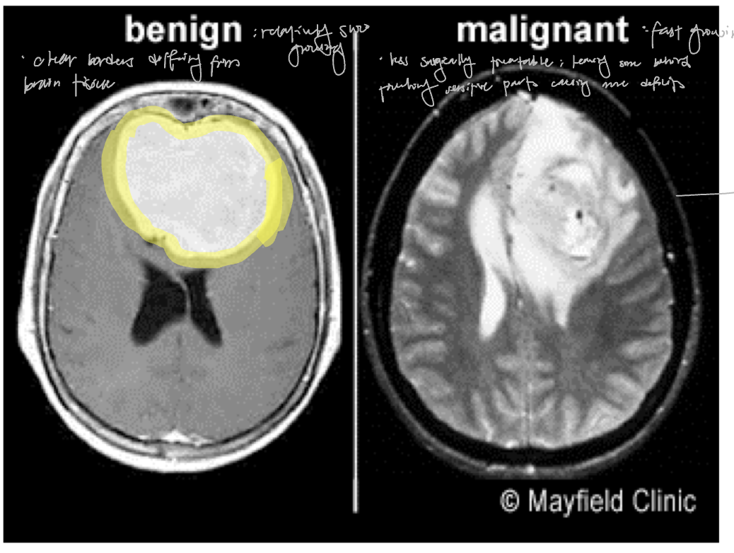
Benign vs. Malignant Brain Tumour
Benign: slow growing, clear borders
Malignant: fast growing, less surgically treatable and likely to come back
Clinical Presentation for Brain Tumour? (3)
gradual onset, months -→ years
depends on location
non-pulsing headache
seizures