Analysis of Urine and Body Fluids Laboratory Summary
 Call Kai
 Learn
Learn Practice Test
 Spaced Repetition
 Match
Match Flashcards
 Knowt Play
1/41

Earn XP
Description and Tags
Comprehensive practice flashcards for the Analysis of Urine and Body Fluids (AUBF) laboratory, covering physical, chemical, and microscopic urine examinations, reagent strip principles, special tests, casts, and crystals.
Name | Mastery | Learn | Test | Matching | Spaced | Call with Kai | Chat |
|---|
No analytics yet
Send a link to your students to track their progress
42 Terms
What is the pigment responsible for the normal amber yellow color of urine, and what does its intensity indicate?
The normal color is due to urochrome, and the intensity of the color is directly proportional to the concentration of the urine specimen.
What clinical conditions or substances cause urine to appear red, red-purple, or red-brown?
Red to red-brown urine can be caused by hemoglobinuria, hematuria, myoglobinuria, porphyrins, beets, aniline dyes, or menstrual contamination.
How are the turbidity levels of urine defined based on visual transparency against printed text?
Clear: No particulates;
Hazy: Few particulates, print easily seen; Cloudy: Many particulates, print blurred; Turbid: Print cannot be seen;
Milky: May precipitate or be clotted.
What indicator system is utilized on reagent strips for measuring urine pH, and what color changes occur across the range?
A double indicator system of methyl red and bromothymol blue is used. Methyl red changes from red to yellow between pH 4 and 6, while bromothymol blue changes from yellow to blue between pH 6 and 9.
What are the common clinical causes of acidic urine versus alkaline urine?
Acidic urine is caused by emphysema, diabetes mellitus, starvation, dehydration, and high protein diets. Alkaline urine is caused by hyperventilation, vomiting, vegetarian diets, and renal tubular acidosis.
What physical principle governs the urinometer, and how is temperature compensation applied?
It works on Archimedes' principle (a body immersed in fluid is buoyed up by a force equal to the weight of fluid displaced). Read the bottom of the meniscus and apply a temperature correction of +0.001 for every 3∘C (5∘F) above the calibration temperature of 60∘F.
How are Isosthenuric, Hyposthenuric, and Hypersthenuric urine defined by specific gravity (SG)?
Isosthenuric urine has an SG of 1.010 (1.008–1.012 range). Hyposthenuric urine has an SG below 1.010. Hypersthenuric urine has an SG above 1.010.
What is the procedure and principle of the Heat and Acetic Acid test for protein in urine?
Principle: Chemical and heat precipitation of protein. Procedure: Fill tube 43 full, heat the upper part to boiling, add 10% (v/v) acetic acid dropwise while boiling, and observe turbidity against a dark background.
What sources of error can affect the Heat and Acetic Acid test for urine protein?
Mucin gives false positives unless removed; Bence Jones proteins may yield false negatives; phosphates and carbonates interfere if urine is not acidic; and over-acidification may dissolve small amounts of albumin.

What principle and main sources of error are associated with the bromphenol blue reagent strip method for protein?
It operates on the 'protein error of indicators' using tetrabromphenol blue at pH 3. False positives occur in decomposed or highly alkaline urine (pH>8) or from contamination with quaternary ammonium compounds, antiseptics, detergents, or chlorhexidine.
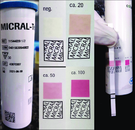
What is the principle, specimen requirement, and sensitivity of the Micral Test?
It is an immunoassay using monoclonal antibody-enzyme conjugate (gold conjugate) fixed to albumin. It requires first morning urine, has a reaction time of 1min, a sensitivity of 20mg/L, and is designed to detect microalbuminuria (20–200mg/L).

What is the principle and reagent composition of Benedict's test for reducing sugars?
Benedict's test is a copper reduction method where reducing sugars reduce Cu2+ to a Cu2O precipitate when heated for 5min in a boiling bath. The reagent contains copper sulfate (CuSO4), sodium carbonate (Na2CO3), and citrate.

How does Clinitest differ from reagent strips in terms of sugar specificity and procedural phenomena?
Clinitest is a self-heating copper reduction tablet (5 drops urine, 10 drops water, 1 tablet) that detects all reducing substances (0.25g/100mL sensitivity) and can show a 'pass-through' phenomenon. Reagent strips use glucose oxidase/peroxidase and are specific for glucose only (75–125mg/dL sensitivity).

What reagents and reaction outcome define Rothera's test for ketone bodies?
Rothera's reagent consists of sodium nitroprusside and ammonium sulfate, overlaid with concentrated ammonium hydroxide (1mL). A positive result is indicated by the formation of a reddish-purple to dark purple ring at the liquid interface within ≤1.5min.

What is the principle and positive end product of Fouchet's test for bile pigments?
It is an oxidation method where urine is mixed with 10% barium chloride (BaCl2) to precipitate bilirubin, filtered, and treated with Fouchet's reagent (trichloroacetic acid and ferric chloride). Bilirubin is oxidized to biliverdin, producing a green color.
How do intact red blood cells vs hemolyzed blood appear on a hemoglobin reagent strip pad?
Intact erythrocytes produce green spots on a yellow pad (non-hemolyzed trace), whereas hemolyzed blood causes a uniform green color change across the test pad after 60sec.

How does Blondheim's test differentiate between hemoglobinuria and myoglobinuria?
Urine is brought to 80% saturation with ammonium sulfate (2.8g in 5mL urine) and filtered. Hemoglobin precipitates out (red precipitate, clear filtrate), whereas myoglobin remains soluble in the supernatant (red precipitate, red filtrate).

What is the principle, reagent composition, and reading time for the Sulkowitch test?
Sulkowitch reagent (oxalic acid, ammonium oxalate, and glacial acetic acid) precipitates calcium as calcium oxalate at pH 5 where phosphates remain soluble. The turbidity degree is read after 2min to estimate urinary calcium concentration.

How is quantitative urine albumin measured using Esbach's method?
Urine is filled to mark 'U' in an albuminometer, and Esbach's reagent (10g picric acid, 20g citric acid per liter) is added to mark 'R'. After standing for 24hours at room temperature, the height of the precipitate is read directly on the tube scale in g/L.

How does the Watson-Schwartz test differentiate urobilinogen from porphobilinogen?
Urine is mixed with Ehrlich's aldehyde reagent and saturated sodium acetate. If a cherry-red complex forms, n-butanol is added. The urobilinogen-aldehyde complex is extractable into the upper n-butanol layer, whereas the porphobilinogen-aldehyde complex is insoluble in n-butanol and remains in the lower aqueous layer.
What are the standard centrifugation parameters and sediment preparation steps for microscopic examination of urine?
Centrifuge 10–15mL (average 12mL) of fresh urine for 5min at 400RCF (1500–2000rpm). Decant supernatant to leave 0.5–1.0mL of sediment, gently resuspend, place 1–2 drops on a glass slide, cover, and examine under LPF (10×) and HPF (40×).
What is the action and function of the Sternheimer-Malbin stain in urine microscopy?
Composed of Crystal Violet and Safranin O, it is the most frequently used stain. It delineates structures and provides contrasting colors for the nucleus and cytoplasm to identify WBCs, epithelial cells, and casts.
What is the primary utility of 2% acetic acid when added to urine sediment on a slide?
It lyses red blood cells and enhances the nuclei of white blood cells, distinguishing RBCs from WBCs, yeast cells, oil droplets, and crystals.
Which stains are used to confirm lipids in urine sediment, and what do they stain?
Oil Red O and Sudan III stain triglycerides and neutral fats orange-red, identifying free fat droplets, lipid-containing cells (oval fat bodies), and fatty casts.
How do red blood cells appear under the microscope in concentrated versus dilute urine specimens?
In concentrated (hypersthenuric) urine, RBCs shrink and become small and crenated. In dilute (hyposthenuric) urine, RBCs absorb water, swell, and lyse, leaving large, faint cell membranes termed 'ghost' cells.
What are 'glitter cells' in urine microscopy, and what conditions cause them?
Glitter cells are neutrophils exposed to hypotonic urine whose cytoplasmic granules display Brownian movement, giving them a sparkling appearance.
What are the three main types of epithelial cells found in urine sediment, and which is the most clinically significant?
- Squamous epithelial cells (largest, normal shedding; 'clue cells' indicate Gardnerella vaginalis). 2. Transitional epithelial cells (caudate/spherical, seen in catheterization). 3. Renal tubular epithelial (RTE) cells, which are the MOST clinically significant because increased numbers indicate renal tubular necrosis or injury.
What are oval fat bodies, and what characteristic patterns do they show under polarized light?
Oval fat bodies are renal tubular epithelial cells that have absorbed lipids. Under polarized light, cholesterol within these cells produces a characteristic 'Maltese cross' formation.
What protein serves as the primary matrix material for all urinary casts, and where are casts formed?
Uromodulin (also known as Tamm-Horsfall protein) forms the major matrix of casts within the lumen of the distal convoluted tubules and collecting ducts.

What microscopic characteristics define Hyaline casts, and what non-pathologic factors cause their elevation?
Hyaline casts are colorless, homogeneous, non-refractile casts (normal 0–2/LPF). They are increased in non-pathologic states such as strenuous exercise, dehydration, emotional stress, and heat exposure.
What do RBC casts indicate clinically in a urinalysis report?
RBC casts signify glomerular bleeding or severe glomerular damage within the nephron, highly characteristic of acute glomerulonephritis.
What is the clinical significance of WBC casts versus bacterial casts?
WBC casts indicate nephron inflammation or pyelonephritis (differentiating upper UTI from lower UTI/cystitis). Bacterial casts contain bacilli bound to protein matrix and specifically indicate acute pyelonephritis.

How do granular casts form, and what are their two morphological types?
Granular casts form from the disintegration of cellular casts, renal tubule cells, or filtered protein aggregates. They are classified into coarsely granular casts and finely granular casts.

What microscopic appearance characterizes waxy casts, and what clinical state do they signify?
Waxy casts appear highly refractile with fragmented, jagged ends and notched sides. They represent extreme urine stasis and signify end-stage chronic renal failure.
What are broad casts, and why are they referred to as 'renal failure casts'?
Broad casts are casts with a wider-than-normal matrix indicating destruction or dilation of the tubular walls (collecting ducts), signifying extreme urine stasis and severe renal failure.
What physical appearance and solubility characteristics define amorphous urates in acidic urine?
Amorphous urates appear as brick-red or pink granules ('brick dust' due to uroerythrin on refrigeration) in acidic urine (pH<5.5). They dissolve upon warming to 60∘C or upon addition of alkali.

What are the common microscopic forms of Calcium Oxalate crystals, and which form is associated with ethylene glycol poisoning?
The dihydrate form appears as colorless 'envelope' octahedral crystals (two pyramids joined at bases). The monohydrate form appears oval or dumbbell-shaped and is associated with ethylene glycol (antifreeze) poisoning.

What is the typical microscopic appearance of Triple Phosphate crystals, and in what urine pH are they found?
Triple phosphate (ammonium magnesium phosphate) crystals appear as prism-shaped 'coffin-lid' crystals found in alkaline urine.

How do Ammonium Biurate crystals appear under the microscope, and in what urine pH are they observed?
Ammonium biurate crystals appear as yellow-brown 'thorn apple' spheres with spicules, observed in alkaline urine.
What appearance characterizes Cystine crystals, and what chemical test is used to confirm their identity?
Cystine crystals appear as colorless, hexagonal plates (thick or thin) in acidic urine. Their identity is confirmed using a positive cyanide-nitroprusside test.
What is the microscopic appearance of Cholesterol crystals, and with what conditions are they clinically associated?
Cholesterol crystals appear as clear rectangular plates with a 'broken window pane' notch at one corner. They are associated with lipiduria, nephrotic syndrome, and hepatic disease.
How do Tyrosine and Leucine crystals differ in microscopic appearance?
Tyrosine crystals appear as fine, colorless to yellow needles arranged in bundles or rosettes.
Leucine crystals appear as yellow-brown spheroids with concentric circles and radial striations.