Test 3 Concepts
 Call Kai
 Learn
Learn Practice Test
 Spaced Repetition
 Match
Match Flashcards
 Knowt Play
1/23

There's no tags or description
Looks like no tags are added yet.
Name | Mastery | Learn | Test | Matching | Spaced | Call with Kai | Chat |
|---|
No analytics yet
Send a link to your students to track their progress
24 Terms
Cytoskeleton and Vesicle Trafficking
What is the cytoskeleton, what is its characteristics, and what are some of its functions
The skeleton of the cell
Network of filaments and tubules in cytosol that provides a structural scaffolding or framework for the cell
Monomers polymerize to form long filaments or tubular structures
It may be very rigid, yet it is very dynamic: constantly changing in most cells
Functions:
Defines cell shape
Supports the plasma membrane
Vesicle movement and organelle localization
Cell movement
Cell division
Cell-extracellular matrix interactions
What are the 3 components of the cytoskeleton and what are their sizes
Microtubules
Subunit: α-β-tubulin heterodimers
Largest part (most wide) of the cytoskeleton
Microfilaments
Subunit: G-actin monomers
Smallest part (most narrow) of the cytoskeleton
Intermediate Filaments
Intermediate in size
Subunit: IF dimer
Variety of proteins that compose intermediate filaments

What has been discovered by studying microtubule polymerization in vitro (in a lab)
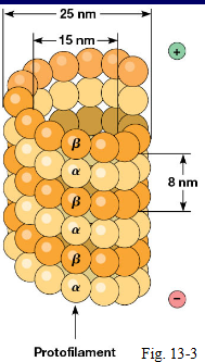
α & β tubulin dimers polymerize to form microtubules
Rate of polymerization depends on the concentration of tubulin dimer
At a high dimer concentration microtubules will polymerize
At a low dimer concentration microtubules will depolymerize
Results in free dimers that can be polymerized again
Causes pattern of shrinking and regrowing
Tubulin dimers are asymmetrical, thus microtubules are asymmetrical
One end looks different than the other end
β tubulin is exposed at one end (plus + end)
α tubulin is exposed at the other end (minus - end)
How does GTP contribute to the growth of microtubules
Both α and β tubulin bind to a molecule of GTP for the dimer to polymerize and join the growing microtubule
Shortly after the dimer joins the microtubule, the GTP bound to β tubulin will be hydrolyzed
When the dimer concentration is high there will be rapid growth and the
microtubule has a GTP capFacilitates the continued addition of tubulin
GTP cap is stable and is a good binding site for more tubulin dimer
Leads to rapid rate to microtubule growth

What is microtubule catastrophe
When the dimer concentration falls too low and rate of addition of tubulin dimer slows down, polymerization ceases
Lower dimer binding allows β tubulin to start hydrolyzing their GTP to GDP
The rate GTP hydrolysis > rate of polymerization
GTP cap is lost
Loss of GTP cap makes the microtubule unstable
GDP bound dimers have lower binding affinity for each other and the microtubule becomes unstable
Depolymerization > polymerization = “microtubule catastrophe”

What is dynamic instability of microtubules
Rapid degradation or growth of microtubules
Catastrophe can be followed by rescue of the microtubule as it alternates between growth and shrinkage based on the concentration of tubulin dimer
What is microtubule nucleation in vivo (in nature)
The beginning of the process of creating microtubules in cells
Start with individual dimers and put them together to start making microtubules
Microtubule nucleation requires γ-tubulin
13 copies of γ-tubulin (shown in green) are organized with other proteins into a structure called the γ-tubulin-ring complex
Serves as the anchor point for the minus (-) end of microtubules
γ-tubulin-ring complexes are found in microtubule organizing centers
The site where microtubules are nucleated (originate) and extend in all directions
Centrosome = where most MTOC is found

What are microtubule organizing centers and where are they mainly found
The site where microtubules are nucleated (originate) and extend in all directions
In most cells, the primary MTOC is located in the centrosome
The centrosome contains pair of centrioles + numerous additional proteins that compose the pericentriolar material
Peri = around, stuff around the centriole
We don’t know what centrioles do; plants have microtubules but no centrioles
Believed that centrioles to contribute to establishing the centrosome
Other MTOC locations
Epithelial cells that form cilia
Basil bodies structure is identical to a single centriole
Basil bodies help form cilia
Nerve cells
Red blood cells
Centrosomes in cell division

Fluorescence microscopy
Microtubules in yellow
MTOC behind the nucleus (bright light in center)
DNA in magenta
Microfilaments in blue

Microtubule toxins
Inhibit polymerization: colchicine, nocodazole, vincristine
Most work by binding up the dimer and preventing the dimer from being added to the microtubule
Used in chemotherapy for cancer treatment
Pulse/chase experiment with colcemid
Inhibit depolymerization: paclitaxel
Stabilizes intact microtubules so that they can never shrink
Also used in chemotherapy
How are microtubule toxins used in cancer treatment
Toxin that inhibit polymerization (colchicine, nocodazole, vincristine):
Prevent microtubules from forming so that mitosis will not occur
Toxins that inhibit depolymerization (paclitaxel):
Prevents mitosis from continuing because microtubules cannot shrink back and pull DNA to opposite ends of the cell
How are microtubule functions regulated in vivo
Regulated by microtubule associated proteins (MAPs)
MAPs either interact with tubular dimer or intact microtubules
Two classes:
Motor MAPS: kinesin & dynein
Non-motor MAPS
What are non-motor MAPs and how do they regulate microtubule function
A class of microtubule associated proteins (MAPs)
Numerous proteins bind to the plus end of microtubules (+TIPs) and promote:
Polymerization
Stabilization
Depolymerization (promote catastrophe)
Some proteins bind laterally to microtubules and promote crosslinking: tau, MAP2 (spacing between microtubules is much larger)

What are the two non-motor MPAs that bind laterally to the microtubules and what is the difference between them
tau and MAP2
MAP2 has greater spacing between the microtubules

How are microfilaments formed and what is its structure
Monomers of globular protein called actin (G-actin) polymerize to form filamentous actin (F-actin or microfilaments)
Rate of polymerization depends on the concentration of G-actin
G-actin is asymmetrical, thus F-actin is asymmetrical (+ end & - end)
G-actin must be bound to ATP in order to be added to the growing microfilament
Shortly after G-actin addition, the ATP bound to it will be hydrolyzed

What are the actin binding proteins and how do they bind to actin to affect microfilament structure (provide examples)
Proteins that bind to actin to affect microfilament structure and structure
Some bind G-actin and regulate:
Polymerization: formin & profilin
Depolymerization: cofilin
Some bind F-actin and regulate:
Filament length:
CapZ
Binding on the end and preventing any more filament addition
Formation of networks
filamin
Bind to filament and creates cross-linked structures of filaments
As filaments cross, they are linked together
Can cause gel-like consistency of cytosol; becomes thicker
Severing F-actin:
gelsolin
Cuts crosslinked filaments into fragments and caps them so they don’t polymerize and grow anymore
Cytosol becomes solvent-like
Branching:
Arp2/3
Allow microfilaments to create a branch point and creates branching filaments
Formation of bundles:
fimbrin, α-actinin
Facilitates parallel formation of bundles

What structure do specialized actin assemblies create and how
Microvilli
Parallel bundles of microfilament that push the membrane out
Common in epithelial cells
Increases the surface area of digestive system cells for transport

How do actin assemblies help with cell motility
Fibroblasts, immune system cells, neutrophils, macrophages, etc.
Filopodium on a leading edge
Parallel bundle structures
Lamellipodium
branched network pushes the whole membrane forward
Cell cortex
in the gel, gives stability for filopodium and lamellipodium
Stress fiber
in contractile bundle that retracts the trailing edge of the cell
Antiparallel structure

How are actin networks formed
Arp2/3
A dimer called actin-related protein
Binds laterally along microfilaments
nucleates branch points on existing filaments
How are the proteins that form actin networks regulated
The Rho family of small G-proteins regulate the formation of different
actin assemblies by regulating proteins the actin binding proteins
Rho, Rac, Cdc42
How do Rho family members contribute to the creation of actin assemblies used in cell motility
Overexpression of ‘dominant active’ forms of each G-protein
Unable to hydrolyze their GTP
Normal cell on the very left
Rho creates stress fibers
Rac creates Lamellipodium
Cdc42 creates Fliopodium

What are some proteins that make intermediate filaments
Lamin proteins provide structure support to the nuclear membrane
Keratin
