Pathology B5
 Call Kai
 Learn
Learn Practice Test
 Spaced Repetition
 Match
Match Flashcards
 Knowt Play
1/34

Earn XP
Description and Tags
Block 5 Brainteasers
Name | Mastery | Learn | Test | Matching | Spaced | Call with Kai | Chat |
|---|
No analytics yet
Send a link to your students to track their progress
35 Terms

Vertebrae from a cow. Name the lesion in the body of the vertebra.
Abscess

Pig femur. 4 months after a traumatic incident. Name the lesions indicated by the 2 asterisks.
1- Fracture
2- Callus

Canine vertebrae. Give the duration of each lesion.
Callus (red arrows) - Chronic

Foal died due to bacterial infection. Give a distribution of this lesion. How did this lesion develop?
Multifocal, hematogenous spread

Skeletal muscle from a calf. Histopathologic review shows necrosis of the myocytes with disruption of the basal lamina. This lesion will most likely:
Repair with extensive fibrosis

Cow. What is the most likely pathology shown in this image?
Bursitis

Horse. Name the lesion and give a morphological diagnosis for this lesion.
Skeletal muscle atrophy, unilateral, subacute to chronic, severe

What is the most likely cause of this lesion?
Neurogenic atrophy
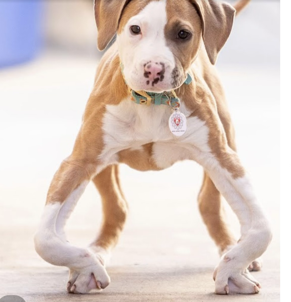
Dog with angular limb deformity. This lesion is more common in older animals (True or False)?
False

Damage to the metaphysis can result in this lesion (True or False)?
False

Name the lesions indicated by the arrows:
Osteophytes

Joint from a rapidly growing dog. A flap of cartilage has separated from the articular surface. Name the disease.
Osteochondritis dessicans

What is the pathophysiological mechanism of this lesion?
Failure of cartilage maturation/mineralization → retention and thickening of cartilage → inadequate vascular penetration → cartilage necrosis → weakened cartilage → fissuring

Lesion in and around the coxofemoral joint of a dog that had an angular limb deformity. Secondary osteoarthritis has developed. Why does this happen?
Abnormal alignment → abnormal mechanical axis → uneven joint loading → focal cartilage overload → chondrocytes respond to abnormal mechanical stress produce matrix metalloproteinases which degrade collagen and proteoglycans → cartilage degeneration → secondary osteoarthritis

Give 2 morphologic diagnoses for these lesions:
Ulcerative keratitis, focal, severe
Chemosis, diffuse, marked

Hereford cow out on the range. What type of ocular cancer is this?
SCCA

Name the scleral lesion.
Scleral injection

Elderly domestic shorthair (DSH) cat with no history of trauma.
Name 2 systemic diseases that could result in this lesion
Hypertension due to chronic renal failure
Hypertension due to hyperthyroidism

Kitten presented at a shelter with ocular lesions and respiratory signs. Name 2 ocular lesions.
Conjunctivitis
Blepharospasm

Which bacteria would most likely be cultured from this lesion?
Fusobacterium necrophorum

Give a morphological diagnosis for this lesion:
Pharynx: Pharyngitis, diffuse, severe, fibronecrotizing

Ventrum of an iguana. Name the lesion.
Dysecdysis
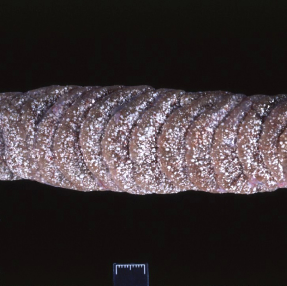
Snake kidney. Name the lesion.
Renal gout.

Feline blood smear: Is this a regenerative or non-regenerative anemia?
Regenerative
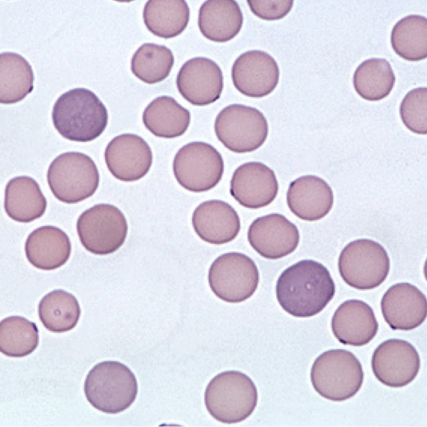
What is the name of the larger bluish cells?
Polychromatophils

Name the disease
Mycoplasma felis

Feline blood smear. Name the cells indicated by the black arrows.
Spherocytes

What do the spherocytes indicate about the status of the bone marrow and this cat’s anemia?
Indicates RBC destruction and are seen in hemolytic anemia

Feline blood smear with an additional special stain. What is the name of the special stain?
Methelyne Blue

What is the name of the blue protuberances on the red cells, and what do they indicate?
Heinz bodies, indicating hemolytic anemia due to oxidative damage

Why are cats RBCs particularly susceptible to oxidative damage?
Feline Hb has a larger number of sulfhydryl groups (8-10) that increases susceptibility to oxidation

Ingestion of what drug could cause this?
Acetaminophen

Avian oviduct. Name the lesion.
Salpingitis
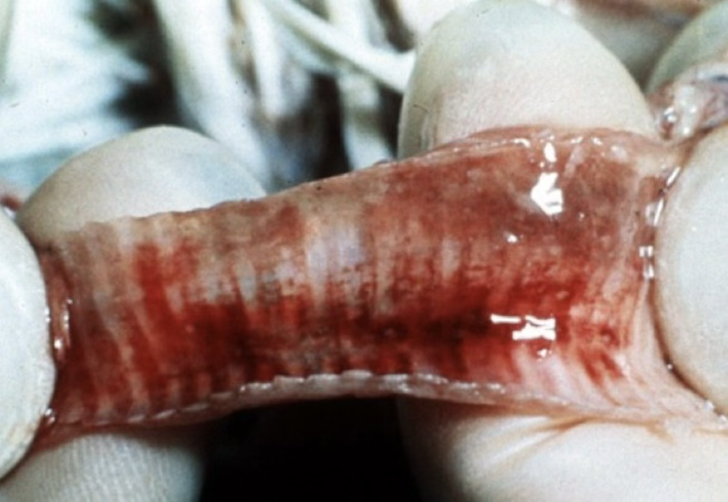
Trachea of a chicken. Which is the correct morphological diagnosis?
Trachea: tracheitis, acute, hemorrhagic, diffuse, severe

Chicken. Name the lesion.
Torticollis