Psych 377 First Midterm
 Call Kai
 Learn
Learn Practice Test
 Spaced Repetition
 Match
Match Flashcards
 Knowt Play
1/175

There's no tags or description
Looks like no tags are added yet.
Name | Mastery | Learn | Test | Matching | Spaced | Call with Kai | Chat |
|---|
No analytics yet
Send a link to your students to track their progress
176 Terms
Nuclei
Groups of Neurons in CNS
Tracts
Groups of axons in CNS
ganglia
cell body grouping axon bundles only in PNS
Nuclei
cell body groups in CNS
Rostral Caudal
Nose to tail axis of the body
synonymous ish with anterior posterior

Dorsal Ventral
Back-to-front axis of the body
synonymous ish with superior-inferior

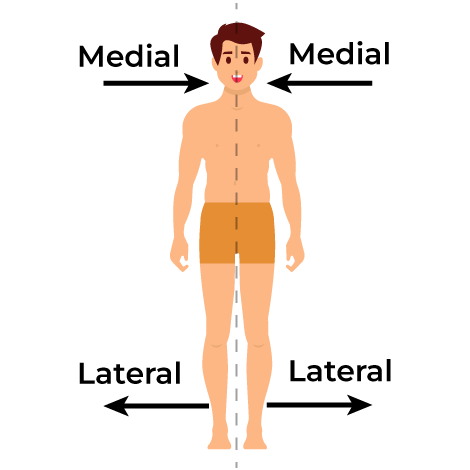
Medial-lateral
Medial: toward the middle
Lateral: toward side
Ipsilateral
Structures on the same side of the body
eg pain receptors, as they are connected to the spinal cord reflex so it needs to be a quick response
Contralateral
structures on the opposite side of the body
Proximal
structures are close together
Distal
Structures are far apart
Afferent
Movement toward the CNS
outside affects body
Efferent
movement away from CNS
body has an effect on the outside
Coronal Section

Transverse(horizontal) Section of Brain

Sagittal Section

Cenbtral Nervous System
Spinal cord and brain
protected by bone
Peripheral Nervous System
Projects out of body
somatic nervous system ineracts with external world
autonomic nervous system is automatic stuff
Sympathetic and parasympathetic branches
Sympathetic branch
PNS, autonomic
fight, flight, freeze, fawn response
sympathetic ganglia are close to the spinal cord
Parasympathetic Branch
PNS, autonomic
rest and digest
Ganglia are near the target organs
Protection of CNS
Bone surrounds tissue in CNS
Mengines surround the CNS to hold it in place
Cerebrospinal fluid fills the spaces in and around the meninges to absorb shocks and carry away waste
Blood-brain barrier provides chemical protection,
Dura Mater
tough outer coating meninges
Arachnoid Mater
web-like structure that connects dura and pia mater
Pia Mater
touch inner layer that adheres to surface of the brain
Blood brain barrier
chemical protection
astroglia hold cells of blood vessels tightly together
regulation of what molecules can exit the blood supply to enter brain
Blood Supply to Brain
multiple arteries supply blood to the brain so that damage to one blood vessel won’t interrupt blood flow to the entire brain
Arteries branch to form smaller arteries
Stroke
interruption of blood supply either because the artery becomes blocked or because the artery bursts
clot (block), hemorrhage (cut)
Anterior Cerebral Artery Location
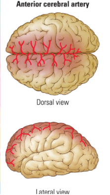
Middle Cerebral Artery Location

Posterior Cerebral Artery Location

Neuroblast cells
Arise from stem cells dividng which gives rise to neuroblast cells which than develop into neurons
Glioblasts
Stem cells dividing into these which differentiate into glia
Sensory Neurons
Transduce information from the environment
bipolar and somatosensory neurons
afferent as they are how the body is affected
Bipolar Neuron
One dendrite and one axon
sensory neuron

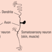
Somatosensory Neuron
one projection from the soma
sensory neuron
Interneurons
Connect sensory and motor neurons within the CNS
will have very extensive branching of dendrites to gather information
Motor Neurons
Found in the brainstem and the spinal cord
project to muscles to carry out movement
efferent as they are how the body has an impact on environment

Ependymal Cell
Small, ovoid; secretes cerebrospinal fluid
Astrocyte
Star-shaped, symmetrical; nutritive and support function
Microglial Cell
Small, mesodermally derived; defensive function
Oligiodendroglial Cell
Asymmetrical; forms insulating myelin around axons in the brain and spinal cord
will wrap around multiple cells to make myelin
Schwann Cell
Asymmetrical; wraps around peripheral nerves to form insulating myelin
can make a guide to reconnect neurons in peripheral nervous system
Gray Matter
Contains cell bodies and capillaries that supply them with blood
typically outer part of the cortex but inner part of the spinal cord
White Matter:
Myelinated axons that connect with other parts of the brain
typically underneath the cortex
eg corpus callosum is only axons that go from one side of the brain to the other
Reticular Matter
Netlike appearance that is a mix of grey and white matter, alternating white and gray matter, so it’s striped
found in brainstem, then the info in processed in cortex and sent to spinal cord
Three Enlargements of the Embryonic Spinal Cord
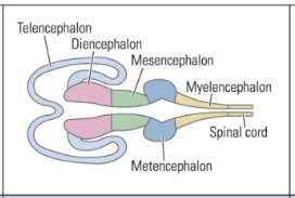
Prosencephalon
Mesencephalon
Rhombencephalon
Prosencephalon
In mammals, this divides to form the telencephalon (cortex and stereotypical brain) and the diencephalon (thalamus and hypothalamus)

Mesencephalon
Remains and becomes the midbrain
Forms: tectum, tegmentum, cerebral aqueduct

Rhombencephalon
Divides to form the metencephalon (pons and cerebellum) and the myelencephalon (medulla oblongata)

Telencephalon
Comes from prosencephalon
Forms neocortex, basal ganglia, limbic system, olfactory bulb, lateral ventricles
endbrain

Diencephalon
Comes from the prosencephalon
between brain
forms; thalamus, hypothalamus, pineal body, third ventricle

Metencephalon
Comes from the rhombencephalon
across brain
forms cerebellum, pons, fourth ventricle
Myelencephalon
spinal brain
Forms from the rhombencephalon
Forms the medulla oblongata and the fourth ventricle

Ventricles
Brain develops as a hollow tube and the hollow parts of the tube develop into the ventricles which are filled with CSF
lateral, third, fourth
Lateral Ventricle
Contained in the telencephalon

Third Ventricle
At the midline of the brain

Fourth Ventricle
between cerebellum and brainstem

Spinal Cord
gray matter surrounded by white matter
nerve roots branch from the cord to carry motor commands to the body and conduct sensory info into the CNS
4 segments: cervical, thoracic, lumbar, sacral
Law of Bell Magnide
afferent sensory info enters the posterior route of the spinal cord, whereas efferent motor neurons exit the anterior
Dermatomes
describe the region of the body surface innervated by each nerve
Cervical, Thoracic, Lumbar, Sacral Numbers
8
12
5
5
Spinal Reflexes
Are basic behaviours that occur without cognitive input
Generated in the spinal cord based on posterior sensory input, making a direct connection to the anterior motor output pathways
More complex reflexes will integrate information across multiple spinal cord segments (eg walking)
Cranial Nerves
twelve pairs of nerves branch from the brain and brainstem to provide sensory and motor innervation to the head, similar to what the spinal nerves do for the body
some are afferent (sensory) and some are efferent (motor), and some are mixed
Need to know: Olfactory, optic, oculomotor, auditory, vestibular, vagus
Brainstem
Extends from where the spinal cord enters the skull to the forebrain
Hindbrain
Cerebellum, pons, medulla, reticular formation
Pons
connects cerebellum with the rest of the brain
Cerebellum
Important for motor control and sensory integration
medulla
regulates functions such as breathing and heart rate
Reticular Formation
spans the pons and medulla, and sends projections to the cortex to maintain alertness and arousal
Up through the spinal cord through the pons and medulla to send info to cortex
damage could lead to a coma
Midbrain Contains
Tectum and Tegmentum
Tectum
Posterior aspect of the midbrain
Superior colliculi relay visual information
Inferior colliculi relay auditory information

Tegmentum
Anterior aspect of the midbrain
Nuclei here are involved in motor control, including the substantia nigra and red nucleus

What surrounds the cerebral Aqueduct?
periaqueductal gray matter and it is important for pain and fear responses
Hypothalamus
Many nuclei influence a wide range of behaviours
Produces and releases hormones that influence the entire body
Hypothalamic-pituitary axis: hormonal release (neural to endocrine system)
Thalamus
sensory relay
Almost all info destined for the cortex passes through the thalamus
it has nuclei that do preprocessing then relay info
relaying sensory info to cortex
relaying info between cortical regions
relaying from cortex to the brainstem
Epithalamus
Nuclei found posterior to the thalamus
Thalamic Projections Anterior
mammillary bodies coming in
signals to cingulate gyrus coming out
Thalamic Projections Ventral Anterior
Basal Ganglia signals coming in
Thalamic Projections Ventral Lateral
Cerebellum
Basal ganglia,
Substantia Niagara signals coming in
Thalamic Projections Ventral Lateral Posterior
Somatosensory signals coming in
Thalamic Projections Lateral and Medial Geniculate Body
Visual and auditory signals coming in respectively
Thalamic Projections Posterior
Areas 17 and 18 Superior colliculus
Thalamic Projections Dorsal Medial
Amygdala, Caudate Nucleus, and Frontal Cortex signals all go in
Basal Ganglia
Are important for motor control and motor learning (higher order)
includes putamen, caudate nucleus, and globus pallidus
Integrates sensory and motor information to produce fluid, skilled movements and is important for motor learning
diseases like Huntington disease with extra movement show hyperkinetic disorders of the basal ganglia
diseases like Parkinson disease show loss of movement which indicates hypokinetic disorders
associative learning(voluntary movement) takes place in the basal ganglia
Limbic System
Important for spatial and emotional functions
Represents an older part of the brain from evolutionary history
amygdala, hippocampus, cingulate cortex
Amygdala
small nuclei in medial temporal lobes
important in emotion and understanding emotion in others

Hippocampus
seahorse-shaped structure in medial and temporal lobes
important in personal memories and navigation
memory

Cingulate Cortex
arcs over the lateral ventricles
involved in decision making and executive functions

Neocortex
outer layers of forebrain
1.5-3mm thick and includes 6 layers of cells
divided into two hemispheres by longitudinal fissure
each of the 4 lobes has different functions
Gyri
ridges in the brain
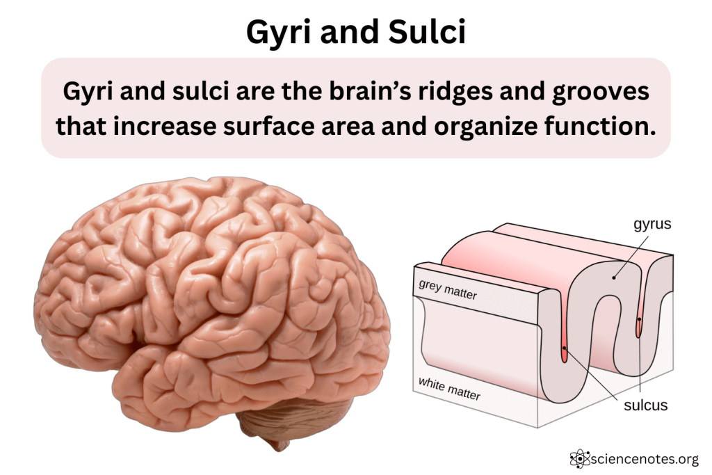
Sulcus
clefts

precentral gyrus

central sulcus

post central gyrus

intraparietal sulcus
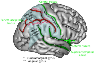
lateral sulcus /sylvian fissure

superior temporal gyrus, medial temporal gyrus, inferior temporal gyrus

superior and inferior temporal sulcus’s

Cingulate Sulcus

cingulate gyrus

Callosal Sulcus
