Blood Vessels and Circulation
 Call Kai
 Learn
 Practice Test
 Spaced Repetition
 Match
 Flashcards
 Knowt Play
1/112

There's no tags or description
Looks like no tags are added yet.
113 Terms
Tunica intima:
endothelium (simple squamous epithelium) plus connective tissue
Some arteries have
an internal elastic lamina
Tunica media:
concentric smooth muscle and connective tissue (elastic fibers
thickest layer in arteries
Tunica externa:
collagen and elastic fibers, a tough covering that anchors the vessel to surrounding tissue; areolar and adipose connective tissue
thickest layer in veins
Vasa vasorum ('vessels of vessels'):
small arteries and veins in the wall of large vessels that supply the vessel itself
Vaso vasorum support the
vessel tissue with its own blood supply
Companion vessels
lie next to each other (an artery and its vein)
Systemic arteries carry
highly oxygenated blood at high pressure
Systemic arteries carry highly oxygenated blood at high pressure, which
pushes blood against the vessel wall (this is why arteries need a thick, elastic media; a damaged artery leaks fast)
Tunica media is thicker in
arteries
Tunica externa is bigger in
veins
In larger veins the tunica intima
folds over to form valves
In larger veins the tunica intima folds over to form valves. The pressure gradient is low, so
valves prevent backflow
A capillary bed has shed the
tunica externa and smooth muscle tissue (only endothelium plus basement membrane remain)
Artery muscle is
thick and tends to hold its round shape; veins often collapse
The artery's inner surface has a
scalloped appearance
The internal diameter is very different: the vein lumen is
much larger
Artery lumen is more:
narrow
Vein lumen is
wider; often looks collapsed in cross section
Artery walls are more:
Thicker than companion vein
Vein walls are more:
Thinner than companion artery
Artery shape:
Keeps circular shape
Vein shape:
Flattens (collapses) when empty
Artery thickest tunic:
Media
Vein thickest tunic:
Externa
Artery elastic and collagen fibers:
More
Vein elastic and collagen fibers:
Fewer
Artery valves:
None
Vein valves:
Present in most veins
Artery blood pressure:
Higher (larger arteries typically above 90 mm Hg)
Vein blood pressure:
Lower (about 2 mm Hg)
Artery blood flow:
Away from heart
Vein blood flow:
Toward heart
Artery blood oxygen:
Systemic arteries high in O2;
pulmonary arteries low in O2
Vein blood oxygen:
Systemic veins low in O2;
pulmonary veins high in O2

Elastic (conducting) arteries:
largest (aorta, pulmonary trunk, carotids)
The elastic lamina runs through
the wall
Elastic fibers stretch with
systole and recoil, smoothing pressure
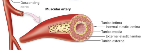
Muscular (distributing) arteries:
medium; internal and external elastic laminae; more smooth muscle for vasoconstriction and vasodilation

Arterioles:
smallest arteries; major control of resistance and blood flow into capillaries

Venules:
no media, sometimes no externa

Small to medium veins have
valves

Large veins have
a thick externa
Thin media and valves formed from the
intima
Venous return is helped by the
skeletal muscle pump and the respiratory pump
Skeletal muscle contraction makes blood flow in one direction because of
the one-way valves (the valve opens above the contracting muscle and closes below it)

Distribution of blood:
30-35% is in the heart, arteries and capillaries
60-65% is in the venous system (veins are the reservoir)
Capillary:
an endothelial tube on a basement membrane
Capillaries lack a
tunica media or externa
Capillaries have a diameter about that of
a red blood cell (about 8 micrometers)
Cells run single file through
the tube, and the wall is size selective
Capillary bed (capillary plexus):
connects an arteriole to a venule; fed by a metarteriole
Precapillary sphincters (smooth muscle rings):
open or close flow to true capillaries
Vasomotion:
pulses of constantly rerouted flow
Collaterals:
multiple arteries that feed one bed
An anastomosis (such as an arteriovenous anastomosis):
lets blood bypass the capillaries
Continuous capillary:
Complete basement membrane; the common type; restricts RBCs and plasma proteins (brain has extra tight junctions)
Continuous capillary example:
Muscle, skin, brain
Fenestrated capillary:
Pores (fenestrations) but a continuous basement membrane; rapid movement across
Fenestrated capillary example:
Kidneys, intestines, endocrine glands
Sinusoid capillary:
Incomplete or no basement membrane, large gaps between cells; proteins and RBCs can cross
Sinusoid capillary example:
Liver, spleen, bone marrow
What is capillary hydrostatic pressure (CHP)?
Pressure that pushes fluid out of the capillary
What is blood colloid osmotic pressure (BCOP)?
Pressure created by plasma proteins that pulls fluid into the blood
What determines net filtration pressure (NFP)?
CHP − BCOP
What is CHP at the arterial end?
35 mm Hg
What is BCOP throughout the capillary?
25 mm Hg
What is NFP at the arterial end?
+10 mm Hg (35 − 25)
What happens at the arterial end of a capillary?
Filtration: fluid moves out into surrounding tissues (~24 L/day)
What is CHP at the capillary midpoint?
25 mm Hg
What is NFP at the capillary midpoint?
0 mm Hg (25 − 25)
What happens at the capillary midpoint?
No net fluid movement
What is CHP at the venous end?
18 mm Hg
What is NFP at the venous end?
−7 mm Hg (18 − 25)
What happens at the venous end of a capillary?
Reabsorption: fluid moves back into the blood (~20.4 L/day)
What causes filtration out of capillaries?
Hydrostatic pressure pushes fluid against and through the capillary wall
What causes reabsorption into capillaries?
Blood colloid osmotic pressure pulls fluid back into the bloodstream
What happens to fluid that is not reabsorbed?
It accumulates in tissues and can cause edema (swelling)
What does the lymphatic system do with excess tissue fluid?
Collects excess fluid, filters it, and supports immune responses
How does increased blood pressure affect filtration?
Increases filtration, causing more fluid to leave capillaries
What does size-selective filtration mean?
The capillary wall allows some substances through while restricting others
What are the three main types of capillaries?
Continuous, fenestrated, and sinusoid
Fluid leaks out of the capillary into the surrounding tissue because
hydrostatic pressure pushes against the capillary wall
Filtration filters the
blood and is size selective
The venule end has
lower pressure, so the net filtration pressure turns negative and fluid moves back in
Osmotic pressure is created by
the blood proteins in plasma; it pulls fluid back into the blood
Fluid that is not recovered causes
swelling (edema)
The lymphatic system picks up the lost fluid,
identifies it and filters it
The lymphatic system is tied to
the immune system: lymph vessels begin with the fluid and the immune response follows
Higher blood pressure means
more filtration, so more fluid is lost into the tissue
Filtration beats reabsorption by about
3.6 L/day
The lymphatic system returns that fluid to
the blood
Placental blood supply:
a pair of umbilical arteries (carry blood to the placenta) and one umbilical vein (brings blood to the fetus)
Umbilical arteries and an umbilical vein drains into the…
ductus venosus and then the inferior vena cava, bypassing the liver
Fetal pulmonary bypass:
the foramen ovale is a right-to-left atrial shunt, and the ductus arteriosus connects the pulmonary trunk to the aorta
Both the foramen ovale and ductus arteriosus let blood…
skip the fetal lungs
The foramen ovale closes at
birth and leaves the fossa ovalis
Low-pressure blood is mixed with
high-pressure oxygenated blood
The ductus arteriosus becomes
tissue (it closes off and shuts down after birth; in the textbook this remnant is the ligamentum arteriosum)
The umbilical cord tears from
the placenta at birth