3. CT Lasers & Imaging
 Call Kai
 Learn
 Practice Test
 Spaced Repetition
 Match
 Flashcards
 Knowt Play
1/25

There's no tags or description
Looks like no tags are added yet.
26 Terms
breast
thorax
tissue usually extends from 2nd to 6th rib
suprasternal notch (SSN)
chest
T2/T3 interspace
angle of louis (sternal angle)
chest
T4/T5 vertebral level
tip of xiphoid process
chest
approximately T10 vertebral level
vertebra prominens
back
C7
base of neck

apex of lung
thorax
ANT level
2-3cm SUP to clavicle
carina (bifurcation of trachea)
thorax
T5
umbilicus
abdomen/pelvis
L3/L4 vertebral level
superior iliac crest
abdomen/pelvis
L4-L5
anterior superior iliac spine (ASIS)
abdomen/pelvis
L5-S1
used to level pelvis

pubic symphysis
abdomen/pelvis
8-10cm INF to ASIS

ischial tuberosity
abdomen/pelvis
4-5cm INF to symphysis pubis

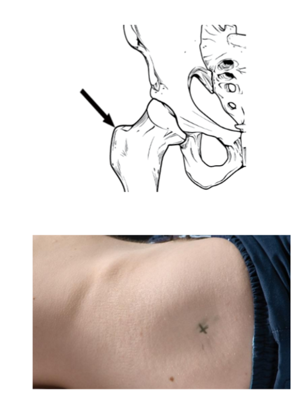
greater trochanter
abdomen/pelvis
~ same level as superior border of symphysis pubis
common landmark for prostate gland
laser alignment
uses surface + relational anatomy landmarks to estimate location of treatment area
starting point for CT sim
departments have site-specific guidelines
lung tumour @ T3
sagittal through midline
coronal at mid sep
transverse 3cm inf of SSN
prostate
sagittal through midline
coronal at mid sep
transverse at greater trochanter
breast in upper quadrant
sagittal through midline
coronal at mid sep
transverse 4cm inf of SSN
ball bearing marks (BBs)
reference marks positioned at intersection of room lasers
small radiopaque markers
material dense enough to show up as white dots on CT image
placed on patient’s skin using stickers/adhesive dots
external + internal purpose
identify location where patient was aligned to room lasers during CT sim
known starting location
location can be reproduced in future
scout image
simple 2D lower-dose x-ray taken just before CT scan begins
ensures scan covers correct anatomy
ensures patient is straight, leveled, centered
straightness
see if midline structures are centered
level
compare paired anatomical structures + ensure they are at same level = not twisted/rotated
centered
assess whether patient is positioned centrally within scanner = not too far left/right
ensures BB marks are in correct place
assess whether BBs are located near intended treatment area
determines if plannes CT scan limits are appropriate
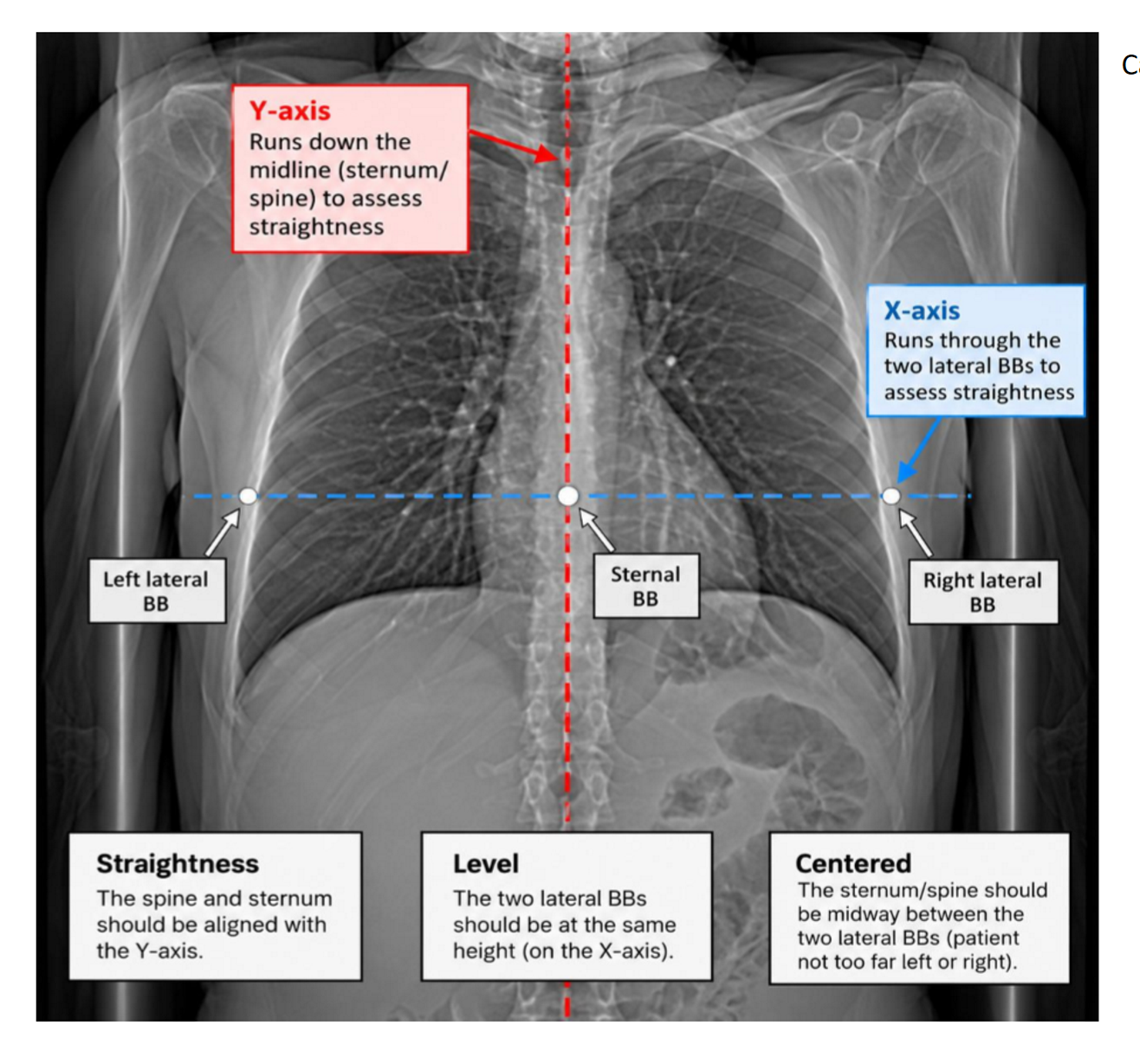
BB marks in scout image
set up patient to external lasers + BBs placed at intersection of lasers (1 anterior + 2 lateral reference marks)
couch moved 500mm into scanner → BBs to center of bore/internal laser
BB marks are 500mm away from center of bore (Z axis)
CT scan automatically moves patient until BBs reach internal bore lasers
BB center to bore = indicates center point of scout image
scout image captures anatomy superior + inferior to this point
SUP + INF margins depend on department, eg. can be 30cm sup + 30cm inf of BBs
scout image used to select CT scan limits → where CT scan starts + finishes
scan limits depend on treatment site
scout image steps
x-ray tube stays in fixed position (unlike CT scan)
couch moves patient through scanner
x-rays pass through patient to detectors
produces single 2D x-ray image
CT scan
3D volumetric image showing anatomy in 3 dimensions → height, width, depth
acquired after scout image confirms correct position, appropriate BBs location, scan limits
CT scan steps
patient lies on couch + RT moves couch through bore
gantry rotates around patient + sends kV x-ray beams through body
detectors on opposite side catch x-rays
computer collects + processes data to build 3D image of inside of body
x-ray interaction
x-rays interact differently w/ various tissues → creates contrast in data collected + allows computer to differentiate b/w different tissues
bone = dense → absorbs more x-rays
white/light grey
soft tissue → absorbs some x-rays
various shades of grey
air (lungs) = not dense → most of x-rays pass through
black
atomic number influence
body made up of different elements
each have atomic number (Z) = # of protons in nucleus
high Z = massive positive charge → pulls electrons tightly to nucleus
harder for x-rays to pass through tightly bound electron barriers
bone x-rays
mainly composed of calcium → high Z = 20
electrons tightly bound to nucleus
x-rays struggle to pass through
appearance = white

lung x-rays
mainly composed of oxygen, carbon, nitrogen → low Z
electrons loosely bound + spread far apart
x-rays pass through easily
appearance = black

soft tissue x-rays
made up of lightweight elements (O, C, N, H) → low Z
electrons loosely bound together but packed closely packed
x-rays pass through easier than bone but not as easily as lungs
appearance = grey

tattoos
small, permanent marks placed at BB locations after CT sim is complete
ensures reproducible position each day during treatment
patient is set up to those points every treatment session
when patient arrives for treatment:
patient is positioned on treatment couch w/ same immobilization equipment documented at CT
treatment room lasers aligned w/ these marks → recreates laser alignment established during CT sim
reproduces position established during CT sim