ANTH 102A Quiz 4 - The Leg
 Call Kai
 Learn
 Practice Test
 Spaced Repetition
 Match
 Flashcards
 Knowt Play
1/43

There's no tags or description
Looks like no tags are added yet.
44 Terms
Os Coxa
Made up of three bones, sometimes called the pelvis.

Ilium
One of the bones making up the Os Coxa. The thin, blade-like portion superior to the hip socket.

Ischium
One of the bones making up the Os Coxa.The massive, blunt, posteroinferior part of the bone that one sits on.

Pubis
One of the bones making up the Os Coxa. The anteroinferior part of the bone that approaches the opposite os coxaea at the midline.

Acetabulum
Feature of the Os Coxa. The laterally facing, hemispherical hollow that forms the socket of the hip, which articulates with the head of the femur.

Acetabular Fossa
Feature of the Os Coxa. The non-articular surface within the acetabulum.

Lunate Surface
Feature of the Os Coxa. The crescent shaped articular surface within the acetabulum.

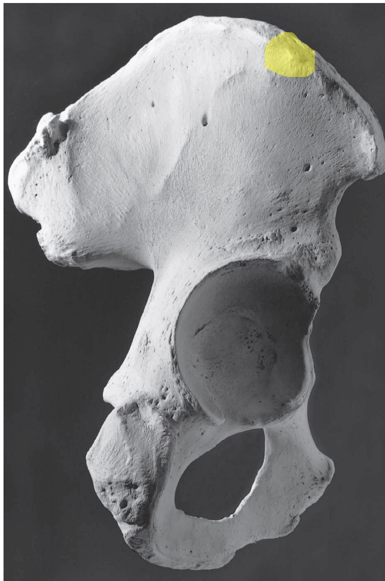
Iliac Pilar
Feature of the Os Coxa. The boney thickening, or buttress, located vertically above the acetabulum on the lateral iliac surface. The pillar extends to the superior margin of the ilium.

Iliac Tubercle
Feature of the Os Coxa. The thickening at the superior terminus of the iliac pillar
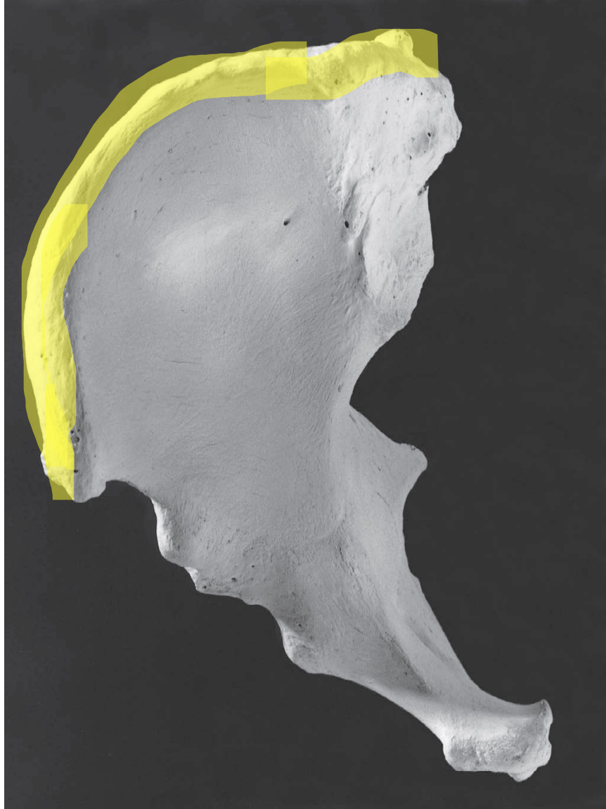
Iliac Crest
Feature of the Os Coxa. The superior border of the ilium
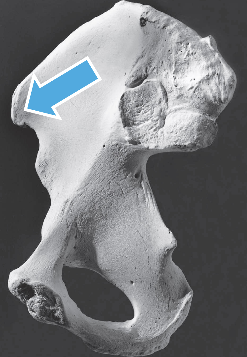
Anterior Superior Iliac Spine
Feature of the Os Coxa. Spine located at the anterior end of iliac crest.
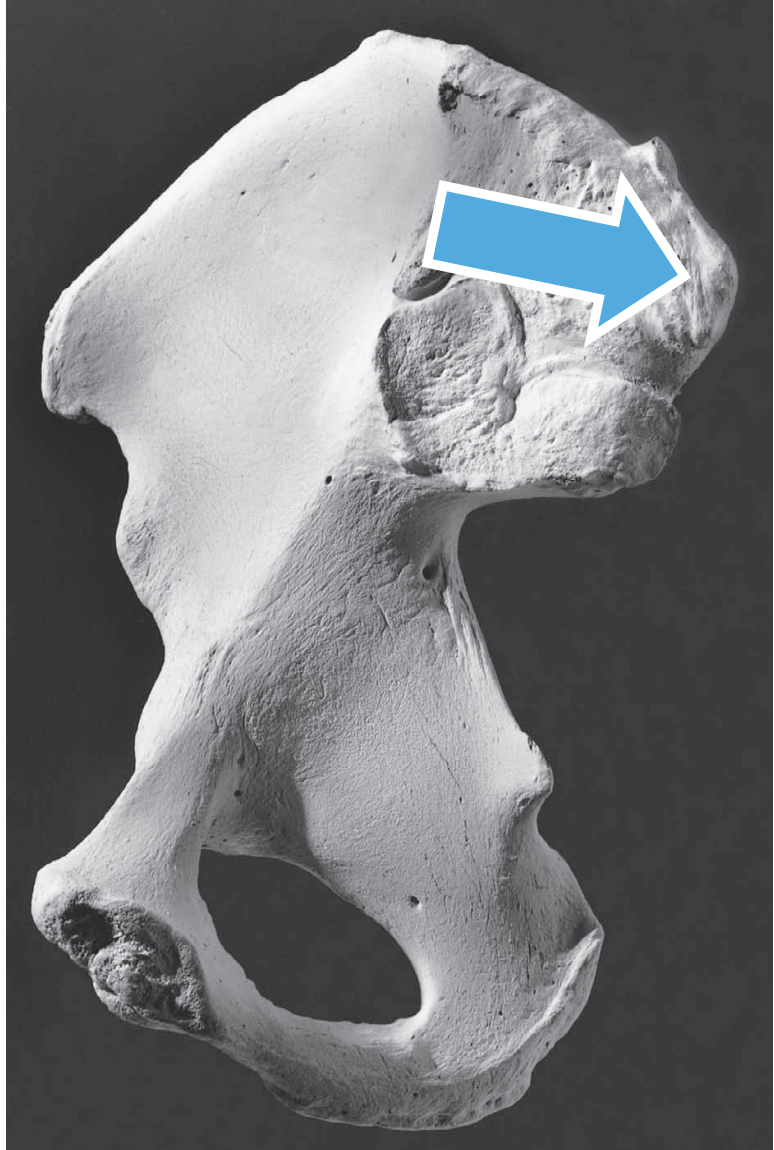
Anterior Inferior Iliac Spine
Feature of the Os Coxa.A blunt projection of the anterior border of the os coxa, just superior to the acetabulum

Posterior Superior Iliac Spine
Feature of the Os Coxa. The posterior terminus of the
iliac crest.
Posterior Inferior Iliac Spine
Feature of the Os Coxa. A sharp projection just posteroinferior to the auricular surface.

Greater Sciatic Notch
Feature of the Os Coxa. The wide notch just inferior to the posterior inferior iliac spine.

Ischial Spine
Feature of the Os Coxa. Located just inferior of the greater sciatic notch.
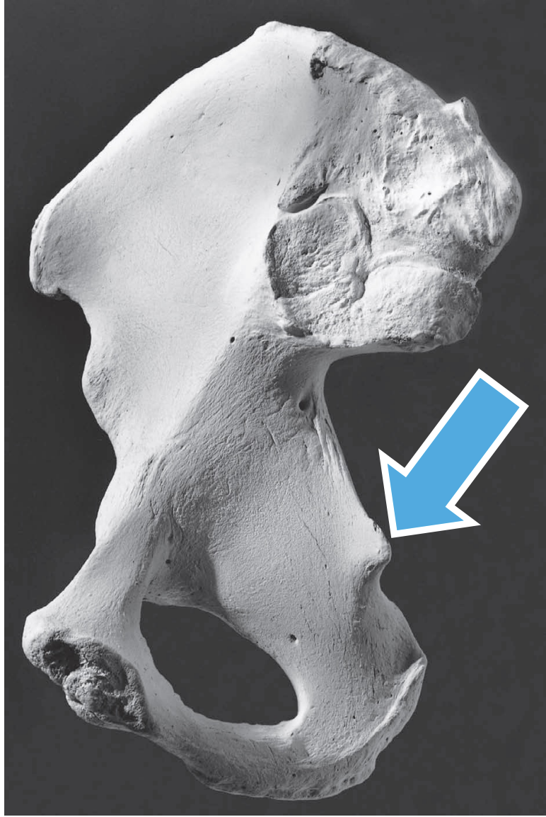
Lesser Sciatic Notch
Feature of the Os Coxa. The notch between the ischial spine and the superiorly and the rest of the ischium inferiorly.

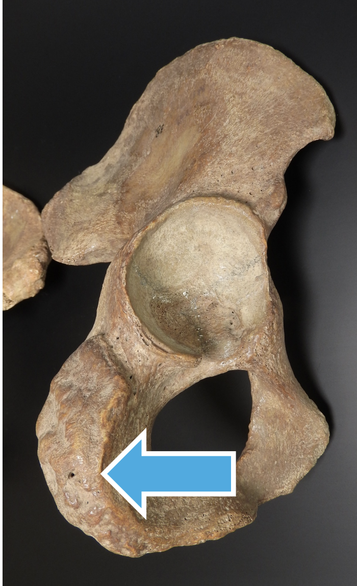
Ischial Tuberosity
Feature of the Os Coxa. The blunt, rough, massive, posteroinferior corner of the Os Coxa.
Auricular Surface
Feature of the Os Coxa. Articulates with the Sacrum.The ear-shaped sacral articulation on the medial surface of the ilium.


Pre-Auricular Sulcus
Feature of the Os Coxa. A variable groove along the anteroinferior edge of the auricular surface.

Iliac Fossa
Feature of the Os Coxa. Anterior. The smooth hollow on the medial surface of the iliac blade.

Arcuate Line
Feature of the Os Coxa. Anterior. An elevation that sweeps anteroinferiorly across the medial surface of the os coxae from the apex of the auricular surface toward the Pubis.


Pubic Symphysis
Feature of the Os Coxa. Where the two Os Coxa meet.

Obturator Foramen
Feature of the Os Coxa. The large foramen encircled by the two pubic rami and the ischium. In life if it mostly closer with a membrane across it.

Femur
Longest, heaviest, and strongest bone in the body.

Head
Feature of the Femur. The rounded proximal part of the bone that fits into the acetabulum. It is more spherical than the more hemispherical humeral head.

Fovea Capitis
Feature of the Femur. The small, non-articular depression near the center of the femoral head.

Neck
Feature of the Femur. Connects the head with the shaft.

Greater Trochanter
Feature of the Femur. The large, blunt, nonarticular prominence of the lateral, proximal part of the femur.

Lesser Trochanter
Feature of the Femur. The blunt, prominent tubercle on the posterior femoral surface just inferior to the point where the neck joins the shaft.

Intertrochanteric Crest
Feature of the Femur. The elevated line on the posterior surface of the proximal femur between the greater and lesser trochanters.

Linea Aspera
Feature of the Femur. Posterior. The long, wide, roughened, and elevated ridge that runs along the posterior shaft surface.

Nutrient Foramen
Feature of the Femur. Posterior. Located about midshaft level on the posterior surface of the bone. The foramen exits the bone distally.

Spiral Line
Feature of the Femur. Spiraling inferior to the lesser trochanter, connects the inferior end of the intertrochanteric line with the medial lip of the linea aspera.

Medial Condyle
Feature of the Femur. M-Shaped. The large, articular knob on the medial side of the distal femur. It’s medial surface bulges away from the axis of the shaft.

Lateral Condyle
Feature of the Femur. L-Shaped. The large, protruding articular knob on the lateral side of the distal femur.
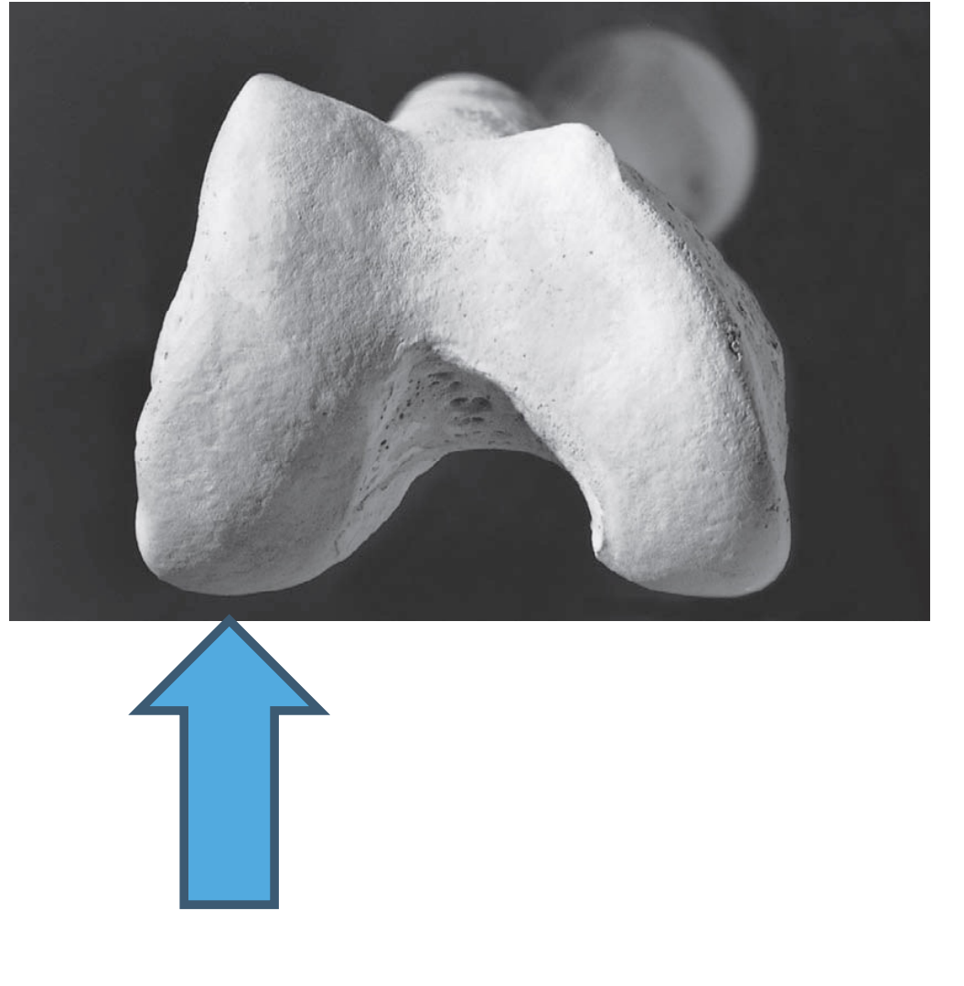
Patellar Surface
Feature of the Femur. Anterior. A notches, articular area on the anterior surface of the distal femur, over which the patella glides during flexion and extension of the knee.

Intercondylar Fossa
Feature of the Femur. Posterior Is a non-articular, excavated surface between the distal and posterior articular surfaces of the condyles.

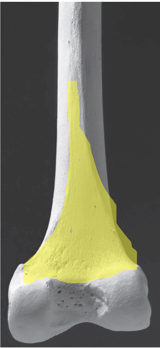
Popliteal Surface
Feature of the Femur. Posterior. A diamondshaped surface on the postero-distal side of the femur.
Popliteal Groove
Feature of the Femur. Posterior. A smooth hollow on the posterolateral side of the lateral condyle.

Patella
The knee cap. The point is down. To side: place on table with point facing away form you, the side it falls on is the side it’s from. Largest sesamoid bone on the body. Posterior side articulates with the femur.

Medial Articular Facet
Feature of the Patella. Posterior. The distal femur faces posteriorly and is smaller than the lateral articular facet.

Lateral Articular Facet
Feature of the Patella. Posterior. The distal femur faces posteriorly and is the largest part of the large articular surface of the patella

Apex
Feature of the Patella. The patella is the non-articular point of the bone. It points distally.
