Lecture 7 Human Neck (Triangle of the Neck, Larynx, and Glands)
 Call Kai
 Learn
Learn Practice Test
 Spaced Repetition
 Match
Match Flashcards
 Knowt Play
1/130

There's no tags or description
Looks like no tags are added yet.
Name | Mastery | Learn | Test | Matching | Spaced | Call with Kai | Chat |
|---|
No analytics yet
Send a link to your students to track their progress
131 Terms
what are the four neck spaces
vertebral compartment, visceral compartment, and two vascular compartments
what does the vertebral compartment contain
cervical vertebrae and postural muscles
which compartment has cervical vertebrae and postural muscles
vertebral compartment
what does the visceral compartment contain
glands (thyroid, parathyroid, and thymus), the larynx, pharynx and trachea
which compartment has glands (thyroid, parathyroid, and thymus), the larynx, pharynx and trachea
visceral compartment
what does the two vascular compartments contain
common carotid artery, internal jugular vein and the vagus nerve, on each side of the neck
which compartment has the common carotid artery, internal jugular vein and the vagus nerve, on each side of the neck
two vascular compartments
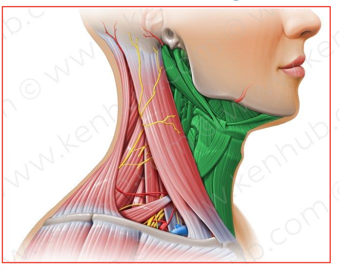
label the structure
anterior triangle

label the structure
posterior triangle
structures in the anterior triangle
submental triangle, submandibular triangle, carotid triangle, muscular triangle
what part of the neck encompasses these structures: submental triangle, submandibular triangle, carotid triangle, muscular triangle
anterior triangle
structures in the posterior triangle
ocipital triangle and subclavian triangle
what part of the neck encompasses these structures: ocipital triangle and subclavian triangle
posterior triangle
nerves of the carotid triangle
ansa cervicalis (cervical spinal nerve 1-3 (CI, CII, CIII)); nerves X, XI, XII; internal and external carotid arteries, internal jugular vein
cervical spinal nerve 1-3 (CI, CII, CIII) are nerves in which part of the neck
nerves of carotid triangle
nerves X, XI, XII are nerves in which part of the neck
nerves of carotid triangle
internal and external carotid arteries are nerves in which part of the neck
nerves of carotid triangle
internal jugular vein is a nerve in which part of the neck
nerves of carotid triangle
what is larynx
a cartilaginous skeleton (part of your respiratory system)
what structure is a cartilaginous skeleton (part of your respiratory system)
larynx
function of the hollow tube of larynx
lets air pass from your throat (pharynx) to your trachea on the way to your lungs
where can we find the vocal cords or voice box in our neck
larynx
functions of larynx
breathing, creating vocal sounds, preventing food and other particles from getting into your trachea, lungs and the rest of your respiratory system
name the structure responsible for: breathing, creating vocal sounds, preventing food and other particles from getting into your trachea, lungs and the rest of your respiratory system
larynx
what is the purpose of the cartilage of larynx
it gives structure
what is the purpose of ligaments in larynx
connect the areas of cartilage and attach your larynx to nearby structures
what is the purpose of membranes in larynx
help hold cartilage together
what is the purpose of muscles in larynx
move your larynx while swallowing, help with breathing and produce vocal sounds
what are the important parts of larynx that serves purpose
cartilage, ligaments, membranes, muscles
name the important ligaments we need to know
Thyrohyoid ligament, Cricotracheal ligament, Cricothyroid ligament
what do you call the suprahyoid muscles in our larynx
elevators
what do you call the infrahyoid muscles in our larynx
depressors
what are the two main subsections of extrinsic muscle in our neck
suprahyoid muscles (elevators) and infrahyoid muscles (depressors)
what is the main elevator and depressor muscle (part of the extrinsic muscle) of the larynx
suprahyoid muscles and infrahyoid muscles
purpose of elevator muscles in larynx
to elevate (raise) the hyoid bone and the larynx
purpose of depressor muscles in larynx
sit below the hyoid bone and act to depress (lower) the hyoid and the larynx
name the suprahyoid muscles
digastric muscle, stylohyoid muscle, geniohyoid muscle, mylohyoid muscle
name the infrahyoid muscles
sternohyoid muscle, sternothyroid muscle, omohyoid muscle
name the intrinsic muscle of the larynx
the thyroarytenoid muscle, vocalis muscle, adductors, abductors
describe the anatomy of the thyroarytenoid muscle
a wide, paired muscle arising from the inner surface of thyroid cartilage
name this structure: a wide, paired muscle arising from the inner surface of thyroid cartilage
the thyroarytenoid muscle
function of the thyroarytenoid muscle
Its fibers pass posterolaterally to insert into the anterolateral surface of the arytenoid cartilage
name the structure with this function: Its fibers pass posterolaterally to insert into the anterolateral surface of the arytenoid cartilage
the thyroarytenoid muscle
describe the anatomy of the vocalis muscle
a small, paired strand-like muscle which sits parallel to the vocal ligament
name this structure: a small, paired strand-like muscle which sits parallel to the vocal ligament
the vocalis muscle
function of the vocalis muscle
controlling and changing the tonal quality of the voice
name the structure with this function: controlling and changing the tonal quality of the voice
the vocalis muscle
what are the adductors of the larynx
Transverse arytenoid muscle, oblique arytenoid muscle, lateral cricoarytenoid muscle
What is the primary action of the transverse arytenoid muscle, oblique arytenoid muscle, lateral cricoarytenoid muscle
Adduction (closing) of the vocal folds
what are the abductors of the larynx
Posterior cricoarytenoid muscle
What is the primary action of posterior cricoarytenoid muscle
Abduction (opening) of the vocal folds
name the glands of the face and neck
thyroid gland, parotid gland, salivary gland anatomy
describe this gland: butterfly shaped, consists of two connected lobes
thyroid gland
where is the thyroid gland located
located at the front of the neck in the lower part of the throat on either side of the trachea
which gland is located at the front of the neck in the lower part of the throat on either side of the trachea
thyroid gland
weight of thyroid gland
20-60 grams on average
the thyroid gland is larger in which gender
women
when does the thyroid gland increase in size for women
during pregnancy
how many parathyroid glands are there
four, two on each side
length of parathryoid gland
6 mm in length and 3-4 mm in width
name the important thyroid hormones
T3, T4, calcitonin
full name for T3
Triiodothyronine
full name for T4
Tetraiodothyronine
name the proper thyroid hormones
T3, T4
where is T3 and T4 made in
follicular epithelial cells of the thyroid gland
what is calcitonin made by
c-cells of thyroid gland
how many % of the thyroid hormones is thyroxine (T4)?
80%
how many % of the thyroid hormones is triiodothyronine (T3)?
20%
what is the building block of T3 and T4?
iodine
what is iodine responsible for
be the building block of T3 and T4
why is dietary intake of iodine important
because this trace mineral is vital to our life
function of the pituitary gland
help regulate thyroid hormone production in the body
main role of thyroid gland
regulate body’s metabolism
main role of parathyroid gland
regulates the calcium level within the blood and the bones
which hormone produce the T3 and T4 hormones
thyroid gland
function of thyroid gland
stimulates body tissues to produce proteins and increase the amount of O2
function of Parathyroid Hormone/Parathormone (pTH)
regulates level of calcium in blood
which hormone does the Parathyroid Hormone/Parathormone (pTH) work with
calcitonin
when is Parathyroid Hormone/Parathormone (pTH) released?
when there is decreased calcium levels and it is released to raise calcium levels
which nerve provides the main parasympathetic and sympathetic fibers
the vagus nerve
the nerves supply of the larynx and thyroid does not play a role in the control of hormonal production or secretion but instead they influence what
vasculature
the superior thyroid artery is a branch of the ?
external carotid artery
inferior thyroid artery is a branch of the ?
subclavian artery
where does the superior and middle veins drain into
the internal jugular vein
where does the inferior veins empty into
brachiocephalic vein
are salivary glands exocrine or endocrine glands
exocrine glands
how are salivary glands classified
based on the nature of the saliva they excrete
functions of salivary glands
To keep the oral mucosa protected and lubricated
Help in the initial stages of digestion during mastication of food
They contribute to amylase that starts the digestion of carbohydrates
what structure is responsible for this function: To keep the oral mucosa protected and lubricated.
salivary glands
what structure is responsible for this function: Help in the initial stages of digestion during mastication of food
salivary glands
what structure is responsible for this function: They contribute to amylase that starts the digestion of carbohydrates
salivary glands
what are the categories of salivary glands based on size
major salivary glands and minor salivary glands
what are major salivary glands
they are large bilaterally paired extra oral glands with extended duct system
what are the three pairs of major salivary glands
parotid, submandibular, sublingual
parotid, submandibular, sublingual glands belong to what type of salivary glands
major salivary glands
where is minor minor salivary glands located
The mucosa & submucosa of the oral cavity with short ducts opening directly onto mucosal surface
name the types of minor salivary glands
Serous glands of Von Ebner; Anterior lingual glands; Lingual, buccal, labial, palatal glands, glossopalatine and retromolar glands
what type of gland are the serous glands of Von Ebner
minor salivary gland
what type of gland are Anterior lingual glands
minor salivary gland
what type of gland are these: Lingual, buccal, labial, palatal glands, glossopalatine and retromolar glands
minor salivary glands