Lesson 10 - Bacterial Cell Envelope

0.0(0)
Studied by 0 people
call kaiCall Kai
learnLearn
examPractice Test
spaced repetitionSpaced Repetition
heart puzzleMatch
flashcardsFlashcards
GameKnowt Play
Card Sorting

1/13

encourage image

There's no tags or description

Looks like no tags are added yet.

Last updated 5:10 AM on 10/16/26
Name
Mastery
Learn
Test
Matching
Spaced
Call with Kai
Chat

No analytics yet

Send a link to your students to track their progress

14 Terms

1
New cards

What are the three major bacterial cell envelope types?

  • Gram-positive bacteria: Thick cell wall.

  • Gram-negative bacteria: Thin cell wall.

  • Mycobacteria: Complex, multilayered cell wall.

  • These additional envelope layers provide structural support and protection.


<ul><li><p>Gram-positive bacteria: Thick cell wall. </p></li><li><p>Gram-negative bacteria: Thin cell wall. </p></li><li><p>Mycobacteria: Complex, multilayered cell wall. </p></li><li><p>These additional envelope layers provide structural support and protection.</p></li></ul><p></p>
2
New cards

What is peptidoglycan, and how does it form the bacterial cell wall?

  • Peptidoglycan (murein) makes up most bacterial cell walls, also called the sacculus.

  • It consists of sugar chains connected by cross-bridges.

  • The interconnected network forms one enormous molecule surrounding the cell.

  • The cell wall is a single molecule

  • It provides structural support and protection.


<ul><li><p>Peptidoglycan (murein) makes up most bacterial cell walls, also called the sacculus.</p></li><li><p>It consists of sugar chains connected by cross-bridges.</p></li><li><p>The interconnected network forms one enormous molecule surrounding the cell.</p></li><li><p><span><strong>The cell wall is a single molecule</strong></span></p></li><li><p>It provides structural support and protection.</p></li></ul><p></p>
3
New cards

What is the structure of bacterial peptidoglycan?

  • Consists of alternating NAG (N-acetylglucosamine) and NAM (N-acetylmuramic acid) sugars.

  • NAM is attached to short peptides containing 4–6 amino acids.

  • Peptides cross-link parallel glycan strands, including through bonds involving D-Alanine and m-Diaminopimelic acid

  • These connections create a strong, interconnected cell wall.


<ul><li><p>Consists of alternating NAG (N-acetylglucosamine) and NAM (N-acetylmuramic acid) sugars. </p></li><li><p>NAM is attached to short peptides containing 4–6 amino acids. </p></li><li><p>Peptides cross-link parallel glycan strands, including through bonds involving D-Alanine and m-<span>Diaminopimelic acid</span> </p></li><li><p>These connections create a strong, interconnected cell wall.</p></li></ul><p></p>
4
New cards

What are the functions of peptidoglycan, and how do penicillin and vancomycin interfere with its formation?

  • Functions:

    • Maintains bacterial cell shape.

    • Helps withstand turgor pressure from the cytoplasm. (osmotic pressure
      arising from the cytoplasm and pressing against the cytoplasmic membrane)

  • Antibiotics:

    • Penicillin inhibits transpeptidase, preventing peptide cross-linking.

    • Vancomycin binds D-Ala–D-Ala, preventing cross-bridge formation.

  • Peptidoglycan is an excellent antibiotic target because it is unique to bacteria.


5
New cards

Where can bacteria insert new peptidoglycan during cell wall growth?

  • Different bacterial species synthesize new peptidoglycan at:

    • Dispersed zones throughout the cell wall.

    • The septum (division site) only.

    • The cell poles (ends of the cell).


<ul><li><p>Different bacterial species synthesize new peptidoglycan at: </p><ul><li><p>Dispersed zones throughout the cell wall. </p></li><li><p>The septum (division site) only. </p></li><li><p>The cell poles (ends of the cell).</p></li></ul></li></ul><p></p>
6
New cards

What characterizes the cell wall of Gram-positive bacteria?

  • Thick peptidoglycan layer.

  • Typically 20–80 nm thick.

  • Consists of many stacked layers, up to 40 in some species.


<ul><li><p>Thick peptidoglycan layer. </p></li><li><p>Typically 20–80 nm thick. </p></li><li><p>Consists of many stacked layers, up to 40 in some species.</p></li></ul><p></p>
7
New cards

What are teichoic acids, and how do wall teichoic acids differ from lipoteichoic acids?

  • Teichoic acids are polymers of glycerol or ribitol (sugars) linked by phosphodiester bonds.

  • They may be decorated with sugars or amino acids.

  • Found throughout the thick peptidoglycan wall of Gram-positive bacteria.

  • Either…

    • Wall teichoic acids (WTA): Linked to peptidoglycan.

    • Lipoteichoic acids (LTA): Anchored to membrane phospholipids.

  • Memory trick: Wall = wall; Lipo = lipid.


<ul><li><p>Teichoic acids are polymers of glycerol or ribitol (sugars) linked by phosphodiester bonds.</p></li><li><p>They may be decorated with sugars or amino acids.</p></li><li><p>Found throughout the thick peptidoglycan wall of Gram-positive bacteria.</p></li><li><p><strong>Either…</strong></p><ul><li><p>Wall teichoic acids (WTA): Linked to peptidoglycan.</p></li><li><p>Lipoteichoic acids (LTA): Anchored to membrane phospholipids.</p></li></ul></li><li><p>Memory trick: Wall = wall; Lipo = lipid.</p></li></ul><p></p>
8
New cards

What are the advantages and disadvantages of Gram-positive bacterial cell envelopes?

  • Advantages:

    • Thick peptidoglycan wall reinforced by teichoic acids provides strength.

    • Protects against osmotic lysis.

    • Stains purple during Gram staining.

  • Disadvantages:

    • Exposed cell wall is susceptible to lysozyme and other wall-targeting substances.

    • Generally more susceptible to antibiotics than Gram-negative bacteria.

    • Examples: Bacillus cereus (rods), Streptococcus pyogenes (chains), Staphylococcus aureus (clusters).


<ul><li><p>Advantages: </p><ul><li><p>Thick peptidoglycan wall reinforced by teichoic acids provides strength. </p></li><li><p>Protects against osmotic lysis. </p></li><li><p>Stains purple during Gram staining. </p></li></ul></li><li><p>Disadvantages: </p><ul><li><p>Exposed cell wall is susceptible to lysozyme and other wall-targeting substances.</p></li><li><p>Generally more susceptible to antibiotics than Gram-negative bacteria. </p></li><li><p>Examples: Bacillus cereus (rods), Streptococcus pyogenes (chains), Staphylococcus aureus (clusters).</p></li></ul></li></ul><p></p>
9
New cards

What are the main structural features of Gram-negative bacterial cell envelopes?

  • Diderm: two membranes separated by the periplasm (space between two membranes).

  • Thin peptidoglycan layer (1–2 sheets) in the periplasm.

  • Outer membrane contains…

    • LPS (lipopolysaccharides) bridged with cations (e.g., Mg2+). Formidable barrier

    • Transmembrane proteins called porins (act as a pore, through which molecules can diffuse)

    • Lipoproteins help anchor peptidoglycan in place.

  • Inner membrane contains proteins with functions distinct from outer membrane proteins.


<ul><li><p>Diderm: two membranes separated by the periplasm (space between two membranes). </p></li><li><p>Thin peptidoglycan layer (1–2 sheets) in the periplasm. </p></li><li><p><strong>Outer membrane contains…</strong></p><ul><li><p>LPS (l<span>ipopolysaccharides</span>) <span>bridged with cations (e.g., Mg2+). Formidable barrier</span></p></li><li><p><span>Transmembrane proteins called porins (act as a pore, through which molecules can diffuse)</span></p></li><li><p>Lipoproteins help anchor peptidoglycan in place. </p></li></ul></li><li><p>Inner membrane contains proteins with functions distinct from outer membrane proteins.</p></li></ul><p></p>
10
New cards

What are the three regions of Gram-negative lipopolysaccharide (LPS)?

  • Reminder: All Gram-negative bacteria have an outer membrane with LPS, major permeability barrier

  • O-antigen: Repeating polysaccharide; composition varies between strains.

  • Core polysaccharide: Relatively conserved carbohydrate region.

  • Lipid A: Anchors LPS in the outer membrane

  • LPS act as endotoxin = molecules that are harmless when pathogen is intact, but become toxic and activate immune response when it is released from a lysed cell

  • LPS also contributes to the outer membrane permeability barrier
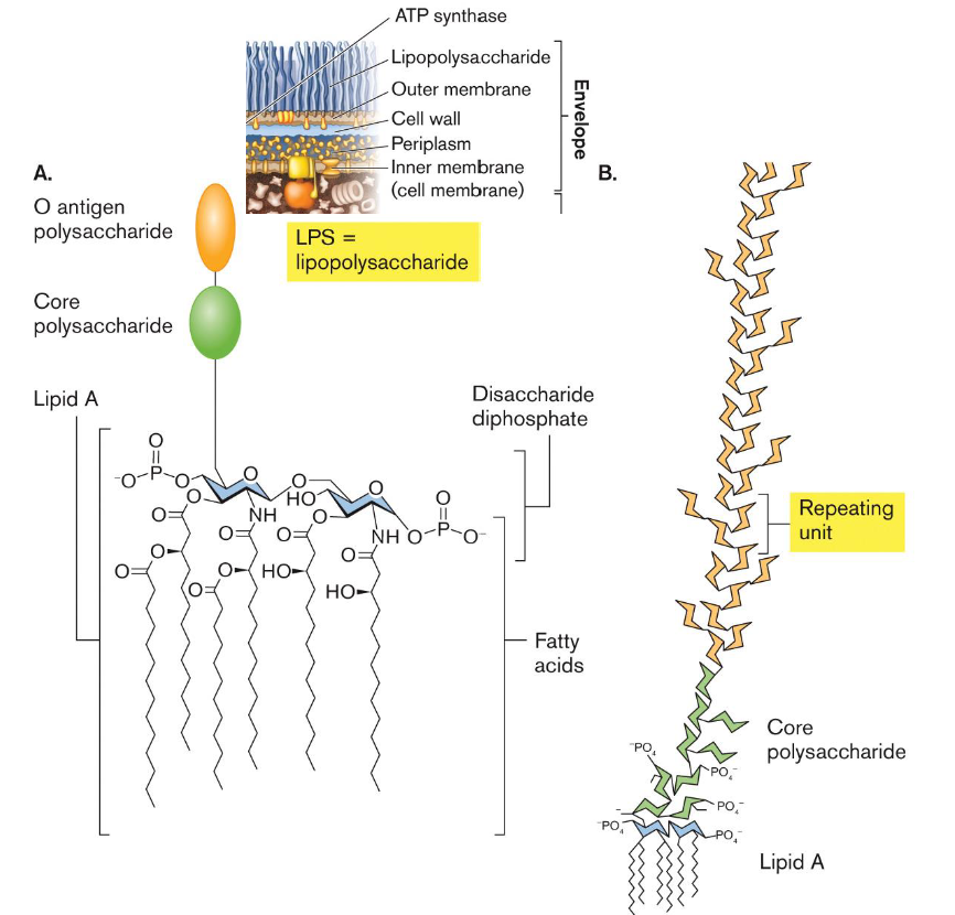

<ul><li><p><strong>Reminder: </strong><span><strong>All Gram-negative bacteria have an outer membrane with LPS, major permeability barrier</strong></span></p></li><li><p>O-antigen: Repeating polysaccharide; composition varies between strains. </p></li><li><p>Core polysaccharide: Relatively conserved carbohydrate region. </p></li><li><p>Lipid A: Anchors LPS in the outer membrane</p></li><li><p><span>LPS act as endotoxin = molecules that are harmless when pathogen is intact, but become toxic and activate immune response when it is released from a lysed cell</span></p></li></ul><ul><li><p>LPS also contributes to the outer membrane permeability barrier</p></li></ul><p></p>
11
New cards

What is serotyping, and how is LPS involved?

  • LPS is one of most structurally variable macromolecules on Earth

  • Serotyping distinguishes bacteria based on differences in surface antigens.

  • Different compositions of LPS among the same species of pathogen = different serovars of the same pathogen

  • Example: E. coli O157:H7.

  • O157 identifies the O-antigen type; H7 identifies the flagellar antigen type.


<ul><li><p>LPS is <span>one of most structurally variable macromolecules on Earth</span></p></li><li><p>Serotyping distinguishes bacteria based on differences in surface antigens. </p></li><li><p><span>Different compositions of LPS among the same species of pathogen = different <strong>serovars </strong>of the same pathogen</span></p></li><li><p>Example: E. coli O157:H7. </p></li><li><p>O157 identifies the O-antigen type; H7 identifies the flagellar antigen type.</p></li></ul><p></p>
12
New cards

Species vs Serovar

  • Species: What type of bacterium is it? (E. coli)

  • Serovar: Which antigenic variant of that species is it? (E. coli O157:H7)

    • Imagine we have three bacteria that all belong to the species E. coli.

    • Even though they're the same species, their LPS structures can differ, particularly in the O-antigen portion.


<ul><li><p>Species: What type of bacterium is it? (E. coli)</p></li><li><p>Serovar: Which antigenic variant of that species is it? (E. coli O157:H7)</p><ul><li><p>Imagine we have three bacteria that all belong to the species E. coli.</p></li><li><p>Even though they're the same species, their LPS structures can differ, particularly in the O-antigen portion.</p></li></ul></li></ul><p></p>
13
New cards

What are the advantages and disadvantages of Gram-negative bacterial cell envelopes?

Advantages:

  • Outer membrane acts as an excellent selective permeability barrier.

  • Protects against a wide range of toxic molecules.

  • Protects the thin peptidoglycan layer.

  • Gram-negative bacteria stain pinkish-red.

Disadvantages:

  • Outer membrane is energetically expensive to build and maintain.

  • Usually have larger genomes containing genes for outer membrane components.


<p>Advantages:</p><ul><li><p>Outer membrane acts as an excellent selective permeability barrier.</p></li><li><p>Protects against a wide range of toxic molecules.</p></li><li><p>Protects the thin peptidoglycan layer.</p></li><li><p>Gram-negative bacteria stain pinkish-red.</p></li></ul><p>Disadvantages:</p><ul><li><p>Outer membrane is energetically expensive to build and maintain.</p></li><li><p>Usually have larger genomes containing genes for outer membrane components.</p></li></ul><p></p>
14
New cards

Review: Hopanoids

  • Membranes also include planar molecules that fill gaps between hydrocarbon chains

  • In eukaryotic membranes, the reinforcing agents are sterols, such as cholesterol

  • In bacteria, the same function is served by hopanoids, or hopanes

  • Prokaryotes regulate membrane fluidity and rigidity by varying the abundance of these different membrane lipids and hopanoids


<ul><li><p><span>Membranes also include planar molecules that fill gaps between hydrocarbon chains</span></p></li><li><p><span>In eukaryotic membranes, the reinforcing agents are sterols, such as cholesterol</span></p></li><li><p><span>In bacteria, the same function is served by hopanoids, or hopanes</span></p></li><li><p><span>Prokaryotes regulate membrane fluidity and rigidity by varying the abundance of these different membrane lipids and hopanoids</span></p></li></ul><p></p>



