PATH 201 - LECTURE 11
 Call Kai
 Learn
 Practice Test
 Spaced Repetition
 Match
 Flashcards
 Knowt Play
1/79

There's no tags or description
Looks like no tags are added yet.
80 Terms
where are the more severe respiratory diseases seen
in the lower respriatory tract
what makes up the lower respiratory tract
trachea
primary bronchi
lungs
what are the 3 main etiologies of common lung pathology’s
inhalation of toxic smoke
inhalation of infectious agents
inhalation of allergens
what are some examples of toxic smoke/noxious agents that are inhaled and commonly cause lung pathologies
tobacco smoke
asbestos
dust
silica
coal
what are the common lung pathologies that are caused by inhalation of toxic substance
chronic bronchitis
emphysema
lung cancer
what are some examples of infectious agents that cause common lung pathologies
bacteria
virus
what are some examples of common lung pathologies caused by inhalation of infectious agents
tubercolosis
pneumonia
influenza
what is an example of pathology due to inhalation of allergens
asthma
what pathologies fall under chronic obstructive pulmonary disease (COPD)
emphysema
chronic bronchitis
they often coexist
what is emphysema
the destruction of the airspace distal to the terminal bronchioles which results in irreversible enlargment
what causes emphysema
causes due to tobacco smoke and the toxins within it breaking down the elastin fibres in the membranes that allow the lungs to expand and contract
what is the most common form of emphysema
centriacinar emphysema
describe centriacinar emphsyema
Respiratory bronchioles are dilated but the distal alveoli remain normal
what is parynchema
lung tissue
what are emphysematous air spaces
dilated respiratory bronchioles
what is anthracosis
holes in the lung parenchyma filled with carbon pigment deposisition due to the tar in cigarrette appear black
what does a centriacinar pattern mean
nromal alveoli exist between areas of emphysema
how does cigarrette smoke cause emphysema
cigarrete smoking produces oxidants whcihc cause accumulation of inflammatory cells
inflammatory cells release oxidants and proteases which destroy the elastic tissue in the walls of airspace
smoking also impairs the acitivty of the antiprotease
what is the naturally occuring antiprotease in lungs that is inhibited by smoking
alpha 1 antitrypsin
what are the main inflammatory cells seen with cigarrete smoke
neutrophils
what are the main proteases released by inflammatory cells that cause emphysema
elastases
why do patients with emphysema have expanded chests
elastin not only allows for stretching, but it keeps the airways patent when we exhale
without it, airways collapse and air gets trapped
trapped air = expanded chest

what is wrong here
emphysema
flattened diaphragm
inflaltion of the apex above the clav
ribs horizontal
decreased bronchovascular markings
why do people with emphysema purse their lips
to prevent airways collapsing during exhalation
what is barrel chest
hyperinflated chest due to trapped air
what do most patients with emphysema also have
some degree of chronic bronchitis since they are both caused by some degree of smoking
how is chronic bronchitis clinically defined
persistent cough with sputum production for east 3 months in atleast 2 consecutive years
what are the symptoms of bronchitis
mucus hypersecretion
inflammation
recurrent infections
fibrosis
what eventually happens to patients with chronic bronchitis
hypoxemia + cyanoses
edema from right heart failure
describe the pathogenesis of chronic bronchitis
cigarrette smoke triggers constant inflammation (chronic)
inflammatory mediators and growth factors change the composition of the bronchus
this results in mucus hypersecretion which causes airway obstruction
movement of cillia is also affected; mucus cannot be cleared
describe the changes in the bronchus wall due to chronic inflammation (chronic bronchitis)
metaplasia of the surface lining > more goblet cells
hypertrophy and hyperplasia of mucus secreting glands in the submucosa > bigger more active glands
what causes fibrosis due to chronic bronchitis
excess mucus causes blockages which means more infections
more inflammation
leads to fibrosis
describe impaired phagocytosis as a consequence of chronic bronchitis
alveolar macrophages start to change their activity
no longer able to effectivley phaocytose bacteria and apoptopic cells
leads to further inflammation>repeat cycle
describe the pathogenesis of lung cancer (from cigarrete smoke)
tobacco smoke contains carcingoens that cause DNA mutations that accumulate
and then induce an inflammtory response
what genes do mutations that cause cancer affect
oncogenes and tumour suppressor genes
what can accumulation of mutations in oncogenes and tumour suppressor genes lead to
dysplasia and carcinogenesis
what is carcinogenesis
process by which normal cells turn into cancer cells
what does the inflammatory response induced by tobacco smoke that leads to cancer contribute to
tumour growth
immune system suprpession
what are the microscopic variants of lung cancer
adenocarcinoma
squamous cell carcinoma
small cell carcinoma
large cell carcinoma (others)
which microscopic lung cancer variant is most strongly assoicated with smoking
small cell carcinoma
which microscopic lung cancer variant is least assoicated with smoking
adenocarcinoma
which microscopic lung cancer variant occurs most commonly in women
adenocarcinoma
what differs between the microscopic lung cancer variants
the cell of origin
where is adenocarcinoma normally seen (in the lungs)
in the respiratory airway in type II alveolar cells
where does small cell carcinoma normally seen/originated
in th conducting airways in neuroendocrine cells
where is squamous cell carcinoma normally seen/originates
in the conducting airways in basal cells
what is dysplasia
Normal cells begin to change in type and number and then we see disordered growth - dysplasia
this is normally driven by genetic mutations due to the carcinogens in tobacco smoke as well as other things
what is carcinoma in situ
when the entire thickness of an epithelium is dysplastic but not yet invaded the basement memrbane
describe the morphology of adenocarcinoma
tumour tissue shows malignant irregular shaped glands surrounded by fibrous tissue
what is desmoplastic storma
the fibrous tissue that surrounds tumourous tissue in adenocarcinoma
describe the genetic mechanism of adencarcinoma
gain of function mutations in EGFR and KRAS
oncogenes
what kind of
describe the morphology of squamous cell carcinoma
squamous dysplasia of bronchial epeithelium
describe the genetic mechanism of squamous cell carcinoma
loss of funciton mutations of tumour suppressor p52 and RB
describe the morphology of small cell carcinoma
small tumour cells
little cytoplasm
dark hyperchromatic nuclei
finely granular chromatin
describe the genetic mechanism of small cell carcinoma
mutations of p53 and BCL2 anti-apoptopic genes
describe the gross morphology of lung carcinoma
gray white colour
hard tumour consistency
anthracosis
heamorrhage and tissue necrosis
what cancer normally has tumours that are hilar/central
squamous or small cell
what cancer normally has tumours that are peripheral and under the pleura
adenocarcinomas
what does lung cancer normally present as
cough
hemoptysis (blood in sputum)
chest pain
dyspnea
weight loss
how is lung cancer investigated
chest x ray
sputum sample examination
CT/MRI
bronchoscopy
tumour biopsy
what is enobronchial biosy
tissue biopsy from a lesion in the bronchial wall or lumen through a bronchoscope
what are some traditional lung cancer therapies
surgery
chemo
radiation
what are some novel therapies for lung cancer
immunotherapy
mutated EGFR inhibitors
in terms of treatment how are lung cancers classified
small cell
non small cell
describe why lung cancer is classified as SCLC and NSCLC
non small cell lung cancer can be localised on diagnosis and surgically cured
SCLC is usually metastatic and usually relapses post radiation and chemo

what is indicated by the red arrows and what else can be interpreted
emphysematous air spaces (dilated respiratory bronchioles due to emphysema)
anthracosis
centriacinar pattern- normal alveoli still exits in between areas of emphysema

what can be seen
this is a nornal lung
R is the respiratory bronchi
AD is alveolar duct
A is alveolus
V is blood vessel
AS is alveolar sac
T is terminal bronchiole

what can be seen
centriacinar emphysema
red is dilated respiratory bronchiles
green is anthracosis
black is normal surrounding alveolar spaces

what can be seen
chronic bronchitis


what can be seen
normal bronchus
top arrow is repiratory epithelium (pseudostratified colunar ciliated epithelium)
second arrow is smooth muscle
third is mucous and serous glands in the submucosa
bottom is cartilage

what am i seeing from top to bottom
chronic bronchitis
goblet cell metaplasia on lining epithelium
smooth muscle hypertrophy from chronic copughing
hyperplasia and hypertrophy of mucus glands
what can be seen
adenocarcinoma
arrows are showing tumour tissue with irregular sized and shaped glands


describe A. B, C, D
A is normal alveolar tissue
B is atypical adenomatous hyperplasia
C is adenocarcinoma in situ
D is lung adenocarcinoma (invasion)
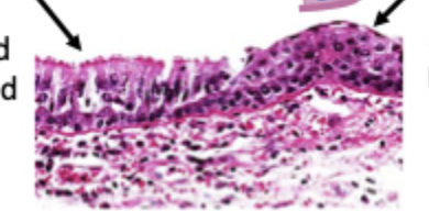
what can be seen
squamous cell carcinoma
The left arrow is pseudostratified coloumnar epithelum
the right arrow is squamous epithelium

what is A, B, C, D, E, F
squamous cell carcinoma
A. goblet cell hyperplasia
B. basal cell hyperplasia
C. squamous metaplasia
D. squamous dysplasia
E. carcinoma in situ
F. invasive squamous carcinoma

what is this
small cell carcinoma

what is this
squamous cell carcinoma

what is this
small cell carcinoma