Physiological Psychology - Exam 2 remaining flashcards
 Call Kai
 Learn
 Practice Test
 Spaced Repetition
 Match
 Flashcards
 Knowt Play
1/28

There's no tags or description
Looks like no tags are added yet.
29 Terms
Retinal visual information pathway summary
A photo of light is absorbed by opsin proteins in photoreceptor outer segments
This causes a decrease in glutamate release between the photoreceptor and bipolar cell (brown)
This light signal is then inverted in ON bipolar cells and passed on to the ON retinal ganglion cells, and then to the brain via the optic nerve in the form of action potentials (opposite in OFF cells)
→ This process is happening on like all the areas of the peripheral retina
The first steps in vision occur in the retina, these steps include phototransduction in photoreceptors and active computations the retina does to extract specific features of the visual scene
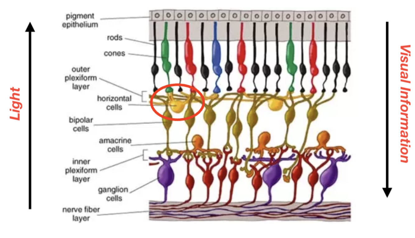
Active retina computations: horizontal cells increase contrast through lateral inhibition
Ganglion → bipolar → light beam activates the retina cells, the blue rods are highly activated, red is activated by some light, but most photons being absorbed by the blue rods
Light hits the retina → Light activates photoreceptors (rods and cones) in the retina
Photoreceptors send signals → The activated photoreceptors send signals to bipolar cells, which then pass the information to ganglion cells (the output neurons of the retina)
Horizontal cells step in → At the same time, horizontal cells, which connect neighboring photoreceptors, detect this activation
Inhibition of neighbors → the horizontal cells release GABA onto adjacent photoreceptors that are less activated–this weakens their signal, making the difference between light and dark areas clearer
Result: Sharper edges → This process enhances contrast, making bright areas appear artificially brighter and dark areas appear darker, which helps us detect edges and fine details more clearly
Horizontal cells (inhibitory, GABA), can sense the difference, they’re wired to specific rods, and through the activity of neurotransmitter release, can tell which rods are getting maximally stimulated and which aren’t
→ If they can identify that the red ones are being maximally stimulated, they will inhibit the surrounding rods(neighbors), release GABA onto these neighbors which inhibits them, making their signal weaker, enhances the acuity so it sharpens edges and enhances contrast, making dark areas appear darker and light areas appear lighter, tells brain specifically where light is coming from in the environment
→ Connect neighboring photoreceptors
→ Lateral inhibition: The phenomenon by which interconnected neurons inhibit their neighbors, enhancing contrast at the edges of regions
Lateral inhibition from horizontal cells increases contrast to enhance edges
Perceived lightness: B & C look more lighter and darker than the rest of the scale
→ but like thats just the retina messing w/us
→ due to lateral inhibition
→ edge detection, enhances visual contrast
Allows us to recognize where a point of darkness begins?
Contrast where it doesn’t actually exist

Central visual pathways
= after light signals leave the retina
White part = nerve made up of the axons of all the retinal ganglion cells, called optic nerve

Visual pathways & binocularity
If staring straight ahead, objects in our left visual hemifield are projected to our right primary visual cortex, and vice versa
Remember this rule: Medial (nasal) fibers of the optic nerves cross at the optic chiasm, while lateral (temporal) fibers stay on the same side
This means that each hemisphere of visual cortex receives input from both eyes
The X → optic tract
Projects all the way to the thalamus, where the retinal ganglion cells make their synapse
→ Thalamus takes inputs from our sensory system and passes it on to visual cortex
*In all vertebrates, some/all of the axons of each optic nerve cross to the opposite cerebral hemisphere
The optic nerves cross the midline at the optic chiasm
(like an X shape)
In humans, axons from the half of the retina toward the nose cross over to the opposite side of the brain
The half of the retina toward the temple projects its axons to its own side of the brain
→ This distribution means that the right side of the brain receives input from the left half of the visual field of both retinas, while the left side of the brain receives input from the right half of the visual field of both retinas
*not all projections from the left eye cross over to the right and vice versa
→ bc we have binocular vision
→ but this is problematic for the circuitry to make sense of info coming in
*some projections cross over but not all
Marble:
Light rays come off of marble and make their way down to the temporal retina (furthest away)
Once here, it does not cross over at the optic chiasm
In the left side (nasal projections cross at the optic chiasm, others don't)
Ends up in right cortex

Right and left visual hemifields & binocularity
We have binocular overlapping → for depth perception → needed for predators and hunting
Rabbits have eyes more spread out, much bigger monocular view, very narrow binocular though
Binocular perception
We use information from both our eyes to process what we are seeing, so we judge the distance of objects by unconsciously comparing inputs from our two eyes
In prey species like rabbits, natural selection sacrificed this to favor instead a broad visual field where the eyes are positioned farther apart, which helps the rabbit detect approaching predators
The lateral geniculate nucleus (LGN) of the thalamus
First place they make a synapse is in the thalamus
Inputs from both eyes project to the thalamus
Most axons of the optic tract in mammals terminate on cells in the visual portion of the thalamus, the lateral geniculate nucleus (LGN)
Visual info from each eye stays segregated there
At some point neurons have to integrate information together
But even in the thalamus its separate
Bands in thalamus that receive projections from either the left or the right eye, not both
While inputs from both eyes project to the thalamus via the optic nerve and optic tract, visual information from each eye stays segregated in the LGN (input from the left vs. right eye project to different LGN layers), and light information from each eye stays segregated all the way to V1
Axons of neurons in the LGN form the optic radiations(don’t need to know) which terminate in primary visual cortex (V1) of the occipital lobe at the back of the brain

Some “vision rules” to remember
Photoreceptors are hyperpolarized by light
“ON-center” bipolar cells have that weird synapse receptor thingy that switches the signal, they express an unusual (sign-inverting) glutamate receptor that turns a hyperpolarization (less glutamate release) into a depolarization (more glutamate release) onto ON-center ganglion cells
For central visual pathways, the medial/nasal retinal axons(closest to the nose) cross over to the opposite side of the brain at the optic chiasm, while lateral/temporal axons do not
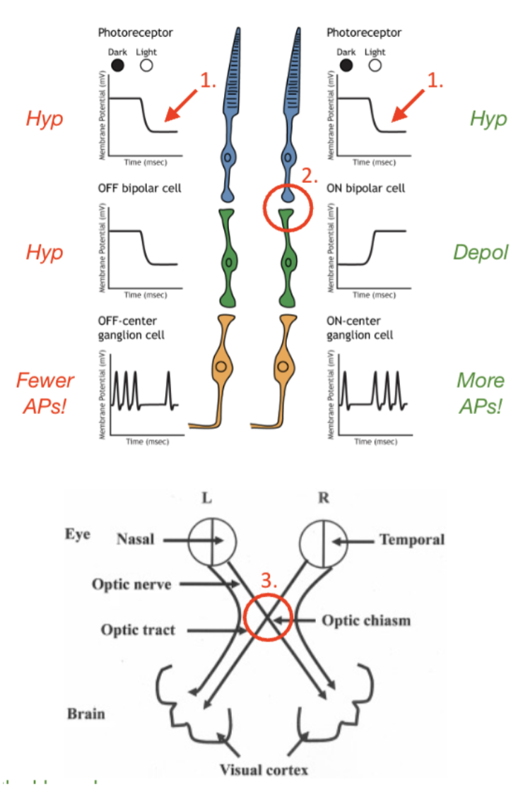
Simple “center-surround” (circular) receptive fields in the retina
Causes it to increase its firing rate
Whatever makes a neuron stimulate the most is its receptive field
Finger pricking example:
AP means its responding to the stimulus, the stimulus is its receptive field
Receptive field: A stimulus (ex. A portion of visual space) that maximally activates/excites a neuron (increasing firing rate)
In the retina and thalamus, receptive fields are very simple concentric “spots” or “circles” of light surrounded by darkness (“center-surround”) that are detected by photoreceptors (think of them as individual pixels) coming from specific locations in the visual field
RFs in the retina and thalamus are simple “spot detectors,” but as we continue up the visual pathway into the primary visual cortex (V1) and beyond, receptive fields of neurons become increasingly more complex

From stars to stripes: “Circular” to “Stripe” receptive fields
Visual system:
Visual field of a RGC would be simple spot detectors, they pick up spots of light, if you shine a bit of light on a retinal cell it will fire, so the receptive field is the spot of light
Neurons in V1 respond to bars/stripes of light, also in specific directions (slanted, diagonal, horizontal, etc.)
Cells:
Retinal ganglion cell RF = center-surround “spots” of light (simple “spot detectors”) coming from specific locations in the visual field
V1 “simple cell” RF = bars of light with specific orientations coming from specific locations in the visual field (more complex)

Primary visual cortex (V1) is in the occipital lobe
In occipital lobe
In back of brain

Cytoarchitecture of primary visual cortex (V1)
Nissl stain shows the cell bodies of the neurons
Layered structure, most of cortex is made up of 6 layers
Visual cortex is made up of 6 layers (known as a “laminar” structure
Inputs come in at layer 4 (down from the surface) in V1 from the thalamus (LGN), make a synapse with neurons there and then project upward towards surface layers of V1
Maybe the information from both eyes integrates at V1?
Info still segregated at layer 4, forming “ocular dominance columns”
Neurons only respond to one eye or the other still
If you move to another column, it'll respond alternately to right eye left eye, right eye, left eye, each column has one of them, pattern
Finally up in the superficial layers in V1, you’ll find neurons responding to light info from both of the eyes
Information from the two eyes begins to converge here
Neurons in other layers (as well as beyond V1) will respond to a visual stimulus independent of which eye the stimulus originates from (“binocular”)
Layers 2/3 of V1 is therefore where light information from the left and right eye are first integrated, above layer 4
Layers 2 + 3 are the superficial layers (upper)
Information from the two eyes converges on cells beyond layer 4 of the primary visual cortex, making binocular three-dimensional vision possible

Lecture 23 Thought Questions
What is lateral inhibition and which cells of the retina are responsible for this phenomenon? How does lateral inhibition work in simple terms and what is lateral inhibition in the retina useful for?
Describe the pathway that light information takes from the retina to the primary visual cortex. Trace out this pathway illustrating the contribution of each eye. Where do pathways from the two eyes cross, and do all axons of the optic nerve cross at this location? What implications does this arrangement have for cortical visual processing?
What is the “receptive field” of a neuron? How do receptive fields change as you move up the visual hierarchy from the retina to V1? What does the receptive field of a retinal ganglion cell look like? What about a V1 neuron? Where, specifically, does light information from the two eyes first begin to become integrated, allowing for binocular perception?
Receptive fields cont. From stars to stripes: “Circular” to “Stripe” receptive fields
Receptive field = some kind of stimulus which will maximally stimulate cells
Retinal ganglion cell RF = center-surround “spots” of light (simple “spot detectors”) coming from specific locations in the visual field (same for the thalamus)
V1 “simple cell” RF = bars of light with specific orientations coming from specific locations in the visual field (more complex “orientation selectivity”)

V1 Neuron “simple cell” receptive field
Used cats, they have good vision
Neurons in primary visual cortex respond to bars of light in specific orientations (known as orientation selectivity) coming from specific locations on the retina
They recorded a single neuron in the primary visual cortex, they were trying to figure out its receptive field, what makes it fire the most? → whatever the stimulus is, is its receptive field
They had an overhead projector, shining circles of light and trying to get that neuron go fire, but the neuron wouldn’t fire/respond, very few APs, so the receptive field of these neurons is different from in the thalamus
They were putting a glass slide over the projector, and the edge made a bar of light and this made the neuron respond (to the bar of light), they did a million experiments and concluded that these neurons respond to bars of light of specific orientations)
*Only talking about one neuron here
As you get more vertical, entirely vertical made that particular neuron fire more
Doesn't respond to the others
But another neuron may respond maximally to the horizontal orientation but not the others
Each neuron responds to different orientations

What this actually looks (or sounds) like

*The video
Simplified schematic of V1 Simple Cell Receptive Field
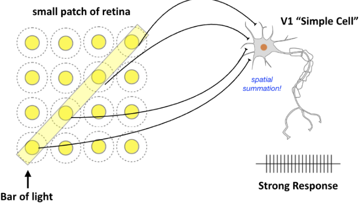
→ This V1 neuron receives input from a row of ON-center ganglion cells and so responds better to a bar of light than to any single spot of light
Each yellow is a spot detector, 16 retinal ganglion cells, each have a spot of light
They might be wired to the V1 simple cell
Those 4 spots connect to the cell, that explains why it is responding
Each one of those spots/axons from those cells, converges on a particular V1 neuron, which responds to
Spatial summation, if receiving those 4 all at once, adds up those convergent inputs to make a strong response
Each spot is like a pixel, v1 neurons are starting to assemble those to create what the image might be
That particular neuron responds to
V1/Primary visual cortex neurons respond best to lines or bars in a particular position and at a particular orientation in the visual field
Simplified schematic of V1 simple cell receptive field: two examples

1st one:
→ It will be a pretty weak response cuz spatial summation is missing here 🙁
→ some response but not as strong
2nd one:

→ No response
→ Conclusion: a given V1 neuron responds to oriented bar of light from specific locations on the retina
Take pixels and turn them into lines, represent contours/edges between light/dark
Individual V1 neurons representation of an image

Simple cells in V1 begin to build an image of the world → good for responding to edges boundaries, transitions between light and dark regions
Starts putting things together, all 4 abcd will put together a little bit when combined but not complete, neurons in V1 do not respond to color
→ This stimulation depicts what attributes of a visual image (greyhound and fence) are represented by responses of V1 neurons tuned to different preferred orientations
Retinotopy: There is a systematic, orderly mapping of orientation selectivity and receptive field position across V1

There is an organized, non random orderly progression of response properties (as you go down)
There's a non organized, orderly progression of response properties (as you go across)
Within each column of V1, each neuron has similar response properties
Across columns, neurons exhibit an orderly progression of response properties
→ Layers 2&3 neurons respond
Essentially, the primary visual cortex has a columnar organization for stimulus orientation
That is, in one orientation column, all the cells may be “tuned” to upright stimuli (at an orientation of 0 degrees)
In an adjacent column, all cells may respond best to another orientation, perhaps at 10 degrees from the vertical, in the next column 25 and so forth
These columns are organized side by side across the cortex
Retinotopy: Systematic variation in orientation preferences in V1
How orientation changes as you go across or down the neuron layers:
As you go down a single column, two things:
All of the neurons in the same column will respond to the same location in space (receptive field position is the same, orientation preference is the same, all respond maximally to the vertical bar of light, non random)
Within each column of V1, each neuron has similar response properties (both RF & OP)
As you go across columns:
Vary in a smooth position
Yes its more variable as you go across, its relatively smooth
Orientation changes (in a logical and repeating), go from horizontal to oblique to vertical, and repeat as you go across V1
Systematic variation of orientation preferences
Within a column, each neuron has similar response abilities
As you go across, its orderly, goes in specific order from hoz → oblique → vertical
Across columns, neurons exhibit an orderly “non-random” progression of response properties

Beyond V1: The dorsal and ventral visual processing streams
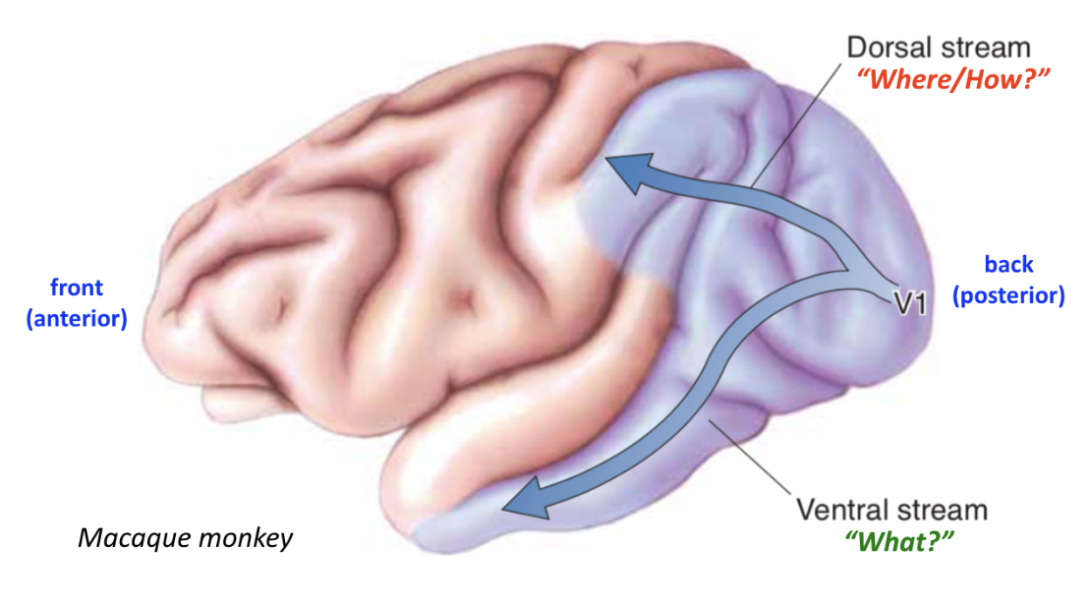
Gets complicated
Visual info is processed in two streams:
Dorsal stream - goes to parietal lobe
Some neurons here respond to visual motion
Where/how pathway
Neurons respond to the movement of our own body and also movement of others
Visual motion analysis and visual control of action
Responsible for perceiving the location of objects and for guiding movement toward them
Ventral stream - goes to temporal lobe
The what pathway
Neurons here don’t care about if an object is moving
Important for object recognition
Might respond to one object differently than another
Complex object recognition
Color recognition
Facial recognition
There’s a particular nucleus here that responds to color
Responsible for visually identifying objects
Higher visual cortices

V2: Responds to similar stimuli as V1 but also contours(illusory, like a triangle with missing parts/boundaries) and important for visual memory,
Area MT (V5): Responds to linear movement, regardless of color, and visual control of action
Neurons here respond to linear movement
Responds to movement of objects
V4: Responds to features of intermediate complexity (ex. geometric shapes) and color
Geometric shape (distinguishing a cube from a sphere…etc.)
IT: Responds to complex objects, including faces! (ex. Fusiform Face Area in IT)
Sub region within it called the FFA(fusiform face area) → these neurons respond to faces
We are experts at recognizing faces
Some expert birders can identify bird faces
Neurons in fusiform face area (within IT) of Macaque Monkeys Respond to Faces

In this example, FFA neurons’ receptive field is faces
If they showed monkey random objects or light, nothing, but if it showed another monkey face it responded
Non social solitary animals don't have this, dont care
Researcher showed own face but monkey kind of responded, not as strong
Without eyes → even less
Smiley face → weaker response
Lines → very weak
→ FFA responds to faces specifically
Why is recognizing important and why are we good at it?

Visual Paradalia: Brains are wired to pick up faces, even from nonhuman things?
The tendency to see faces in inanimate objects, shadows, clouds, abstract patterns, etc.
There’s several theories as to why we’re good at this as a species, but the prevailing hypothesis is that it reflects our social evolution–human survival depends on others (whether we need their help or fear their violence); our brains are likely wired to quickly detect others
Studies in humans and other primates have shown, unsurprisingly, that the Fusiform Face Area (FFA) in Inferotemporal Cortex (IT) is involved in this phenomenon
Auditory paradalia: hearing voices from a white noise machine for example
From single neurons to perception: receptive field hierarchy

How is visual perception–the task of assigning meaning to objects in space–integrated and where?
As we move up the receptive field hierarchy, we encounter neurons that respond to increasingly complex features
Does this mean that somewhere in your brain there are highly selectively neurons that only respond to, for example, the face of your grandmother?
Probably not–they’ve never really been found and it’s too risky!
Receptive field hierarchy gets more complex
There is an upper limit to complexity
Other theorists: higher-order circuits of cells could detect any possible form
A neuron could be constructed that would enable a person to recognize his or her grandmother, and such hypothetical “grandmother cells” were frequently mentioned in the literature
According to this view, whenever such a cell was excited, up would pop a mental picture of one’s grandmother
This hypothesis was given as a possible explanation for facial recognition
Neurons are an orchestra, not solo musicians
brain uses many different neurons to respond to different objects, motion, stimuli, noises
→ visual perception is across all of those ensembles of neurons
→ like an ensemble orchestra
→ each responsible for a different aspect of perception

Summary/review of visual system processing
The retina transduces light information into action potentials and also extracts information about contrast and some other visual features (active retinal computations)
Receptive fields of the retina are simple “spot detectors”
The optic nerve synapses in the LGN of the thalamus–the LGN relays light information to primary visual cortex (V1)
Receptive fields of LGN neurons are also simple “spot detectors”
“Simple cells” in V1 process this information, noting edges, orientation preferences and variations in contrast, among other features
Receptive fields of V1 simple cells are bars of light with specific orientation preferences
Information from the V1 is then passed to higher visual cortices where visual perception is further developed–elements of visual perception are spread widely in cortex throughout the occipital, parietal and temporal lobes (ex. dorsal vs. ventral parallel processing streams)
Receptive fields of higher visual cortices are specific the visual brian region–some neurons respond to linear motion, while others respond to color, shapes, faces
How all of this information is then stitched back together by the brain, through fundamental to our conscious visual experience, remains for the most part a mystery
But we are learning more about how this process works all the time!
Lecture 24 Thought Questions:
What is the “receptive field” of a neuron? How do receptive fields change as you move up the visual hierarchy from the retina to the thalamus to V1 and beyond? What does the receptive field of a retinal ganglion cell look like? What about a neuron in the thalamus (LGN)? What about a V1 neuron? What about a neuron in IT? What hints does this give us about how the brain reconstructs our visual world?
What is “retinotopy” and how does it compare to the concept of “tonotopy” in the cochlea and A1? Describe the nature of the variation of light bar orientation preferences across V1 columns.
What are the dorsal and ventral visual processing streams? Which visual nuclei are found in each of these streams, and what aspects of visual perception is each region thought to contribute to?