Unit 4 Exam Lecture Powerpoints (Elimination, Fluid and electrolytes)
 Call Kai
 Learn
 Practice Test
 Spaced Repetition
 Match
 Flashcards
 Knowt Play
1/84

There's no tags or description
Looks like no tags are added yet.
85 Terms
Elimination definition
excretion of waste from the body
Bowel elimination
the passage and expelling of stool through the intestinal tract by means of intestinal smooth muscle contraction
Urinary elimination
the passage of urine out of the urinary tract through the urinary sphincter
Key Terms
•Defecation
•Micturition
•Voiding
•Continence
•Incontinence
•Retention
Defecation
passing of bowel, bowel movement
Micturition
passing of urine, urination
voiding
passing of urine, urination
continence
purposeful control of urine or fecal elimination
incontinence
loss of control of elimination of urine or feces or both
retention
keeping materials within the body that are normally expelled

Scope of concept
-Excretion of waste products: Feces/ stool & Urine
-Scope: Waste formation and Waste excretion
-2 types of issues: control and retention
-Includes the normal or expected physiological process of waste formation and excretion as well as problems associated with the process
-Urine consists of Electrolytes
-Includes impairment of urinary and bowel elimination
Urine consists of what Electrolytes:
potassium, sodium, chloride, calcium, hydrogen, urea, creatinine, ammonia, and drugs
-The amount of water and electrolytes reabsorbed into the blood or excreted in renal tubules is controlled by aldosterone, anti-diuretic hormone, parathyroid hormone, and renin

Variations & Context
•Impaired elimination refers to one or more problems associated with the elimination process.
•terms associated with impaired urinary elimination:
-anuria (absence of urine)
-dysuria (painful urination)
-polyuria (multiple episodes of urination, as with diabetes)
-urinary frequency (multiple episodes of urination with little urine produced in a short period of time)
-urinary hesitancy (the urge to urinate exists, but the person has difficulty starting the urine stream)
The GI/GU System

Structures and Functions of Urinary System

Upper urinary system
•2 kidneys
–Forms urine
•2 ureters
–Drains urine
Lower urinary system
•Urinary bladder
–Stores urine
•Urethra
–Urine leaves body
What does the upper urinary system consist of?
2 kidneys, 2 ureters
Kidneys do what?
Form urine
-2
-Upper urinary system
ureters do what?
drains urine
-2
-upper urinary system
What does the lower urinary system consist of?
urinary bladder, urethra
Urinary bladder does what?
stores urine
-lower urinary system
Urethra does what?
urine leaves the body
-lower urinary system
Urinary elimination

The Kidneys

Urinary System: Kidneys
Principal organs
Primary functions of the kidneys
•Primary functions
–Regulate volume and composition of extracellular fluid (ECF)
–Excrete waste products from the body
•Other functions
–Control BP
–Make erythropoietin
–Activate vitamin D
–Regulate acid-base balance
-Red blood cell (RBC) production: Erythropoietin—hormone secreted in response to hypoxia and decreased renal blood flow
-BP regulation: Renin-angiotensin-aldosterone system. Prostaglandin synthesis—vasodilation; decreased BP and SVR
Function of the Kidneys

Physiology of urine formation
–Outcome of complex, multistep process
•Filtration
•Reabsorption
•Secretion
•Excretion of water, electrolytes, and metabolic waste products
Primary function of the kidneys
–Filter the blood
–Maintain internal homeostasis
Urine Electrolytes:
K+, Sodium Chloride, Calcium Hydrogen, urea, creatinine, ammonia, and drugs
-The amount of water and electrolytes reabsorbed into the blood or excreted in renal tubules is controlled by aldosterone, ADH, Parathyroid hormone, and renin.
Urine formation continued..
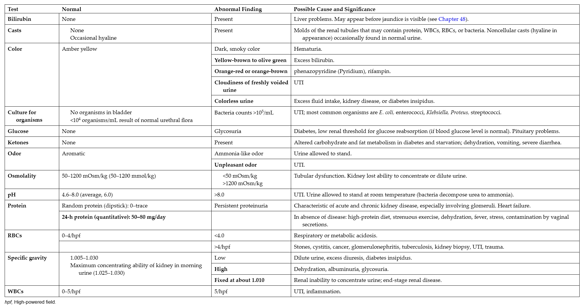
Specific gravity (concentration of urine) 1.005 - 1.025
Acidity of the urine (pH) 4.5 - 8
Protein in the urine (mainly albumin)
Glucose in the urine (sugar)
Ketones (products of fat metabolism)
Hemoglobin/blood in the urine
Leukocyte esterase (suggestive of white blood cells in urine)
Nitrite (suggestive of bacteria in urine)
Bilirubin (possible liver disease or red blood cell breakdown)
Urobilinogen (possible liver disease)
Elimination Across the Lifespan: Newborn
•The feces from a newborn is sterile, dark greenish, black, tarry and called meconium, viscous and consists of amniotic fluid, bilirubin, and cells.
•Most healthy term babies pass this within 12-24 hours of life.
Elimination Across the Lifespan: Toddler
•When is a toddler ready to potty train? Infants and toddlers initially lack control over the sphincters and muscles that control urination and bowel elimination, but control is attained during early childhood as a normal developmental milestone.
•They are usually between 18-24 months before they can recognize the urge to urinate and defecate.
•Potty training can begin when they can have a dry diaper for two hours straight around 2-3 years of age.
Elimination Across the Lifespan: Pregnancy
•As the fetus grows, increased pressure is placed on the bladder, and frequent urination occurs.
•Larger volumes of urine are produced because the blood volume circulating is larger.
•The growing fetus can also obstruct peristalsis causing constipation.
•Taking prenatal vitamins with iron can also contribute to constipation.
Elimination Across the Lifespan: Older adult
#1 Contributing Factor to Elimination Problems
•Atrophy of smooth muscle layers in the colon and reduced mucous secretions affect bowel elimination.
•Although the bladder retains tone with age, the volume of urine that can be held reduces, leading to urinary frequency.
•In many older adults, muscles around the urethra become weak, thus increasing the risk of incontinence.
•Reduced tone of the internal and external sphincter as well as reduced neural impulses (reducing the sensation of bowel evacuation) can make the older adult more susceptible to constipation or incontinence.
Populations at Risk
•All individuals, regardless of age, gender, or race, are potentially at risk
•Populations at greatest risk for problems with bowel and urinary elimination are
–Children
–Pregnant women
–Older adults
Aging and the GU System: Everything Decreases (what is decreased)
• Kidney size
• Glomerular filtration rate
• Renal blood flow
• Bladder size
• Ability to withhold urine
• Urethra thickness
• Muscles (thin and weaken) control of sphincters
• Urine output

Types of Urinary Incontinence
1. D-Stress incontinence-Sneeze- is the leakage of small amounts of urine during physical movement (e.g., coughing, sneezing, exercising). An increase in intraabdominal pressure, related to specific activities, can precipitate stress incontinence.
2. H-Urge incontinence-Midnight Tsunami- is the leakage of large amounts of urine at unexpected times, including during sleep. These differ in the amount of urine lost from the bladder and in the timing.
3. B-Overactive bladder-Frequency- refers to urinary frequency and urgency, with or without urge incontinence.
4. F-Functional incontinence-Germophobe- is untimely urination because of a physical disability, external obstacles, or cognitive problems that prevent the person from reaching toilet. It is the result of problems other than those of the urinary system.
5. C-Overflow incontinence-Spillage- is the unexpected leakage of small amounts of urine because of a full bladder. Similar to stress incontinence, the loss of small amounts of urine is involved but, unlike stress incontinence, the bladder must be full.
6. E-Mixed incontinence-Cough Flood- is typically the occurrence of stress and urge incontinence together.
7. A-Transient incontinence-Temporary- is leakage that occurs temporarily because of a situation that will pass (e.g., infection, taking a new medication, coughing with colds)
8. G-Nocturnal Enuresis-Rubber Sheet- is bed wetting and is usually due to the child just not waking up because of exhaustion, they hold their urine during the day, or their bladder just isn’t big enough to hold their urine. Make sure it isn’t from diabetes, UTI, pinworm infection, or neurological/emotional problems.


Consequences
urinary retention, bowel retention
Urinary Retention Cause:
External sphincter does not open for release of urine or blockage of urethra
Urinary Retention leads to:
•Leads to increased urine volume and bladder distention
–Backflow to the upper urinary tract
–Dilation of the ureters and renal pelvis
–Pyelonephritis and renal atrophy
–Incontinence
Bowel retention causes:
Ignoring the “urge to go” or decreased peristalsis
Bowel retention leads to:
•Leads to retention of stool in the rectum
–Stool dries and hardens
–Constipation
–Impaction
–Flatulence
–Hemorrhoids
–Diarrhea
Assessment Elimination
History & Physical
Assessment Techniques
Inspection
Auscultation
Palpation
Percussion
Diagnostics
Subjective data
Objective data
Assessment Elimination (History)
History: Conducting a history is the first step to understanding problems associated with elimination.
*A symptom analysis approach is helpful to fully understand the symptom.
•Patterns of urinary and bowel elimination, including frequency, appearance of stool and urine, and associated symptoms.
•Common symptoms directly related to elimination include alterations in elimination patterns; changes in the appearance, frequency, or quality of stool or urine; or discomfort and difficulty associated with elimination.
•Determine if there have been changes in diet, recent changes in health status (e.g., cognition, mobility, functional ability, or other medical conditions), and if there are new medications or changes in medications. Voluntary or involuntary (incontinence) emptying of the bowel or bladder also must be assessed to determine the impact of the condition on the individual.
•Adults of all ages should be asked about urinary continence because it is an underreported phenomenon.
Assessment Elimination (Assessment techniques)
Assessment Techniques
•Inspection: The abdomen is inspected for contour; abdominal or bladder distention is an abnormal finding. The genitalia are also inspected to examine the urinary meatus for evidence of redness, lesions, or discharge. Discharge from the urinary meatus is an abnormal finding and may suggest infection, especially if the patient reports pain with voiding. Observe the perianal area. The area should be free from redness or lesions; the presence of hemorrhoids or an anal fistula may be observed if the patient complains of pain with defecation. Inspection also includes looking at a stool or urine sample if available. Urine should be clear and yellow with mild odor. Very dark urine may be indicative of dehydration, may be a side effect of medication, or may indicate the presence of blood. Stool should be brown and formed. Stools that are black and tarry in appearance often signal gastrointestinal bleeding. Loose stools or diarrhea may be associated with diet, inflammation, or infection.
•Auscultation: Auscultation is limited to the abdomen to listen to bowel sounds. Bowel sounds should be heard in all four quadrants. An absence of bowel sounds may be associated with paralytic ileus; hyperactive bowel sounds may be noted with gastrointestinal inflammation or with an intestinal obstruction.15 Auscultation is not indicated in urinary assessment.
•Palpation: Abdominal palpation is a physical examination technique for both urinary and bowel elimination. The abdomen should be soft and nontender with palpation over the entire abdomen and over the urinary bladder. Abdominal or urinary distention is considered an abnormal finding and may be associated with reduced peristalsis or retention of stool or urine. Rectal palpation is done to assess the rectal sphincter and to examine for the presence of masses, lesions, or impacted stool. Digital palpation is also part of the prostate examination. Although this is not directly an examination associated with elimination, prostate enlargement can result from a tumor (benign or malignant) or inflammation; both can contribute to urinary retention. Bladder holds approximately 300-500mL before pressure increases to signal the need to void.
•Percussion: Abdominal percussion may be performed to help identify masses or excessive intestinal gas. Direct fist percussion over the costovertebral angle (over the kidney) should be painless; if sharp pain is produced, it could signal the presence of a kidney infection.
Assessment Elimination (examination techniques)
Bowel elimination: Abnormal clinical findings can include abdominal distention; hypoactive, hyperactive, or absent bowel sounds; and hemorrhoids.
Urinary elimination: Abnormal clinical findings can include bladder distention, discharge from the urinary meatus, redness or lesions of the urinary meatus, and dark or bloody urine.
Assessment Elimination (diagnostic tests)
Diagnostic Tests
•Laboratory tests
Urinalysis, blood urea nitrogen (BUN), creatinine,
culture, occult blood, Glomerular Filtration rate (GFR)
•Radiography
Bladder scan, X-rays, computed tomography (CT) scans, magnetic resonance imaging (MRI), ultrasound
•Direct observation tests
Colonoscopy, cystoscopy, uroscopy
•Other tests
Bladder stress testing, urine flow studies
Assessment Bladder
•Inspection
•Palpation
•Auscultation
•Bladder holds approximately 300-500mL before pressure increases to signal the need to void
Assessment of Urinary System Subjective Data
•Important health information
–Health history
•Kidney disease or urologic problems
•Health problems that affect kidney function
–Medications
•OTC, prescription, herbals
•Nephrotoxic
•Alter quantity and character of urine
–Surgery or other treatments
•Hospitalizations related to renal or urologic problems; radiation or chemotherapy
•Functional health patterns
–Health perception–Health management pattern
–Nutritional–metabolic pattern
–Elimination pattern
•General manifestations, edema, pain, patterns of urination, urine output and urine composition
–Activity–exercise pattern
–Sleep–rest pattern
•Nocturia
–Cognitive–perceptual pattern
•Mobility, visual acuity, dexterity, incontinence, pain
–Self‑Perception–self‑concept pattern
–Role–relationship pattern
–Sexuality–reproductive pattern
•Incontinence is not directly related to sexual problems, but can have devastating effects on self-esteem and intimate relationships
Assessment of Urinary System Objective Data
•Physical examination
–Inspection
•Skin, mouth, face and extremities, abdomen
•Weight, general state of health
–Palpation
•Costovertebral angle (CVA)
•Left kidney rarely palpable
•Right kidney—lower pole
•Bladder
–Percussion
•Kidney punch
–Normally not painful
–If tender, may indicate infection or polycystic kidney disease
•Bladder
–Does not percuss until it contains at least 150 mL of urine
•Auscultate bowel sounds
–Use diaphragm of stethoscope
•Focused assessment
–Evaluate status of previously identified urinary system problems, monitor for signs of new problems
•Documentation
–Normal physical assessment
–Assessment abnormalities
•Changes in older adult
–Age-related changes
Diagnostic Studies of Urinary System
•Urine studies
–Urinalysis
•First morning void; examine specimen within 1 hour
–Creatinine clearance—most accurate indicator of renal function
•Collect 24-hour urine specimen and blood specimen
•Creatinine clearance closely approximates GFR
•Urodynamic studies
–Urinary tract function
•Radiologic studies—bowel prep to enhance visualization
–Magnesium citrate and Fleet enema—do not use with kidney failure
–Iodine-based contrast can cause contrast-induced kidney injury (CIN) and allergic reactions; maintain hydration and administer N-acetylcysteine to reduce CIN
–NPO—avoid dehydration; insulin adjustment for diabetics

Diagnostics urinary system
Specific gravity (concentration of urine) 1.005 - 1.025
Acidity of the urine (pH) 4.5 - 8
Protein in the urine (mainly albumin)
Glucose in the urine (sugar)
Ketones (products of fat metabolism)
Hemoglobin/blood in the urine
Leukocyte esterase (suggestive of white blood cells in urine)
Nitrite (suggestive of bacteria in urine)
Bilirubin (possible liver disease or red blood cell breakdown)
Urobilinogen (possible liver disease)
Elimination PRIMARY prevention
•Hydration
•Adequate dietary fiber
•Regular toileting practices
•Regular exercise
•Avoidance of environmental contamination
Elimination SECONDARY prevention
General discussion questions:
Who should undergo these screening procedures?
How often should each be done?
What factors inhibit persons from having these screening procedures done?
•Colonoscopy screening
•Occult blood screening
•Prostate cancer screening
•Urinalysis
Colonoscopy-Because colonoscopy testing is highly accurate and colorectal cancer tends to grow slowly, most experts recommend that people at average risk should have a baseline colonoscopy at age 50, then repeat the exam every 10 years.
Fecal Occult Blood Test (FOBT) or Fecal Immunochemical Test (FIT) Annually Screens for colon cancer. It tests for hidden blood in the stool, which can be an early sign of cancer. FIT only detects human blood from the lower intestines. Medicines and food do not interfere with the test. So it tends to be more accurate and have fewer false positive results than other tests.
Prostate Cancer-Age 50 for men who are at average risk of prostate cancer and are expected to live at least 10 more years.
Age 45 for men at high risk of developing prostate cancer. This includes African Americans and men who have a first-degree relative (father or brother) diagnosed with prostate cancer at an early age (younger than age 65).
Age 40 for men at even higher risk (those with more than one first-degree relative who had prostate cancer at an early age).
Collaborative interventions: Pharmacologic agents: antibiotics, antispasmodics, agents to manage stool retention, analgesics
Incontinence management: Invasive procedures and surgical interventions: catheterization, urethral stents, urinary diversion, lithotripsy, nephrectomy, prostate surgeries
The recently revised recommendation is for periodic prostate-specific antigen (PSA)-based screening of men ages 55 to 69 to be an individualized decision, after weighing risks and benefits associated with screening with their healthcare provider (recommendation grade C). The USPSTF recommends against screening for prostate cancer in men over the age of 70.20 The USPSTF reports insufficient evidence to support routine screening for bladder
2010 ACS Guideline for the Early Detection of Prostate Cancer: American Cancer Society Recommendations- EBP
The informed decision-making discussion should begin:
At age 50 for men who are at average risk of getting prostate cancer and have at least a 10- year life expectancy
At age 45 for men at high risk of developing prostate cancer
• African American men,
Men who have a first-degree relative (father, brother, or son) diagnosed with prostate cancer at an early age
(younger than age 65)
At age 40 for men at even higher risk (those with several first-degree relatives who had prostate cancer at an early age
Function of the Large Intestine: Absorbs water and vitamins. Converts digested food into feces or waste excretion.
Feces- contains water, electrolytes, bile, intestinal secretions, bacteria, calcium
Fiber Rich Foods

FLUID AND ELECTROLYTES
Fluids and electrolytes play a vital role in homeostasis within the body by regulating various bodily functions including cardiac, neuro, oxygen delivery and acid-base balance and much more
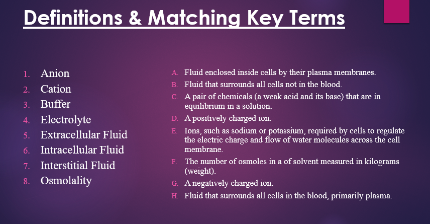
Anion-G
Cation-D
Buffer- C
Electrolyte-E
Extracellular Fluid H
Intracellular Fluid A
Interstitial Fluid B
Osmolality F


The maintenance of physiological balance of body fluid and electrolytes is a dynamic interplay between three processes: intake and absorption, distribution, and output. Intake and absorption refers to the addition of fluid and electrolytes to the body (intake) and their movement into the blood (absorption). Distribution is the process of moving fluid and electrolytes between the various body fluid compartments.
The scope covers, as above, with optimal balance or an imbalances to the sides. Fluid and electrolyte imbalances can be too little, too much, or misplaced.
Conceptually, fluid balance has two aspects: extracellular volume (ECV)(Fluid that surrounds all cells in the blood, primarily plasma) and osmolality (The number of osmoles in a of solvent measured in kilograms (weight)).
Electrolyte balance requires separate consideration. Extracellular fluid volume (ECV), body fluid osmolality, and plasma electrolyte concentrations can each be visualized as a continuum with three categories: optimal balance and two types of imbalances high or low
Fluid and electrolyte balance is a dynamic interplay because fluid and electrolyte output occurs continuously. Intake of fluid and electrolytes influences output to some degree, but intake can easily become less than or more than output. Fluid and electrolyte distribution can shift rapidly when conditions change.

Review of Physiology Fluid and electrolyte
The three physiological processes whose interplay creates fluid and electrolyte balance were defined previously:
intake and absorption, distribution, and output.
Optimal balance occurs when both of the following characteristics are present:
• Intake and absorption of fluid and electrolytes matches the output of fluid and electrolytes.
• Volume, osmolality, and electrolyte concentrations of fluid in the various body fluid compartments are within their normal ranges.
(Under each of the three scales volume, osmolality and electrolytes are balanced)
To maintain optimal fluid and electrolyte balance, output must be matched by appropriate intake, and the intake must be absorbed
Some things to consider about physiology with intake and absorption and distribution, and output
( WHAT COMES IN BALANCES WHAT GOES OUT)
The most common route of fluid and electrolyte intake is oral.
Fluid and electrolytes that enter the body by all routes except IV must be absorbed into the bloodstream
If fluid and electrolytes are not absorbed, they remain in the GI tract and leave the body in the feces.
After fluid and electrolytes enter the blood, they are distributed to various body compartments.
Normal fluid and electrolyte distribution is necessary for optimal function.
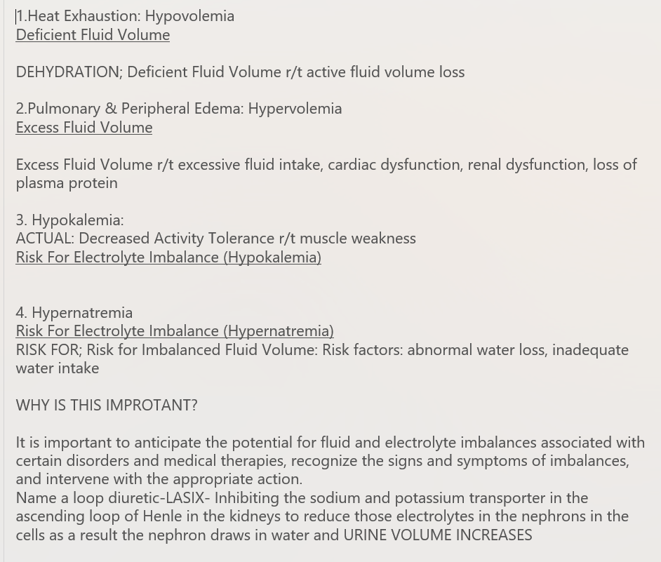
It is important to anticipate the potential for fluid and electrolyte imbalances associated with certain disorders and medical therapies, recognize the signs and symptoms of imbalances, and intervene with the appropriate action.
Balance

Body Water Over the Life Span
The body is composed primarily of water.
It accounts for about 50% to 60% of body weight in the adult.
Water content varies with body mass, gender, and age
Lean body mass has a higher percentage of water, while adipose tissue has a lesser percentage of water.
So, the more fat present in the body, the less the total water content.
Women generally have a lower percentage of body water because they tend to have less lean body mass than men.
Older adults also tend to have less lean body mass, resulting in a lower percentage of body water when compared to younger adults.
SO knowing the allows us to care for the patients accordingly.
In older adults, body water content averages 45% to 50% of body weight. This places them at a higher risk for fluid-related problems than young adults.
Neonates and infants are not able to communicate thirst except by crying, which can affect their fluid intake if their caregivers are not vigilant. They also have a high percentage of body weight as water and more ECF than intracellular fluid.
Because both neonates and infants have large body and lung surface areas compared with body mass, they have higher fluid exchange ratios than any other age group.
In addition, neonates and infants have immature kidneys that have little reserve capacity, reaching maturation only during childhood.
Body Fluid Compartments
uIntracellular fluid (ICF) Inside the cells
uExtracellular fluid (ECF) Outside the cells Main Components are:
uInterstitial: The fluid in the spaces between cells
uIntravascular (plasma)
uTranscellular : Cerebrospinal fluid; fluid in the gastrointestinal (GI) tract and joint spaces; and pleural, peritoneal, intraocular, and pericardial fluid

Mechanisms Controlling Fluid and Electrolyte Movement


Diffusion is the movement of molecules from an area of high concentration to an area of low concentration. Eventually, the sugar molecules are evenly distributed.
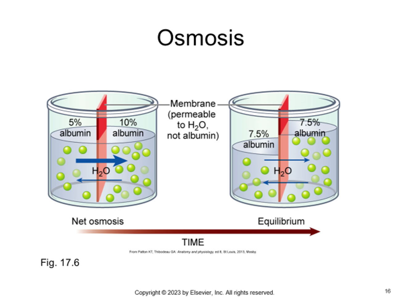
Osmosis is the process of water movement through a semipermeable membrane from an area of low solute concentration to an area of high solute concentration.
If you add albumin to one side, water will move from the less concentrated side (has more water) to the more concentrated side of the chamber water (has less water) until the concentrations are equal
•Whenever dissolved substances are contained in a space with a semipermeable membrane, they can pull water into the space by osmosis. The concentration of the solution determines the strength of the osmotic pull. The higher the concentration, the greater a solution's pull, or osmotic pressure. Osmotic pressure is measured in milliosmoles (mOsm). It may be expressed as either fluid osmolarity or fluid osmolality. Although you will often see the terms osmolarity and osmolality used interchangeably, they are different measurements. Osmolarity measures the total milliosmoles per liter of solution, or the concentration of molecules per volume of solution (mOsm/L). Osmolality 273measures the number of milliosmoles per kilogram of water, or the concentration of molecules per weight of water. Osmolality is the preferred measure to evaluate the concentration of plasma, urine, and body fluids.

Body fluids can be discussed in terms of their specific fluid compartment, a location that is largely separate from another compartment by some form of a physical barrier.
The 2 fluid compartments in the body are the intracellular space (inside the cells)(ICF)(YELLOW) and the extracellular space (outside the cells) (Fig. 17.2). About two-thirds of body water is found within cells ( represented above with 28 L). It is the intracellular fluid (ICF)(YELLOW)
The fluid in the extracellular space is extracellular fluid (ECF). The 2 main compartments containing ECF are the interstitial fluid (IF)(BLUE), the fluid in the spaces between cells, and the intravascular fluid or plasma, the liquid part of blood. (RED)
GIDDENS REMINDS US
After fluid and electrolytes enter the blood, they are distributed to various body compartments. Normal fluid and electrolyte distribution is necessary for optimal function
______________________________________________________________________________________________________________________________________________
Other ECF compartments include lymph and transcellular fluids. Transcellular fluids include cerebrospinal fluid; fluid in the gastrointestinal (GI) tract and joint spaces; and pleural, peritoneal, intraocular, and pericardial fluid. ECF makes up about one third of the body water; this amounts to about 14 L in a 70-kg man.


Homeostasis
•Body fluids and electrolytes play an important role in maintaining homeostasis, the body's stable internal environment.
•Body fluids are in constant motion transporting nutrients, electrolytes, and oxygen to cells and carrying waste products away from cells.
•The body uses a number of adaptive responses to keep the composition and volume of fluids and electrolytes within narrow limits to maintain homeostasis and promote health
•Many diseases and their treatments affect fluid and electrolyte balance.
•
•It is important to anticipate the potential for fluid and electrolyte imbalances associated with certain disorders and medical therapies, recognize the signs and symptoms of imbalances, and intervene with the appropriate action.
•Fluid Shift: If capillary or interstitial pressures change, fluid may abnormally shift from one compartment to another, resulting in edema or dehydration

Causes of Fluid
and Electrolyte Disturbances
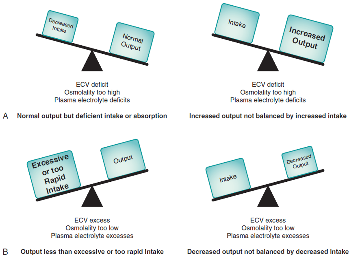
A- Normal output but deficient intake or absorption- When intake is less than normal output, the fluid imbalance a person develops depends on the type of fluid that is deficient. For example, people who do not drink enough water to match their water output can make their body fluids too concentrated (osmolality too high).
-Increased output not balanced by increased intake- Another situation in which fluid and electrolyte output becomes greater than intake arises when output increases but intake does not increase enough to balance it. For example, diarrhea increases output of Na+, water, and K+. During diarrhea, an intake of Na+, water, and K+ that does not balance the increased output (DIARRHEA)
B-Output less than excessive or too rapid intake-Individuals with normal renal function may overwhelm their output capacity by excessive or very rapid intake of fluid and electrolytes. This situation most commonly occurs with IV infusions. Any person receiving IV fluid is at risk for developing fluid and electrolyte imbalances from excessive or too rapid infusion of the solution, even if the infusion is intended to replace a fluid or electrolyte deficit. (IV)
-Decreased output not balanced by decreased intake-People who have oliguria/ decreased output of fluid and electrolytes; they must decrease their intake to prevent imbalances.
Once we understand the cause we can understand the intervention
Many diseases and their treatments affect fluid and electrolyte balance. Chemotherapy used to treat the cancer may result in nausea and vomiting and, subsequently, dehydration and acid-base imbalances. When correcting dehydration with IV fluids, the patient requires close monitoring to prevent fluid overload and electrolyte imbalance.

Risk Factors:
Populations at Greatest Risk
Infants are at great risk for fluid and electrolyte imbalance because they have a high metabolic rate, which increases fluid loss and results in a rapid turnover in fluid. Infants also have immature kidneys that are limited in their ability to conserve water. Insensible fluid loss is greater because of the more rapid respiratory rate in infants and their proportionately greater body surface area.
Similar to the very young, the very old are also at great risk for fluid and electrolyte imbalance for a number of reasons. Thirst sensation is often blunted. The kidneys are less able to respond to ADH, and this, along with increased levels of atrial natriuretic factor, impairs their ability to conserve water. These age-related changes create a risk of dehydration and, when placed in the context of common chronic heart and kidney diseases of aging, further increase the likelihood of fluid and electrolyte imbalance.
Infants and often the very old are dependent on others to provide foods and fluids, a fact that adds to the risk of dehydration.


Assessment of Skin Turgor

•Assessment of skin turgor. (A and B) When normal skin is pinched, it resumes shape in seconds. (C) If the skin stays wrinkled for 20 to 30 seconds, the patient has poor skin turgor.

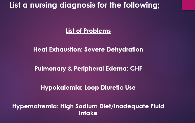
Interrelated concepts (fluid, electrolytes)
Fluid and electrolytes and Acid–Base Balance are closely related; changes in one can cause changes in the other, and situations such as vomiting and diarrhea can lead to both. Hormonal Regulation has a powerful influence on fluid and electrolytes because of the significant role hormones play in maintaining balance. Nutrition greatly influences fluid and electrolyte intake. Elimination creates fluid and electrolyte output; changes in elimination, such as oliguria and diarrhea, can disrupt fluid and electrolyte balance. Fluid and electrolyte imbalances can influence Perfusion and Gas Exchange (ECV imbalances), Cognition (osmolality imbalances), and Mobility (electrolyte imbalances that cause muscle weakness). These interrelationships are illustrated in Fig. 8.5.

Connecting Acid Base Balance to Fluid And Electrolytes

Compensation mechanisms

GLUCOSE REGULATION AND NUTRITION ADD