2.4 - Thoracic wall
 Call Kai
 Learn
Learn Practice Test
 Spaced Repetition
 Match
Match Flashcards
 Knowt Play
1/84

There's no tags or description
Looks like no tags are added yet.
Name | Mastery | Learn | Test | Matching | Spaced | Call with Kai | Chat |
|---|
No analytics yet
Send a link to your students to track their progress
85 Terms
thorax
The cranial trunk between the neck and abdomen
Thoracic cavity
Space protected by the rib cage and limited caudally by the diaphragm
Rib cage
Bony structure that protects the thoracic cavity and part of the abdominal cavity
Ribs, thoracic vertebrae, and sternum
What 3 structures make up the entirety of the rib cage
36 ribs
How many ribs does a horse have?
26 ribs
How many ribs does a dog have?
26 ribs
How many ribs does a goat have?
28-30 ribs
How many ribs does a pig have?
Cartilage of manubrium
A

Sternebrae
B

Xiphoid cartilage
C

Tubercle
A

Head
B

Neck
C

Body
D

Costal cartilage
E

Intervertebral joints
Identify the joint of the rib cage - 1

Costovertebral joints
Identify the joint of the rib cage - 2

Costochondral joints
Identify the joint of the rib cage -3
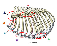
Sternocostal joints
Identify the joint of the rib cage -4

Sternal joints
Identify the joint of the rib cage -5

Intercostal joints
Identify the joint of the rib cage -6

Supraspinous ligament
Identify the intervertebral ligaments - 1

Interspinous ligament
Identify the intervertebral ligaments - 2

Dorsal intervertebral ligament
Identify the intervertebral ligaments - 3

Ventral intervertebral ligament
Identify the intervertebral ligaments - 4

Ligament of costal tubercle
Identify the costovertebral ligament - 6

superficial radiate costal ligament
Identify the costovertebral ligament - 7

Radiate ligament
Identify the costovertebral ligament - 8

Costotransverse ligament
Identify the costovertebral ligament - 9

Lateral costotransverse ligament
Identify the costovertebral ligament - 10

Periosteum and perichondrium
In the costochondral joints, what part of the rib and which part of the costal cartilage attach to one another
Costochondral joint
1

Sternal rib
2

Asternal ribs
3

Floating rib
4

Radiate sternocostal ligament
Identify the red box

Sternal ligament
Identify

Calcifies
What happens to the sternal cartilages to make it one bony piece
True
T/F - species to species, the contribution of the superficial muscles to the thoracic wall varies
Deep
Is the serratus ventralis deep or superficial to the latissimus dorsi
Latissimus dorsi

External abdominal olique
2

Latissimus dorsi
1

Serratus ventralis
1

Levatores costarum
Identify the muscle

External intercostals
Identify the muscle

Caudal borders of the ribs
Origin of external intercostals

Cranial borders and lateral surfaces of the ribs
Insertion of external intercostals

Dorsal Rami, intercostal nerves
What innervates the levatores costarum

Intercostal nerves
What innervates the external intercostals

Internal intercostals
Identify the muscle

Cranial border of the ribs
Origin of internal intercostals

Caudal borders of next ribs
Insertion of internal intercostals

Intercostal nerves
Innervation of internal intercostals

to pull ribs caudally during expiration
Action of internal intercostals

Retractor costae
Identify

Transversus thoracis
Identify

Rectus thoracis
Identify

Pectoralis superficialis
Identify

Pectoralis profundus
Identify

Diaphragm
Broad unpaired muscle that divides the thoracic and abdominal cavities
Costal, sternal,and lumbar regions of the sternum and ribs
Origin of the diaphragm
Central tendon
Insertion of diaphragm
Phrenic nerve
innervation of diaphragm
Increase the volume of thoracic cavity during respiration
action of diaphragm
Central tendon
A

Costal region
B

Sternal region
C

Lumbar region
D

Aortic hiatus
opening in the diaphragm allowing the aorta, azygos vein, and thoracic duct to pass from the thorax to the abdomen
Esophageal hiatus
where the esophagus passes through the diaphragm
Foramen vena cavae
Allows the caudal vena cava to pass from the abdomen to the thorax
Aortic hiatus
A
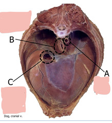
Esophageal hiatus
B

Foramen vena cavae
C

dorsal intercostal branches off the aorta
What is the dorsal blood supply to the thoracic wall
Ventral intercostal branches off the internal thoracic
What is the ventral blood supply to the thoracic wall
Internal thoracic vein
What does thoracic wall blood drain to ventrally
azygous vein
Where does thoracic wall blood drain to dorsally
Ventral rami
What innervates the lateral thoracic wall and the cutaneous innervation
Intercostal artery, vein, and nerve
Artery, vein, and nerve found caudal to the ribs and cranial to the intercostal space
Intercostal collateral artery, vein, and nerve
Artery, vein, and nerve found cranial to the ribs and caudal to the intercostal space
Thoracotomy
surgical incision into the chest cavity

Thoracic lavage
Uses saline to clear out debris or infection from the chest cavity