Vital Signs : BP
 Call Kai
 Learn
Learn Practice Test
 Spaced Repetition
 Match
Match Flashcards
 Knowt Play
1/103

There's no tags or description
Looks like no tags are added yet.
Name | Mastery | Learn | Test | Matching | Spaced | Call with Kai | Chat |
|---|
No analytics yet
Send a link to your students to track their progress
104 Terms
The pupil’s ability to change size (constrict and dilate) in response to changes in focusing on near objects
accommodation
Patient lying on their back with the head of the bed elevated about 15–30°
Low Fowler’s position
Patient lying on their back with the head of the bed elevated about 30–45°
Semi-Fowler’s position
Patient lying on their back with the head of the bed elevated about 60–90°
High Fowler’s position
PERRLA
Pupils Equal, Round, Reactive to Light and Accommodation.
[Think: Light = flashlight. Accommodation = near object.]
Functions that support adequate cardiac output and transport oxygenated blood blood to the body tissues.
Perfusion
6 Rights of Medication Administration
Right client,
Right Medication,
Right dose,
right route,
right time
right documentation
EMR
Electronic Medical Record
Measurements that reflect the body's physiological functions, including pulse, temperature, blood pressure, respiratory rate, oxygen saturation, and sometimes pain level
Vital signs
To cough up or spit out phlegm, mucus, or sputum from the throat and lungs
Expectorate
A measurement of the amount of pressure exerted by the blood within the circulatory system, expressed in mmHg
Blood pressure
The maximum amount of pressure exerted when the heart contracts and forces blood into the aorta during systole (represented by the top number in a blood pressure measurement)
Systolic blood pressure
The minimum amount of pressure exerted when the heart is relaxed during diastole (represented by the bottom number in a blood pressure measurement)
Diastolic blood pressure
Cardiac output, blood volume, blood viscosity, vascular elasticity, and peripheral vascular resistance
Five direct physiological determinants of blood pressure
The amount of blood pumped into the circulatory system by the heart within 1min
Cardiac output
calculated as CO=SV×HR
where SV is stroke volume and HR is heart rate.
The amount of blood ejected by a ventricle during one heart contraction
Stroke volume

Identify the Right Coronary Artery


Identify the Aorta


identify the Left Main Coronary Artery

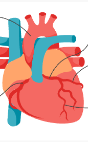
Identify the Circumflex Coronary Artery
Identify the


Identify the Left Descending Coronary Artery

Blood viscosity
The resistance of a liquid to flow.
Vascular elasticity
The ability of blood vessels to stretch and compress, then return to their original shape.
Peripheral vascular resistance
The total resistance to flow of blood in the vascular bed.
the resistance to blood flow,
Think of it like a faucet:
🚰 Vessels constrict (narrow) → resistance increases → BP increases
🚰 Vessels dilate (widen) → resistance decreases → BP decreases
Contractility
The heart's ability to contract efficiently, measured by the ejection fraction.
*do not confuse with heart beating*
Ejection fraction
The percentage of blood ejected from the ventricles with each contraction, generally measured in the left ventricle via an echocardiogram.
Ejection fraction = (Stroke Volume ÷ End-Diastolic Volume) × 100
Preload
The amount of blood inside the ventricles immediately before they contract. (fill)
Afterload
The amount of resistance or constriction that the heart must overcome to eject blood into systemic circulation.
When the ventricle contracts, it tries to push that blood into the aorta.
Afterload = the resistance/pressure the ventricle has to push against
→ Mainly the pressure in the arteries (especially the aorta)
Effect of decreased vessel elasticity on blood pressure
Increases the rigidity of the vessel wall, which in turn increases blood pressure.
Less elasticity → stiffer blood vessels → higher blood pressure.
Effect of increased afterload on cardiac muscle
Leads to cardiac hypertrophy, which ultimately decreases contractility.
atherosclerosis
hardening of the arteries
consists of a cuff with an inflatable bladder. The cuff is connected to a manometer that indicates the amount of pressure in the cuff.
Sphygmomanometer
How to document palpated blood pressure?
120/p
p=palp
Palpated blood pressure only gives you a systolic reading
This is the palpable estimated systolic blood pressure.
[Blood Pressure Assessment] Routine measurement age criteria in health care settings
Clients older than 3years of age, or younger clients with a preexisting medical condition.
[Blood Pressure Assessment] Clinical importance of accurate measurement
Many treatment decisions are directly based on a client's blood pressure.
[Blood Pressure - Patient Preparation] Requirement regarding client clothing at the cuff site
Bulky or layered clothing over the measurement site must be removed prior to applying the cuff.
[Blood Pressure - Patient Preparation] Proper leg and foot placement for a seated client
Legs uncrossed with feet flat on the floor.
[Blood Pressure - Patient Preparation] Proper positioning and height of the client's arm
Supported at the level of the heart with the palm facing upward.
[Blood Pressure - Cuff Placement] Correct placement location relative to the antecubital fossa
Applied snugly approximately 1inch above the antecubital fossa with the artery indicator aligned over the brachial artery.
[Blood Pressure - Auscultation] Stethoscope placement location on the upper arm
The bell or diaphragm is placed directly over the brachial artery.
[Blood Pressure - Palpation Method] Clinical indications for use
Used when Korotkoff sounds are difficult to hear during auscultation or when a digital monitor fails to detect blood pressure.
[Blood Pressure - Palpation Method] Indicator of estimated systolic blood pressure during cuff deflation
The pressure reading on the manometer when a pulsatile thrill is first felt.
[Blood Pressure - Auscultation Method] Target cuff inflation level above expected systolic pressure
≈30mmHg above the expected systolic pressure value.
[Blood Pressure - Auscultation Method] Recommended cuff deflation rate
Approximately 2mmHg per second.
[Blood Pressure - Auscultation Method] Manometer indicator of systolic blood pressure
The point at which the first Korotkoff sound is heard.
[Blood Pressure - Auscultation Method] Manometer indicator of diastolic blood pressure
The point at which Korotkoff sounds are no longer audible.
A series sounds created by movement of blood through a partially compressed vessel during a manual blood pressure assessment.
Korotkoff sounds
An IV catheter placed in a large vein to give medications, fluids, nutrition, or blood.
Central intravenous line
A connection between an artery and vein that provides access for hemodialysis.
Arteriovenous (AV) shunt
Alternative blood pressure measurement methods for clients with an arm circumference greater than 50cm
Using a forearm cuff, thigh cuff, or conical-shaped cuff.
Extrinsic factors affecting blood pressure
External and modifiable factors that a client can control to some extent, such as weight, caffeine or nicotine intake, medications, sodium intake, stress, activity level, anxiety, pain, and fever.
Intrinsic factors affecting blood pressure
Non-modifiable internal factors including age, ethnicity, genetics, and natural hormonal variations.
Effect of heart failure on blood pressure
Causes a decrease in blood pressure.
Structural arterial change in adults that frequently causes hypertension
Thickening of arterial vessel walls and a decrease in vessel elasticity, which increases peripheral vascular resistance.
Measurement requirement for a diagnosis of hypertension
At least two elevated readings taken on two or more separate occasions.
Blood pressure criteria for Normal classification in adults
Systolic <120mmHg and diastolic <80mmHg
Blood pressure criteria for Elevated classification in adults
Systolic 120 to 129mmHg and diastolic <80mmHg
Blood pressure criteria for Stage 1 Hypertension in adults
Systolic 130 to 139mmHg or diastolic 80 to 89mmHg
Blood pressure criteria for Stage 2 Hypertension in adults
Systolic ≥140mmHg or diastolic ≥90mmHg
Blood pressure criteria for Hypertensive crisis
Systolic ≥180mmHg and/or diastolic ≥120mmHg
Sensory receptors in blood vessels that respond to pressure and stretching
Baroreceptors
Three hormones released or triggered via baroreceptor response to regulate blood pressure
Antidiuretic hormone (ADH), renin, and aldosterone
Physiological mechanism by which antidiuretic hormone (ADH) elevates blood pressure
Secreted by the hypothalamus, ADH increases water reabsorption in the kidneys (raising plasma volume) and causes mild vasoconstriction at higher levels.
Three triggers for juxtaglomerular cells in the kidneys to release renin
Decreased blood pressure
Increased sympathetic nervous system activity
Decreased sodium levels in the distal convoluted tubules
Substrate converted by renin in the renin-angiotensin-aldosterone system
Angiotensinogen (produced by the liver), which renin converts into angiotensin I.
Enzyme that converts angiotensin I to angiotensin II and the resulting effects
Angiotensin-converting enzyme (ACE) converts angiotensin I into —> angiotensin II, which increases blood pressure through vasoconstriction and improved reabsorption of water and sodium.
The adrenal gland; aldosterone enhances the activity of the sodium-potassium pump, increasing sodium reabsorption and potassium excretion.
Gland that releases aldosterone and its mechanism for raising blood pressure
An idiopathic form of high blood pressure, commonly linked to genetics and salt sensitivity.
Primary (essential) hypertension
High blood pressure resulting from identifiable underlying conditions, such as endocrine disorders, vascular issues, kidney problems, medications, or sleep apnea.
Secondary hypertension
Vascular and cardiac complications associated with prolonged hypertension
Damage to arterial walls, development of atherosclerosis (plaque buildup), stroke, and heart failure.
64/41mmHg
Expected blood pressure (50th percentile) for a newborn (birth to 4 weeks)
85/50mmHg
Expected blood pressure (50th percentile) for an infant (1 to 12 months)
Expected blood pressure ranges (50th percentile) for toddlers (1 to 2 years)
Male: 85 to 91/37 to 46mmHg\n- Female: 86 to 89/40 to 49mmHg
Male: Greater than 103 to 109/56 to 65mmHg\n- Female: Greater than 104 to 107/58 to 67mmHg
Expected blood pressure ranges (95th percentile) for toddlers (1 to 2 years)
Male: 91 to 98/46 to 53mmHg\n- Female: 89 to 93/49 to 54mmHg
Expected blood pressure ranges (50th percentile) for preschoolers (3 to 5 years)
Male: Greater than 109 to 112/65 to 72mmHg\n- Female: Greater than 107 to 110/67 to 72mmHg
Expected blood pressure ranges (95th percentile) for preschoolers (3 to 5 years)
Male: 96 to 106/55 to 62mmHg\n- Female: 94 to 105/56 to 62mmHg
Expected blood pressure ranges (50th percentile) for school-aged children (6 to 12 years)
Male: Greater than 114 to 123/74 to 81mmHg\n- Female: Greater than 111 to 123/74 to 80mmHg
Expected blood pressure ranges (95th percentile) for school-aged children (6 to 12 years)
complications of Hypertension
Hypertension increases a client’s risk of heart attack, stroke, blood vessel damage (atherosclerosis) , kidney failure, and vision loss (hypertensive retinopathy).
hypotension in an adult.
a systolic pressure less than 90 mm Hg or a diastolic pressure less than 60 mm Hg
Manifestations of hypotension
dizziness, nausea, blurred vision, increased pulse, and fatigue.
Standard adult blood pressure threshold for hypotension without baseline data
Systolic <90mmHg or diastolic <60mmHg
Common causes of low blood pressure (hypotension)
Dehydration, blood loss, shock, and significant illness such as sepsis
Clinical manifestations of hypotension
Dizziness, nausea, blurred vision, increased pulse rate, and fatigue
Two common underlying causes of shock resulting from extreme hypotension
Significant blood loss and heart failure
Clinical manifestations of shock secondary to extreme hypotension
Cold, pale skin, rapid breathing rate, and a weak, rapid pulse
Immediate nursing interventions for a client experiencing shock
Rapid infusion of IV fluids or blood products and administration of medications to increase blood pressure and cardiac contractility
Orthostatic hypotension
A drop in blood pressure that occurs when a client rises to a sitting or standing position.
Common underlying causes of orthostatic hypotension
Dehydration, baseline hypotension, heart failure, or central nervous system disorders.
Primary safety risk associated with orthostatic hypotension
Feelings of faintness or dizziness, which significantly increase the client's risk for falls.
Assessment sequence used to evaluate a client for orthostatic hypotension
Measuring blood pressure sequentially while the client is lying, sitting, and then standing.
Diagnostic blood pressure criteria for orthostatic hypotension
A drop in systolic pressure of at least 20mmHg or a drop in diastolic pressure of at least 10mmHg within 1min of position change
Timeframe to recheck blood pressure for clients reporting symptoms during position changes [Orthostatic hypotension]
3min after moving to a sitting or standing position.
Client education and nursing interventions to minimize orthostatic hypotension
Instruct the client to change positions slowly, slightly elevate the head of the bed while sleeping, avoid prolonged sitting or lying, wear compression stockings as prescribed, stay well hydrated, and avoid temperature extremes.