Pathophysiology 1 Exam 2
 Call Kai
 Learn
Learn Practice Test
 Spaced Repetition
 Match
Match Flashcards
 Knowt Play
1/102

There's no tags or description
Looks like no tags are added yet.
Name | Mastery | Learn | Test | Matching | Spaced | Call with Kai | Chat |
|---|
No analytics yet
Send a link to your students to track their progress
103 Terms
What is the total blood volume for male and female?
Total blood volume
o 75.5 mL/kg in men
o 66.5 mL/kg in women
7% to 8% of body weight
5 to 6 L
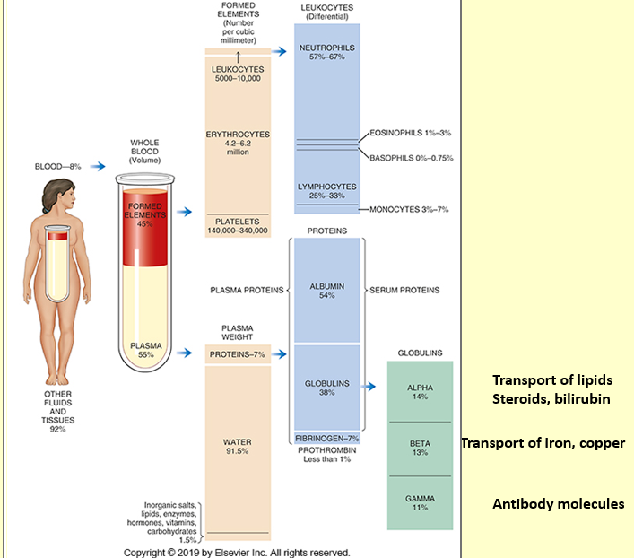
Blood cells make up ~ 40-45% of blood volume
RBCs
Most numerous
4.2 to 6.2 x 10⁹ cells/mL
Transport oxygen to tissues
Remove carbon dioxide from the tissues
Buffer blood pH
oContain carbonic anhydrase
Plasma makes up what percent of blood volume?
Plasma ~55-60% of blood volume
Plasma is composed of what?
Composed of 92% water, 7% plasma proteins
True or False: Red blood cells have a nucleus
False
No nucleus or cytoplasmic organelle
What is erythropoietin?
Hormone from kidney that stimulates erythrocyte production
RBCs are produced through Hematopoiesis
Two-stage process involving:
o Mitotic division (proliferation)
o Maturation (differentiation)
Erythropoiesis is controlled by a system sensitive to the concentration of hemoglobin in the blood

Per one hemoglobin molecule, how many oxygen molecules can it bind to?
Hemoglobin carrying oxygen: oxyhemoglobin; each Hb molecule can bind 4 oxygen molecules
What do hemoglobin carry?
Hemoglobin: oxygen-carrying protein in mature RBCs
Where does the production of hemoglobin take place?
Immature RBCs: factory for hemoglobin
90% of RBC dry weight
What is required for hemoglobin synthesis?
67% of total body iron is bound to heme and 30% is stored bound to ferritin. Remaining 3%
is lost in daily urine. IRON
What nutritional requirements are needed for normal RBC development?
Protein and Vitamins (B12, Folate, B6, Riboflavin, Niacin, Vit E and C)
RBC Development
Large nucleated cells change to small reticulocytes that have no nucleus
Reticulocyte leaves the marrow and enters the blood stream
Matures into erythrocyte (RBC) in 24-48 hrs.
Mitochondria and ribosomes disappear
Mature RBC can no longer make hemoglobin
RBC lifespan: ~120 days in adults
Red Blood cells + bone marrow precursors=
Erythron
A decrease in hemoglobin decreases what in kidneys?
A decrease in Hb level decreases the tissue oxygen tension in the kidney
In response to this hypoxia, the kidney secretes the hormone, erythropoietin (EPO) that stimulates stem cells in the marrow to differentiate to make RBC

What do RBCs need energy for?
to survive ~120 days
Oxygen transport and exchange processes are passive (don’t need energy)
RBC need energy for operate membrane pumps for maintaining
o High intracellular potassium ion [K+]
o Low intracellular sodium ion [Na+]
o Very low intracellular calcium ion [Ca++]
RBC rely on glycolysis to produce the energy, because they don’t have mitochondria, nuclei and other subcellular organelles
Where do RBC get digested by macrophages?
80-90% of RBC get digested by macrophages in spleen and liver. 10-20% inside blood vessels
Red Cell Destruction
As RBCs age, enzyme activities decrease
Amount of membrane lipids decrease
Cell loses ability to deform and becomes fragile
Hb A levels increase
Heme is reduced to bilirubin
Globin and iron portions are conserved and reused
Bilirubin is then degraded to urobilinogen and excreted primarily in the feces and in the urine.
Increased levels of bilirubin give the skin a yellowish tone (jaundice).

True or False: The 3% that is dissolved in plasma is measured as PO2
TRUE
What does the oxygen-hemoglobin dissociation curve describe?
Describes the relationship between PO2 (pressure) and SO2 (saturation)
Gas Transport and Acid-Base Balance
Oxygen Transport
97% of oxygen transported in blood is bound to Hb within the RBC
PaO2 = PO2 in arterial blood = 80-100 mmHg
PvO2 = PO2 in venous blood = 35-40 mmHg

What three forms is CO2 transported as?
Transported in 3 forms
1. As dissolved gas (5%)
2. As bicarbonate ion (HCO3- ,75%)
3. In association with hemoglobin forming carbaminohemoglobin (20%)
In body tissues, CO2 inside the cells diffuses into the blood and attaches to hemoglobin as oxygen is released to the tissues
RBC contain the enzyme carbonic anhydrase which greatly increases the conversion of water and CO2 into HCO3- and H+

True or False: Carbaminohemoglobin release CO2 in the lungs which we exhale out
TRUE
What are the RBC disorders?
Alterations of Oxygen transport
Decreased oxygen reaching the tissues results in secretion of erythropoietin (EPO) and increase in the production of RBCs.
Factors that decrease hemoglobin mass (anemia) or decrease arterial saturation (hypoxia) lead to increased release of erythropoietin.
Red blood cell disorders
Anemia: Deficit of RBC
o Patient has tissue hypoxia due to low-oxygen-carrying capacity of blood
General Effects of Anemia
Reduction in oxygen-carrying capacity- Tissue hypoxia (low oxygen)
Compensatory mechanism to restore tissue oxygenation
• Increased heart rate
• Increased cardiac output,
• Preferential increased flow to vital organs
• Increase in erythropoietin activity
Mild anemia
• Usually, no clinical symptoms
• Elderly with cardiovascular, pulmonary disease may have symptoms
Mild to moderate anemia
• Fatigue,
• generalized weakness
• loss of stamina, followed by tachycardia and exertional dyspnea
Moderate to severe anemia
o Decrease in blood pressure: orthostatic (after standing) and generalized hypotension
o Vasoconstriction
o pallor
o Tachypnea (rapid breathing), dyspnea (labored breathing)
o Tachycardia (rapid heart rate), transient murmurs, angina pectoris, heart failure
o Intermittent claudication, night cramps in muscles
o Headache, lightheadedness and faintness
o Tinnitus, roaring in the ears
Polycythemia: Excess of RBC
o Patient has increased blood viscosity and volume due to the increase in RBC numbers
What is the pathogenesis of aplastic anemia?
Etiology and Pathogenesis
o Stem cell disorder characterized by reduction of hematopoietic tissue, fatty marrow replacement
o Caused by toxic, radiant, or immunologic injury to the bone marrow stem cells
Laboratory features
o Pancytopenia (low RBC, WBC and platelets)
o Low WBC is important for prognosis (low leukocytes = susceptibility to infections)
o Diagnosed with bone marrow biopsy
Clinical Manifestation
• Insidious onset of symptoms
• Late symptoms include weakness, fatigue, lethargy, pallor, dyspnea, palpitations, transient murmurs and tachycardia related to low RBCs.
• Thrombocytopenia (low platelets; prone to bleeding)
• Neutropenia (low WBC; prone to infections)
• Disease of the young (15 to 25) or old (>60)
Treatment
• Determine efficacy of bone marrow transplantation.
• Administer immunosuppressive therapy or stimulate hematopoiesis and bone marrow regeneration.
Prognosis
• Fatal unless bone marrow transplant successful
• Bone Marrow transplantation is highly successful and curative for 80-85% of un-transfused patient and 55-60% of patients with multiple previous transfusions
What does anemia chronic renal failure cause?
Etiology and Pathogenesis
o Primarily from failure of the renal endocrine function, which causes impaired erythropoietin (EPO) production
o Secondarily from failure of renal excretory function
o Leads to hemolysis, bone marrow cell depression and blood loss
Laboratory features and clinical manifestations
o Low RBC count
o Low hematocrit
o Low hemoglobin
o Some grossly deformed RBC
o General signs and symptoms of anemia usually manifest when hematocrit decreases to < 20%
Treatment
Dialysis
Administration of erythropoietin
o Erythropoietin is only used until Hgb is 12 g/dL.
Replacement of iron, folate, and B12 due to dietary restrictions and anorexia
Prognosis
o 95% respond to erythropoietin therapy.
Anemia in relation to vitamin B12 or Folate deficiency causes a disruption in what?
Etiology and Pathogenesis
Disruption in DNA synthesis of blast cells produces megaloblasts (macrocytic)
Pernicious anemia is caused by lack of intrinsic factor leading to vitamin B12 deficiency. Problem is with B12 absorption rather than “deficiency”.
Folate deficiencies from dietary deficiencies, alcoholism, cirrhosis, pregnancy, or infancy
• Low folate levels associated with neural tube deficits
Laboratory Features
o Low RBC, WBC, and platelet counts; megaloblastic dysplasia
o Macrocytic and hyper-segmented neutrophils
Clinical Manifestation
o Clinical features of vitamin B12 deficiency:
o Paranoia, dementia, cognitive dysfunction, delusions, hallucinations
o Peripheral nerve degeneration and memory impairment
o Clinical features of both B12 and folate deficiency:
o Edema in the feet/ Tachypnea/ Weight loss
Treatment
o Administer B12 and folate parenterally or orally
Prognosis
o Majority respond well to treatment.
o Reversibility of the neurologic damage is slow
What is the most common cause of anemia?
Iron Deficiency Anemia
Etiology and Pathogenesis
o Most common cause anemia (iron deficiency is the most common nutritional deficiency in the world)
o Results in the unavailability of iron for hemoglobin synthesis
Possible causes
o Low iron intake
o Diminished absorption
o Increased requirement (e.g. pregnancy)
o Excessive iron loss (e.g. hemorrhage)
o Renal failure
o Hemodialysis
o GI bleeding
o Menorrhagia
Laboratory features
o Smaller and paler RBC
o Low red cell indices
o Decreased serum ferritin
Treatment and Prognosis
o Oral administration of ferrous sulfate or intravenous ferric gluconate (only in severe cases); continue for 4 to 6 months
o Treat underlying cause
o Prognosis: Usually very good
True or False: Thalassemia has increased RBC destruction referred to as hemolysis
TRUE
Thalassemia
Etiology and pathogenesis
o Increased RBC destruction (hemolysis) resulting in decreased RBC survival rates
o Associated with mutant genes that suppress the rate of globin chain synthesis
o Classified by the polypeptide chain(s) with deficient synthesis
• α-thalassemia or β-thalassemia
o Most clinically severe form: thalassemia major (homozygous);
Thalassemia minor (heterozygous) is less severe
Laboratory Features
o Hypochromic, microcytic RBCs
o RBC indices are low
o Erythroblastic hyperplasia (bone marrow)
INHERITED
For hemolytic newborns, what is clinically relevant?
Anemia related to extrinsic red cell destruction or loss
Hemolytic disease of the newborn
Etiology and pathogenesis
o Fetal RBCs cross placenta, stimulate production of maternal antibodies against antigen on fetal
RBC not inherited from mother.
o Maternal antibodies cross into the fetal circulation causing destruction of fetal cells in subsequent pregnancies.
Laboratory features
o Elevated serum bilirubin due to hemolysis
o Bilirubin in amniotic fluid is an indicator
Clinical manifestation
o Jaundice
o Hepatomegaly
o Splenomegaly
o Diffuse intravascular coagulation
Treatment and prognosis
o Standard dose of anti-Rh immune globulin (RhoGAM) is given to the mother before or after delivery.
o Severe cases, in utero blood transfusion and early delivery
o Prognosis: Death, possible retardation, or barely perceptible hemolytic process
Polycythemia can be defined as?
Excess RBC results in increased blood viscosity, leading to clinical symptoms such as hypertension.
Types of polycythemia are categorized by the cause.
1. Polycythemia Vera
o Neoplastic transformation of bone marrow stem cells
2. Secondary Polycythemia
o Caused by chronic hypoxemia with resultant increase in erythropoietin
production
3. Relative Polycythemia
o Caused by dehydration with spurious increase in RBC production
What are the three types of polycythemia?
Polycythemia Vera
Absolute increase in RBC mass, leukocytosis, thrombocytosis; increased uric acid because of excess proliferation; oxygen saturation normal.
Signs and symptoms from increased viscosity
o Hypertension
o Thrombosis
o Congested spleen and liver
Treatment
o Reducing increased blood volume, viscosity, RBC mass, and platelet count with phlebotomy,
radioactive phosphorus, and chemotherapeutic agents
Prognosis
o Poor unless properly managed
o No cure
Secondary Polycythemia
Increased RBC production without increase in WBCs or platelets
Treatment: Identify and manage underlying cause of hypoxemia; phlebotomy may be used to decrease cardiovascular workload
Prognosis: depends on underlying condition
Relative Polycythemia
Elevated hematocrit, hemoglobin, and RBC count
Two groups
o Disturbed fluid balance
o Stress polycythemia (long-standing state of low plasma volume)
Treatment
• Recognize and manage underlying cause
• Fluid administration with management of long-term conditions
Prognosis: Excellent if managed properly
Chapter 15: Alterations in Blood Flow:
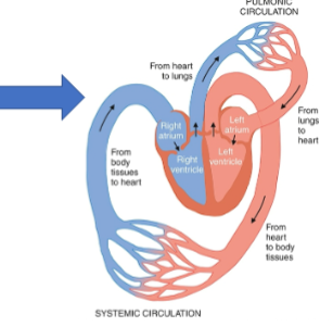
What does the circulatory circuit do?
Absorption and delivery of nutrients
Oxygen uptake and delivery
Removal of waste products
Lymphatic circulation
o Specialized system of channels and tissues (nodes)
o Collect the excess fluid (2-4 liters/day) that leaks from vascular network into the interstitium and returns it to the general circulation.
How long does it take to move 5 liters of blood through the entire circuit?
The complete process moving ~5 liters of blood through the entire circuit takes only ~1 minute
What do arteries contain in comparison to veins?
Three microscopically distinct layers (tunicae)
o Intima: endothelial cells in direct contact with the blood as it flows through the vessel
• Periodically, the intima layer protrudes into the lumen, creating valves that prevent the backflow of blood
o Media: smooth muscle tissue (thickest section in arteries)
o Adventitia: collagenous connective tissue (thickest section in veins)
Arteries only contain elastic tissue, veins only have elastic tissue in large veins

What does the intima layer function in?
endothelial cells in direct contact with the blood as it flows through the vessel
• Periodically, the intima layer protrudes into the lumen, creating valves that prevent the backflow
True or False: Blood flow is measured as a given number of liters or milliliters per
second, minute, or hour
TRUE
Includes the concepts of
• Pressure
• Resistance
• Velocity
• Turbulent flow
• Wall tension
Describe how pressure works
Blood flow—movement along a pressure gradient within the vascular bed
Pressure—blood moves from areas of higher pressure (arteries) to an area of lower pressure (veins); the greater the pressure difference, the greater the blood flow
Resistance—opposing forces that deter blood flow; as resistance increases, blood flow decreases
Physical laws govern the flow of blood though vessels
Important Laws:
• Flow = Pressure/Resistance
• Blood pressure = flow (cardiac output) x Resistance
• Resistance = Pressure/Flow

What are the determinants of vascular resistance?
Determinants of Vascular Resistance
1. Vessel length
2. Vessel radius
3. Blood viscosity
also includes elastic flexibility of tube
At a constant pressure gradient:
The longer the blood vessel, the higher the resistance and the lower the flow
The narrower the blood vessel, the higher the resistance, the lower the flow
True or False: the longer the blood vessels, the higher the resistance and lower the flow
TRUE
Turbulent flow is generated where?
Turbulent flow generated at a vessel bifurcation
Velocity and Laminar and Turbulent Flow
• Laminar flow: Fastest blood flow in the center of stream
• Turbulent flow is an interruption in the forward current of blood flow by crosswise flow.
Turbulent flow may manifest as
• Bruit (audible vascular sound)
• Thrill (vibratory sensation on the skin over a turbulent flow)
• Thrombus formation (blood clot affecting flow)
• Example of pathologic cause of turbulence

What is clinically important in capillaries?
Dynamics in the Microcirculation (Capillaries)
Essence of the entire circulatory system
Exchange of nutrients and gases takes place
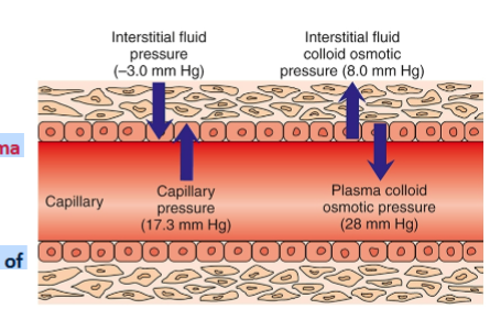
Clinically, The capillary fluid pressure and plasma colloid osmotic pressure are most important
Capillary fluid pressure is the blood pressure inside the capillary (force that pushes fluid out of
vessel)
Pathologic conditions resulting in increases in BP or resistance to flow, can alter this force, increasing it and propelling more fluid into the interstitial space, resulting in the formation of edema
What are the controls of blood flow?
By Extrinsic mechanisms (Central control)
All blood vessels except small venules and capillaries contain smooth muscle cells.
Arteries have much more smooth muscle than veins
Smooth muscle cells are innervated by nerve fibers from the sympathetic nervous system (SNS)
Smooth muscle cells have α1 adrenergic receptors that respond to the SNS neurotransmitter, noradrenaline resulting in vasoconstriction
Skeletal muscle blood vessels contain β2 adrenergic receptors which respond to epinephrine (adrenaline) resulting in vasodilation
By intrinsic mechanisms (Local control)
Autoregulation is ability of blood vessels within organs to maintain a relatively constant blood flow regardless of changes in arterial pressure
oWhen vascular smooth muscle is stretched, it contracts in response causing vasoconstriction
oEndothelial cells also play an important role in vessel dilation and constriction
• Produce nitric oxide (NO) gas which causes dilation by relaxing neighboring smooth muscle cells
What happens when systemic vascular resistance is increased?
It causes the heart to work harder to meet metabolic demands of the body
Thrombosis is initiated by alterations in what?
Thrombus is a stationary blood clot formed within a vessel or a chamber of the heart
• Clot is composed of aggregated platelets, clotting factors and fibrin that adhere to the vessel wall
Etiology: Thrombosis is Initiated by alterations in one or more of the following:
• Blood flow: slow or turbulent flow
• Blood vessel wall: damage or inflammation to the intimal wall of vessel
• Blood coagulability: emergence of a hypercoagulable state
Can occur in arteries (arterial thrombosis) and veins (venous thrombosis)
Oral contraceptives may increase tendency for thrombosis
Arterial thrombosis is classified as?
Arterial thrombosis
• Clot within an artery reduces flow and increases turbulence which enhances thrombus enlargement and formation of more thrombi
• Decreased distal flow can cause ischemia which can cause
Arterial occlusion
Myocardial infarction (heart attack)
Stroke
True or False: Venous thrombosis symptoms may be absent or maybe life- threatening secondary to pulmonary embolism
TRUE
Venous thrombosis
• Clot in a vein alters venous return impairing the removal of metabolic waste and producing swelling
• Inflammation that occurs in a vein is called phlebitis and when it is accompanied with a clot, it is called thrombophlebitis
What is the major reason for arterial disease?
Athclerosis
Means hardening of the arteries
Pathologic origin for the vast majority of arterial disease
Ultimately the leading cause of death in U.S and western Europe
Develops mostly in large and medium size arteries
o Coronary (associated with most mortality; coronary artery disease [CAD])
o Cerebral
o Carotid
o Femoral
o Aorta
o Other organs
Modifiable Risk Factors
• Smoking
• Elevated blood pressure
• Glucose intolerance (e.g. diabetes)
• Elevated cholesterol
• Decreased physical activity
• Ineffective stress management
• Depression
Nonmodifiable Risk Factors
• Age
• Gender
• Heredity (Family history)
What is atherosclerosis an underlying condition of?
hypertension , renal disease, cardiac disease, and peripheral arterial disease
Where are aneurysms mostly found?
Localized arterial dilations, bulge outward
Classified as true or false aneurysms
True aneurysms include
• Saccular: one-sided balloon
• Fusiform: both sides balloon out
• Berry: balloon has a stem/neck
Frequently found in cerebral circulation and thoracic and abdominal aorta

Alterations in venous flow can be accompanied with?
Incompetent valves (e.g. in obesity, pregnancy, right heart failure, prolonged standing) producing
o Varicose veins- Superficial veins affected
o Chronic venous insufficiency- Deep veins affected
o Deep vein thrombosis- Could lead to life-threatening pulmonary emboli
Accompanied by edema, venous stasis, inflammation, ulcers, and pain
True or False: Lymphedema is most common in the U.S because of lymph node removal,
and radiation
TRUE
Alterations in Lymphatic Flow
Lymphedema
Occurs when normal flow is obstructed or altered in some fashion
Primary lymphedema results from congenital anomaly or dysfunction of the lymphatic system.
Secondary lymphedema associated with a disease process or iatrogenic
oMost common in United States because of lymph node removal, radiation
Chapter 16: Alterations of Blood Pressure
Systemic arterial blood pressure is the result of what?
Provides momentum for the flow of blood around the body
Arterial blood pressure is produced by the force of the left ventricle contraction overcoming the resistance of the aorta to open the aortic valve
• Pressure is maintained in the arterial system throughout the cardiac cycle
It is the pressure difference between the left and right sides of the heart that produces the gradient allowing this systemic movement of blood
The systemic arterial blood pressure is the physiologic result of:
cardiac output and resistance to the ejection of blood from the heart
The resistance to ejection into arterial circulation is known as the systemic vascular resistance (SVR) and is determined by:
• Radius of arteries
• Degree of vessel compliance
• SVR is also known as cardiac afterload
SVR can be altered by constricting or relaxing (dilating) arterial smooth muscle
What is the difference between systolic and diastolic pressure?
Cardiac Function
Cardiac output (CO)
• Blood ejected by a ventricle in 1 minute
• CO = SV (Stroke volume) x HR (heart rate)
Stroke volume (SV)
• Volume of blood pumped out of ventricle with each contraction
Blood Pressure
Systolic pressure: exerted when blood is ejected from ventricles (high)
Diastolic pressure: sustained pressure when ventricles relax (lower)
BP is directly proportional to blood volume and is controlled by hormones
What is used clinically as part of cardiovascular assessment?
Mean Arterial Pressure Map (MAP) is the calculated average pressure within the circulatory system
(2 x Diastolic P) + Systolic P] / 3
Pulse Pressure = Systolic - Diastolic
Out of the Korokthoff sounds, which is classified as the systolic and diastolic pressure
I. Initiation of a clear Tapping Sound (Systolic P)
II. Murmuring or swishing sounds
III. Increase in intensity and crispness of sound
IV. Muffling of sound
V. Disappearance of sound (Diastolic P)
According to American Heart Association, what are the guidelines for hypertension
grade 1 and 2 for both pressures?

What is the short term regulation mechanism mediated by?
Short Term Regulation Mechanisms
Rapid adjustments in response to
• Position changes
• Exercise
• Emotional changes (e.g. fear/anxiety)
• Physiologic changes (e.g. volume depletion)
Mediated by the sympathetic branch of the autonomic nervous system
Long Term Regulation Mechanisms
o Week after week and month after month
o Interplay of neural, hormonal, and renal interactions
• Intimately connected with the body’s fluid volume
What activates the vasomotor center directly and indirectly?
Short-Term Regulation of Blood Pressure
Regulated by the vasomotor center (located in medulla of brainstem)
Vasomotor center directly activated by various stimuli or indirectly via pressure-sensitive baroreceptors ( which monitor arterial pressure (MAP) variations)
Stimulation of SNS results in the release of neurotransmitters noradrenaline and adrenaline
Activates α1 receptors on smooth muscle of arterioles, causing vasoconstriction (↑ Systemic
vascular resistance, [SVR])
• Activates β1 receptors of the heart (↑ heart rate)
• Receptor responsiveness decline with age (age-related arterial stiffening)
Chemoreceptors stimulate the medullary vasomotor center to increase SNS activity.
• Located in carotid and aortic arterials
• Activated only when BP extremely low
Beta receptors of the heart increase or decrease heart rate?
Increase
When the kidney is stimulated by low arterial pressure, what happens?
Long-Term Regulation of Blood Pressure: The Renin –Angiotensin- Aldosterone System (RAAS)
Juxtaglomerular cells (in the kidney when stimulated by low arterial pressure release renin activates angiotensinogen to angiotensin I.
Angiotensin I when in contact with ACE is converted to angiotensin II, a potent vasoconstrictor and stimulates release of aldosterone by the adrenal cortex.
Aldosterone, a hormone, causes reabsorption of sodium and water passively follows.

What is the function of the aldosterone?
Aldosterone, a hormone, causes reabsorption of sodium and water passively follows.
What is the most common primary diagnosis in the U.S?
Hypertension (High Blood Pressure)
Most common primary diagnosis in the U.S
Most common risk factor for cardiovascular disease worldwide
Increases morbidity and mortality associated with heart disease, kidney disease, peripheral vascular disease, and stroke
> 40% of adults 25 years or older are diagnosed
Responsible for an annual worldwide death rate of 7.6 million
Areas most frequently damaged by hypertension
• Kidneys
• Heart
• Brain
• Retina
Predisposing factors
• Incidence increases with age
• Men affected more frequently and more severely
• Incidence in women increases after middle age
• Genetic factors
• Sodium intake, excessive alcohol intake, obesity, smoking, prolonged or recurrent stress

Describe hypertensive crisis?
Hypertensive crisis: Systolic over 180 and/or diastolic over 120, with patients needing prompt changes in medication if there are no other indications of problems, or immediate hospitalization if there are signs of organ damage.
True or False: Primary essential hypertension, majority of the cases are caused unknown
TRUE
Essential hypertension – idiopathic (Majority of cases: 90 -95% of hypertensive patients)
• Blood pressure consistently above 130/90 mm Hg
Also called essential hypertension
Rare prior to the age of 10
Incidence in children is escalating
Systolic BP: major risk factor for cardiovascular disease
Isolated systolic hypertension: systolic BP is ≥130 mm Hg while diastolic pressure remains <90 mm Hg
Isolated diastolic hypertension: diastolic pressure is ≥90 mm Hg with a systolic pressure of <130 mm Hg
Combined systolic and diastolic hypertension: both systolic and diastolic exceed prehypertension levels
Nonmodifiable
o Family history
o Age
o Ethnicity/Genetics
Modifiable
o Dietary factors
o Sedentary lifestyle
o Obesity/weight gain
o Metabolic syndrome
• Elevated circulating insulin and lipid levels
o Elevated blood glucose levels/diabetes
o Elevated total cholesterol
o Alcohol and smoking
What does the silent killer refer to?
Primary Hypertension: Outcomes
Sometimes called the “silent killer” as damage has already occurred to organs before diagnosis is made
o End-organ damage: a function of stage and duration of hypertension
• Renal failure, stroke, heart disease
• Damage to arterial system and acceleration of atherosclerosis lead to cardiovascular disease
• Increased myocardial work results in heart failure.
• Glomerular damage results in kidney failure.
• Affects microcirculation of the eyes
• Increased pressure in cerebral vasculature can result in hemorrhage (stroke, Brain
What constitutes a hypertensive emergency?
Hypertensive emergency: sudden increase in either or both systolic or diastolic blood pressure with evidence of end-organ damage
o Rapid but controlled reduction of blood pressure using parenteral antihypertensive agents under close monitoring (typically in ICU setting)
o Responsible for 25% of emergency visits
Hypertensive urgency: similar blood pressure elevation without evidence of end-organ damage
o Oral medications to bring blood pressure under control over 24 to 48 hours
Chapter 17: Cardiac Function
63. Describe the Circulatory System
Left sided heart chambers produce the force to propel blood through the vessels of the systemic (body) circulation
The left atrium receives oxygenated blood from the lungs by the way of pulmonary veins and delivers it to the left ventricle
The oxygenated blood is pumped by the left ventricle into the aorta which supplies the arteries of the systemic circulation
Venous blood is collected from the capillary networks of the body and is returned to the right atrium by the way of vena cava
Blood from the head returns via the inferior vena cava

The blood supplied to the heart muscle is provided by?
The Coronary Arteries
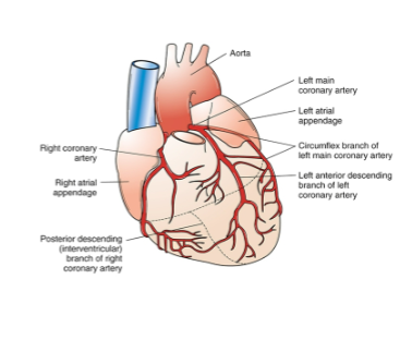
The blood supply of the heart muscle is provided by the coronary arteries
1. Right coronary artery
2. Left circumflex artery
3. Left anterior descending
Describe the cardiac cycle
Each heartbeat is composed of a period of ventricular contraction (systole) followed
a period of relaxation diastole.
What does blood flow equal to?
Blood flow = Pressure/Resistance
What are the two determinants of coronary vascular resistance?
Coronary vascular resistance has 2 major determinants
1. Artery diameter
2. Varying degrees of external compression by myocardial contraction and relaxation
Coronary artery diameter is continuously adjusted to maintain blood flow at a level adequate for myocardial demands
Vessel dilation (vasodilation) occurs in response to increased tissue metabolism
Vessel constriction (vasoconstriction) in response to decreased metabolic activity
A disruption in cardiac blood flow (ischemia) generally results in some degree of pump failure and damage to cardiac tissues.
Reduction in coronary blood flow
Increased myocardial demand for oxygen
What are the two general cardiac myocytes?
Two general type
1. Working cells (mechanical pumping functions)
2. Electrical cells (transmit electrical impulses)
Both types are excitable and unable to proliferate
How do the heart cells store excess ATP?
Like other tissues, the heart utilizes from ATP generated by glycolysis and oxidative reactions
Under conditions of ATP excess, heart cells are able to store the excess ATP as creatine phosphate (CP) by the enzyme creatine kinase (CK)
The enzyme CK is useful in the diagnosis of myocardial infarction (heart attack) because it leaks into the blood stream
Both cardiac contraction and relaxation require energy
True or False: Both cardiac contraction and relaxation require energy
TRUE
Describe the determinants of stroke volume
Volume of blood in the heart (preload)
• Resistance to ejection from the ventricle (afterload)
Chapter 18: Alterations of Heart Function
72. What is cardiac output(CO)?
Cardiac Output (CO) = Stroke Volume (SV) x heart rate (HR)
CO depends on:
Determinants of heart rate
• Under the control of sympathetic and parasympathetic nervous system
Determinants of Stroke Volume
• Volume of blood in the heart (preload)
• Resistance to ejection from the ventricle (afterload)
What is stroke volume(SV)?
The amount of blood ejected from the ventricle with each contraction
What are the determinants of stroke volume?
Determinants of Stroke Volume
• Volume of blood in the heart (preload)
• Resistance to ejection from the ventricle (afterload)
Preload, contractility, afterload
True or False: Coronary Heart Disease (CHD) is responsible for approx 50% of deaths by
CVD
TRUE
What is CHD characterized by?
Also called ischemic heart disease or coronary artery disease (CAD)
Accounts for 31% of all deaths
Coronary Hearty Disease (CHD) is responsible for ~50% of deaths by CVD
Stroke, high blood pressure, heart failure and others claim the remainder
16 million in the US have history of CHD
Men and women equally represented
• Characterized by insufficient delivery of oxygenated blood to the myocardium caused by
atherosclerotic coronary arteries
• Sequelae of CHD includes:
• Angina pectoris
• Myocardial infarction
• Dysrhythmias
• Heart failure
• Sudden cardiac death
Lipid Processing Basics
Chylomicrons from dietary fat absorption are taken up by the liver and resynthesized into
• VLDL
• LDL
• HDL
HDL (“good cholesterol”) circulates to the tissues and takes up excess free cholesterol and takes it back to the liver
Triglycerides are removed from VLDL for tissue use
More triglyceride removal leads to formation of LDL
LDL (“bad cholesterol”) is absorbed by the tissues and 70% returns to the liver
What are the known risk factors for CHD?
Known risk factors:
• Atherosclerosis: the source of nearly all CHD
• Possible microcirculation abnormalities
Atherosclerosis causes narrowing of the arterial lumen that can lead to cardiac ischemia through
• thrombus formation.
• coronary vasospasm.
• endothelial cell dysfunction.
Risk factors of CHD
Nonmodifiable
o Age: >45yrs in men; >55 yrs for women
o Family history: MI or sudden cardiac death in male first degree relative (< 55 yrs old; female < 65)
o Gender: male (earlier onset)
Lipid Risk factors
o Total cholesterol (>200 mg/dL)
o LDL cholesterol (>130 mg/dL)
o Triglycerides (>150 mg/dL)
o HDL cholesterol (< 40 mg/dL)
Nonlipid Risk factors
o Hypertension
o Cigarette smoking
o Thrombogenic state
o Diabetes
o Obesity
o Physical inactivity
o Poor diet
Probable (emerging) Risk factors
o Chronic inflammation
What is defined as good and bad cholesterol?
HDL (“good cholesterol”) circulates to the tissues and takes up excess free cholesterol and takes it back to the liver
LDL (“bad cholesterol”) is absorbed by the tissues and 70% returns to the liver
What are lipids transported via?
Mechanisms of CHD
Lipids are transported via apoproteins.
o Lipid + protein = Lipoprotein
Lipoproteins associated with a greater risk of atherosclerosis
High-density lipoproteins (HDL) transport cholesterol from peripheral tissue back to the liver, clearing atheromatous plaque.
LDL = “Bad cholesterol”
HDL = “Good cholesterol
What does HDL do?
HDL circulates to the tissues and takes up excess free cholesterol and takes it back to the liver
What does LDL do?
LDL is absorbed by tissues and 70% is returned to the liver
What is atherosclerotic plaque formation initiated by?
Atherosclerotic plaque formation initiated by injury to coronary artery endothelium
Endothelium becomes dysfunctional, permeable and recruits leukocytes
LDL leakage into the vessel wall occurs with oxidation by endothelial cells and macrophages.
Oxidized lipids are damaging to endothelial and smooth muscle cells and stimulate recruitment of macrophages into the vessel.
Macrophages (and smooth muscle cells) engulf the lipids becoming foam cells (lipid-filled macrophages) which will release inflammatory mediators and growth factors, attracting more
leukocytes and stimulating smooth muscle proliferation.
Excess lipid and debris accumulate within vessel wall and coalesce into lipid core.
Vulnerable plaques may rupture or become eroded, which stimulates clot formation on the plaque.
What are the differences between vulnerable and stable plaques?
Pathogenesis of Coronary Atherosclerosis
Vulnerable plaques have
• large lipid core.
• thin cap.
• high shear stress.
• Inflammation within
Stable plaques have
• more collagen and fibrin.
• stable cap
Plaques increase in size overtime
Often all three coronary arteries are Simultaneously affected
What can ischemia result in?
It can result in chronic or acute coronary syndromes.
Ischemia results from oxygen supply insufficient to meet metabolic demands
Chronic Occlusion of a coronary vessel is associated with clinical syndrome of stable angina
Acute occlusion is associated with plaque disruption (rupture) and thrombus (clot) formation and results in acute coronary syndrome (unstable angina or MI)
Ischemia can also be caused by vasospasm
• Usually occur in areas of atherosclerotic plaque
• In response to some drugs (e.g. cocaine)
• Inflammation
True or False: Ischemia results in oxygen supply insufficient to meet metabolic demands.
TRUE
The rate of coronary perfusion can be altered by?
1. Large, stable atherosclerotic 2. Acute platelet aggregation and thrombosis 3.
Vasospasm 4. Failure of autoregulation by microcirculation 5. Poor perfusion
pressure
Describe the pathophysiology of chronic and acute ischemia
Chronic occlusion of a coronary vessel is associated with clinical syndrome of stable
angina. Acute occlusion is associated with plaque disruption (rupture) and
thrombus (clot) formation and results in acute coronary syndrome (unstable angina
or MI)
Acute Coronary Syndromes
Associated with acute changes in plaque morphology and thrombosis (clot formation) leading to sudden obstruction of coronary artery
Unstable Angina
Myocardial Infarction (MI)
Sudden cardiac arrest
Abrupt onset and life-threatening consequences
Any of the coronary heart syndromes (chronic or acute) may precipitate Sudden Cardiac Arrest and associated Dysrhythmias!
True or False: Acute Coronary Syndrome is associated with acute changes in plaque
morphology and thrombosis (clot formation), which causes a sudden obstruction of
coronary artery.
TRUE
True or False: Chronic or acute coronary heart syndromes may precipitate sudden cardiac
arrest and associated dysrhythmias.
TRUE
True or False: Stable angina cannot be relieved by rest.
FALSE
Stable angina (Classic or Typical Angina)
• Most common (also called typical or classic angina)
• Narrow atherosclerotic coronary vessels
• Coronary perfusion is inadequate during physical exertion
• Onset of pain is predictable and relieved by rest
What factors of stable angina may upset the balance?
Factors that decrease coronary supply OR increase myocardial oxygen demand can
upset the balance and lead to ischemia and anginal pain
True or False: MI occlusion is complete and the thrombus lasts long enough to cause
irreversible damage.
TRUE
Acute Coronary Syndrome
Unstable Angina and MI are difficult to distinguish on the basis of clinical manifestation and are lumped together as ACS
Chest pain usually more severe and lasts longer than typical angina
In both cases plaque rupture with acute thrombus development
Unstable angina: occlusion is partial, or the clot is dissolved before myocardial tissue damage
MI: occlusion is complete, and thrombus lasts long enough to causes irreversible damage
o Necrosis of myocardial cells
ECG and biomarkers used for diagnosis
What is an accurate diagnosis of ACS?
Accurate Diagnosis of ACS: Serum Biomarkers
The appearance of certain proteins in the blood after myocardial cell death is a sensitive and reliable indicator of MI
Elevated levels indicate suggest leakage from fatally damaged heart cells
• Myoglobin
• Troponins I and T (Test of choice)
• Lactate dehydrogenase
• Creatine kinase (CK-MB; specific for heart muscle necrosis)
1.Signs and symptoms
2.ECG changes
3.Biomarkers
MI is a result of prolonged or total disruption of blood flow
• More than 125,000 deaths/year in U.S.)
Acute occlusion causes a range of cellular events, depending on
• availability and adequacy of collateral blood flow.
• relative workload.
• length of time that flow is interrupted.
Ultimate size of the infarcted tissue depends on the extent, duration, and severity of ischemia.
Nearly all (>97%) infarcts are located in the left ventricular walls
7-day old Infarction
After 18 to 24 hours: area of infarction becomes paler than surrounding tissues
5 to 7 days: turns yellowish and soft with a rim of red vascular connective tissue
At 1 to 2 weeks: necrotic tissue progressively degraded and cleared away; infarcted myocardium weakened and susceptible to rupture
By 6 weeks: necrotic tissue replaced by tough fibrous scar tissue

What does chronic ischemic cardiomyopathy refer to?
Refers to a disorder in which heart failure develops insidiously (slowly) because of progressive ischemic myocardial damage.
Patients typically have history of angina or MI
Appears to be a consequence of slow, progressive apoptotic death of myocytes from chronic ischemia
oScattered throughout the myocardium
More common in older adults
Mitral valve is between which atrium and ventricle?
Mitral valve (valve between the left atrium and left ventricle)
Disorders of the Heart Valves
Heart valves may be damaged by
• inflammation and scarring.
• calcification.
• congenital malformations.
Murmurs are common with valvular disorders
Mitral Valve Stenosis and Regurgitation
Stenosis
Blood flow from left atrium to left ventricle impaired during ventricular diastole
Increased pressure of the left atrium leads to atrial chamber enlargement and hypertrophy.
Can lead to chronic pulmonary hypertension, right ventricular hypertrophy, and right-sided heart failure
Low-pitched, rumbling diastolic murmur; open snap; atrial dysrhythmias, atrial clots; exertional dyspnea
Regurgitation
Backflow of blood from the left ventricle to the left atrium during ventricular systole
Left atrium and ventricle dilate and hypertrophy cause by extra volume
May lead to left-sided heart failure
High-pitched, pansystolic, blowing murmur; giant V waves; chronic weakness and fatigue
What is the function of the aortic valve?
Aortic valve (valve allowing outflow from left ventricle to the aorta)
Aortic Valve Stenosis and Regurgitation
Stenosis
Results in obstruction of aortic outflow from the left ventricle into the aorta during systole
Predominant cause is age-related calcium deposits on the aortic cusps.
Left ventricle hypertrophy may result in ischemia and left-sided heart failure.
Regurgitation
Incompetent aortic valve allows blood to leak back from the aorta into the left ventricle during diastole.
Causes: abnormal aortic valve or aortic root dilation
Leads to left ventricle hypertrophy and dilation with eventual left-sided heart failure
High-pitched blowing murmur during ventricular diastole; high systolic blood pressure; diastolic blood pressure usually low; palpitations
What is Stenosis?
Stenosis: failure of the valve to open completely results in extra pressure work for the heart
What is regurgitation?
Regurgitation (insufficiency): inability of a valve to close completely results in extra volume work for the heart
What defines rheumatic heart disease?
Diseases of the Endocardium
• Acute inflammatory disease that follows infection with group A β- hemolytic streptococci
• Damage is caused by immune attack on the individual’s own tissues.
• Antibodies against the streptococcal antigens damage connective tissue in joints, heart, skin.
o Cause valve swelling and damage
• Occurs mainly in children
• Fever; sore throat; joint inflammation; involuntary movements (Sydenham chorea); and a distinctive truncal rash
What defines infective endocarditis?
Diseases of the Endocardium
• Invasion and colonization of endocardial structures by microorganisms with resulting inflammation—vegetations
• Most common bacteria
o Streptococcus
o Staphylococcus
• Acute infective endocarditis
o Prognosis poor
o Intravenous drug users susceptible
• Subacute infective endocarditis
o Onset is insidious
o Risk factor for acute endocarditis