Exposure & Techniques
 Call Kai
 Learn
 Practice Test
 Spaced Repetition
 Match
 Flashcards
 Knowt Play
1/83

There's no tags or description
Looks like no tags are added yet.
84 Terms
which Image Receptor (IR) is used in computer radiography?
Photostimuable Phosphor
This IR uses an imaging plate (IP) to trap electrons in higher energy states that give off light that is then converted into an xray image
Photostimuable Phosphor
Which IR still requires 2 steps?
Photostimuable Phosphor
this IR converts xray beams into an electronic signal and produces an xray image made by a matrix of pixels?
Digital Detector
IR that uses an image intensifier to convert xray energy into an electron beam that is then converted into visible light and recorded by a video camera.
Fluoroscopic Screen
what do grids help to capture when taking an xray?
scatter radiation
what is scatter radiation
parts of the primary beam that bounce off body tissue, bone, and room surfaces in different directions
what are grids made of?
lead and plastic
in the context of xray imaging, what is milliampere in mAs?
amount of electrons in the xray tube current
in the context of xray imaging, what is seconds in mAs?
exposure time
the number of xray photons is directionally proportional to ____?
the product of tube current and time
in the context of xray imaging, what is kilovolts?
the force acting on electrons in the xray tube
in the context of xray imaging, what is peak?
the amount of forces pulling on the electrons (voltage)
what is used to control how much the beam spreads when taking an xray?
collimation
what does collimation mean?
trim
Source to Image Receptro Distance is defined as ____?
the distance from the anode inside the xray tube to the IR
What can cause your xray beam to spread out?
Increased SID
Increased SID can ____ the intensity of your xray beam
decrease
which factor best counteracts SID’s affect on the intensity of the xray beam?
mAs
how does mAs help counteract SIDs affect on xray beam density?
it increases the amount of xray photons sent from the xray tube meaning there are more photons to cover a larger surface area
SID and the Inverse Square Law
the intensity of an X-ray beam is inversely proportional to the square of the source-to-image distance (SID)
in the context of xray, what is brightness?
the appearance of the image on a display montior
what primarily controls the brightness of the image?
system software
what does window width on the system software control?
contrast
increased window width with means _____
more shades of grey
decrease window width means ____
less shades of grey
window level on the system software controls what?
brightness and darkness
increased window level mean ___?
darker image
decrease window level means __?
brighter image
what is contrast?
the range of the gray scale on an xray image
contrast helps with seeing anatomical structures because?
if they are not a different color gray from what is surrounding them, we are unable to see them
if an image has low contrast then there is a _____ grey scale meaning there are ______ present
longer, more shades of grey
if an image his high contrast then there is a _____ grey scale, meaning there are _____ present
shorter, less shades of grey
it’s easier to the see the differences in anatomical structures when an image has _____ contrast
high contrast
is high contrast inherently better than low contrast in an xray image?
no, it depends on what body part/tissue is being examined
what is signal?
remnant beam strength
what is remnant beam?
the part of the primary beam that makes it through the patient and is captured by the IR
if you increase mAs, ____ and ____ increases
signal, dose
if you increase kVp, _____, _____, and ______ increases
signal, penetration, scatter
signal and dose are ________ to each other
directly proportional
signal, penetration, scatter, are to each other
directly proportional
what can increase noise/randomness in the image and cause it to look muddy/grainy?
scatter
which factor affects the amount of radiation exposure a patient receives the most?
mAs
What factor automatically terminates the exposure whenit’s hit by a certain number of photons?
Automatic Exposure Control (AEC)
which factor is inversely proportional to intensity?
SID
which factor is used by tech to absorb scatter xray protons (and sometimes primary photons)
grid
the most consistent/accurate way to measure part thickness is by ____
using a caliper
which factor must be increased for thicker objects?
kVp
whether body parts are made up of muscle, bone, fat, or air refers to_____
part composition
body parts that are composed mostly of bone require more _____ than those composed of air
mAs
since pneumothorax creates thinner tissue because it collapses the lung, a(n) _______ in technique is required
decrease
since there is more air in a emphysema lung than a health lung, a(n) ______ in technique is required
decrease
since emaciation is thinning due to starving, a(n) ______ in technique is required
decrease
since atrophy is when muscles lose size from not being used enough, a(n) _____ in technique is required
decrease
if a patient has pneumonia that means the air in the lungs is replaced by fluid, so a(n) _______ in technique is required
increase
a patient with pleural effusion has fluid build up in the lungs, so a(n) ______ in technique is required
increase
hydrocephalus is when fluid builds up in the brain, so a(n) ______ in technique is required
increase
edema involves soft tissue swelling and fluid build up, so a(n) _____ in technique is required
increase
if a patient has ascites, fluid has gathered in their peritoneal cavity, so a(n) _______ in technique is required
increase
what is the key to quality in digital imaging?
attention to detail
______ kVp yield better image than ______ kVp
higher, lower
______ and ______ work to lessen the effects of scatter on the final image in digital imaging
grid, software
in digital imaging, one the exposure is taken the xray images goes from being ______ to _______
photons, data(pixels)
what’s the optimal technique that will produce the lowest dose for patients?
high kVp, low mAs
why is it better to increase kVp over mAs?
kVp doesn’t directly increase the dose to patient, but mAs does
if your kVp is too low it will _______ and cause your xray image to be a white silhouette
under penetrate
if your mAs is too low that will cause ______
quantum mottle
quantum mottle occurs when random pixels don’t receive enough _____ so the image doesn’t know what to broadcast
photons
since the computer in digital systems constantly manipulates the image, there is no guarantee that _____ nor _____ is due to radiographic technique
brightness, contrast
why are radiographers unable to receive obvious feedback that excessive technique is used for an image?
computer manipulation of image contrast and brightness
What’s the lowest % you can come in at, according to the manufacturer's technique recommendations, without seeing quantum mottle?
75%
At what % of under-exposure will quantum mottle definitely show?
near 50%
if a radiographer was to over expose the patient, the software is able to ____ pixel intensities
rescale
the software’s ability to _________ pixels is why radiographer are unable to tell when they overexpose the image
automatically rescale
exposure indicators do not report _____
patient dose
exposure indicators report the strength of your
remnant beam signal
if the exposure indicator shows that your remnant beam strength is higher than recommended that means ____
your patient dose was also higher than recommended
this is caused by insufficent techniques that result in low exposure indicatiors
mottle noise
mottle noise is present the same as ____ but is caused by ______
scatter, insufficient technique

on this graph, exposure indicator is the ___
midpoint between C-B and represents anatomic exposure

on this graph, point C represents ___
prosthetics, bright white metallic objects
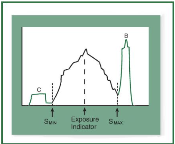
on this graph, point B represents ___
primary radiation, is jet black

in this graph, most anatomy falls _____
in between point C and B, is a range of grays