BIOL 118 Exam 2
 Call Kai
 Learn
 Practice Test
 Spaced Repetition
 Match
 Flashcards
 Knowt Play
1/66

Earn XP
Description and Tags
Nervous system
67 Terms
How is a signal transmitted through a reflex arc
5 main steps
Receptor - detects a stimulus, such as touching something hot
Sensory neuron (afferent neuron) - carries the signal from the receptor to the spinal cord (nerves carry the signal)
Association neuron/interneuron - relays the signal from the sensory neuron, passing through the spinal cord and traveling through the motor neuron
Motor neuron (efferent(exit)neuron)- carries signal to effector;muscles or glands
Effector - the muscle or gland that produces a response/movement, such as muscle contracting to pull your hand away
what is homunculus and how does it differ from the sensory homunculus?
is a map-like representation of the body on the cerebral cortex. The sensory homunculus specifically maps body sensations to areas of the somatosensory cortex
What is the posterior column pathway how does it bring sensory signals to the brain?
a sensory pathway that carries fine touch, vibration, pressure, and proprioception from the body up to the brain.
What is decussation?
the crossing of a neural pathway from one side of the CNS to the opposite side; in the posterior column pathway, the 2nd order neuron crosses in the brainstem
What is the sensory homunculus? What is the motor homunculus?
Sensory homunculus maps body parts to the somatosensory cortex for sensation
Motor homunculus maps body parts to the motor cortex for movement
Where is the primary motor cortex?
in the frontal lobe; precentral gyrus
Where is the primary sensory cortex?
in the parietal lobe; postcentral gyrus
Where is dopamine produced in the brain?
in the substantia nigra of the midbrain and helps regulate movement
Which neurological disorder is associated with destruction/damage of dopaminergic neurons?
Parkinson disease
How is information transmitted through the corticospinal pathway? What is the role of an upper motor neuron? What is the role of a lower motor neuron?
by two motor neurons, an upper motor neuron carries the signal from the primary motor cortex to the spinal cord, and a lower motor neuron carries the signal from the spinal cord to the skeletal muscle
note: these two motor neurons bring motor signals from the brain to the skeletal muscles in the corticospinal pathway
What are the major symptoms of Parkinson’s disease?
resting tremor - shaking movement in the muscle when it is relaxed and not intentionally moving
slowed movements
stiff muscles
trouble with balance and posture
How does exercise reduce symptoms and the progression of Parkinson’s disease?
it improves immune response, stimulates neurongensis (making of new neurons), and strengthens the synpatic strength
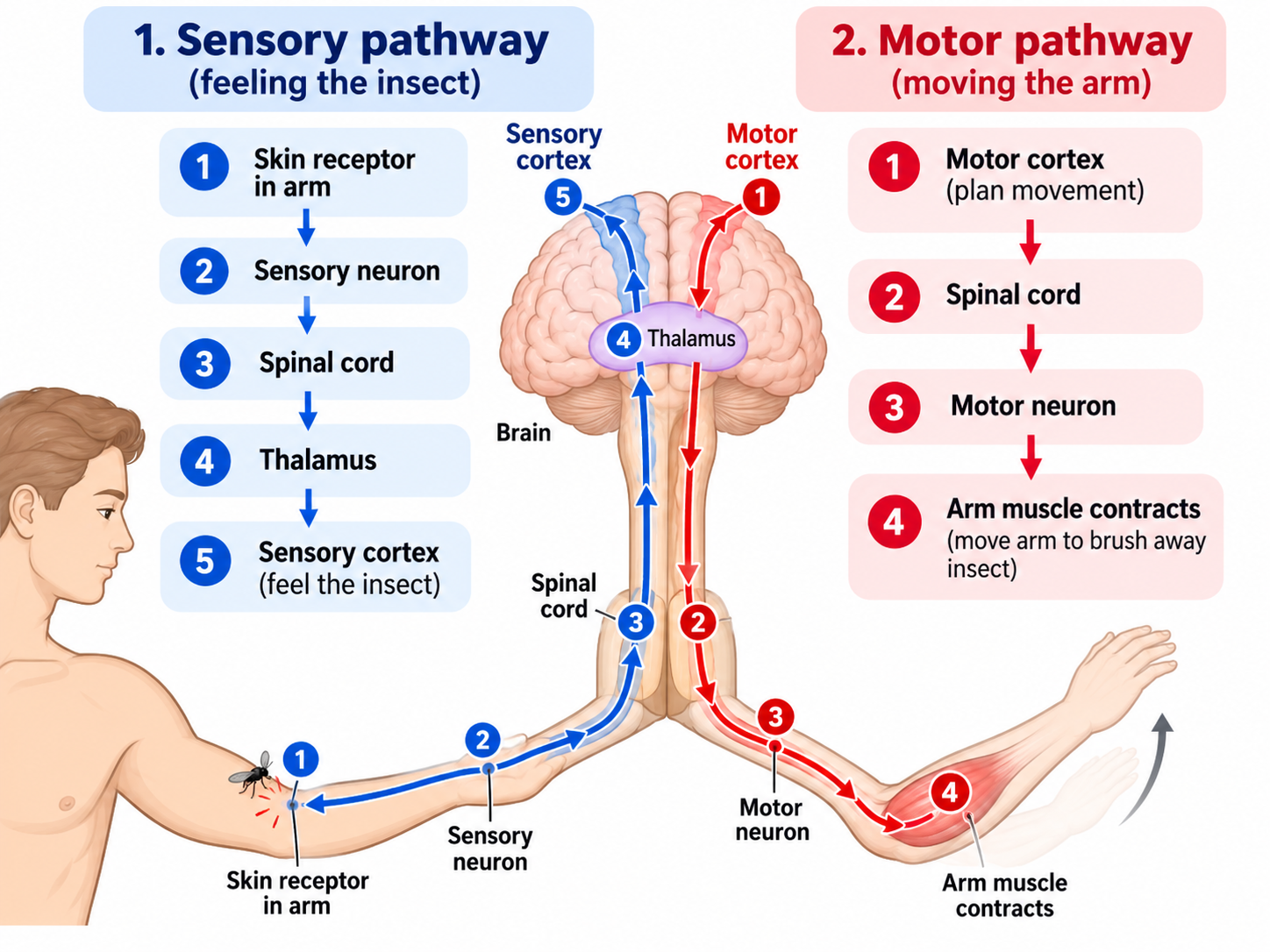
Draw out the pathway showing the brain and spinal cord areas responsible for consciously moving your arm in response to an insect landing on your arm. Show the pathway of sensory information and the pathway of motor information
What are the different structures of a neuron and what happens at each?
dendrites, cell body, axon hillock, axon, myelin sheath, nodes of Ranvier, and axon terminals
Dendrites receive signals from other cells
cell body contains the nucleus and intergrates incoming signals
axon hillock is where an action potential is triggered if threshold is reached
axon carries the action potential down from the cell body
meylin sheath insulates the axon and speeds signal conduction (bounces between diff. regions of axon)
nodes of Ranvier are gaps in myelin where the action potential is regenerated
axon terminal releases neurotransmitters to communicate with the next target cell
What is flux and how does it influence movement of substances within a cell?
flux is the movement of a substance between compartments/locations; it determines how quickly a substance moves between compartments
greater flux means faster movement
higher flux = more of the substance moves
lower flux = less of the substance moves
What is an electrochemical gradient and how does it influence movement of material?
it is the combined concentration and electrical forces that drive ions across a membrane; influences movement by determining which direction ions tend to move and how strongly they move across the membrane
What are the conditions on a resting neuron’s membrane: distribution of (Na+, K+, Cl- & proteins) inside & outside of the neuron, and charge distribution across the membrane?
For a resting neuron ion distribution:
outside: high Na+ and high Cl- (cell is positive)
inside: high K+, high negative proteins (cell negative)
charge: outside of cell is more positive, inside is more negative
membrane is polarized
*K+ is positive; the negative proteins inside the cell are what contribute strongly to the negative interior*
What is depolarization and hyperpolarization?
depolarization is making the inside of a cell more positive (moving it closer to positive)
hyperpoloarizatixn is going to make the inside of the cell more negative (moving further from pos numbers and closer to neg)
Why is a resting membrane said to be “polarized”?
The two sides of the membrane have different electrical charges: the outside is positive, and the inside is negative (-70 mV)
K+ leak channels allow positive K+ to leak out of the neuron, while large negative proteins remain trapped inside, adding to the inside being negative
What is a typical resting membrane potential?
about -70mV
What is the role of the sodium-potassium pump in the membrane of cells?
its role is to maintain the ion gradients across the cell membrane—keeping Na+ and K+ unevenly distributed (3 Na+ out of cell and 2 K+ into cell) the pump uses ATP to make these moves
this helps maintain the resting membrane potential and prepares the neuron for future action potentials
How does the sodium-potassium pump contribute to membrane potential in cells?
it pumps 3 Na+ out and 2 K+ in, moving a net positive charge out of the cell and helping keep the inside negative while maintaining Na+ and K+ gradients
What are similarities and differences between leak, voltage-gated, and ligand-gated ion channels?
All three are protein ion channels in the cell membrane that allow ions to move across it. The main difference is what they transport and the rate of them opening or closing.
leak channel: always open
voltage-gated channels: open or close when the membrane voltage changes—Na+, K+, Ca2+ - fast
ex: when the inside of a cell becomes more positive, the channel will open—allowing Na+ to rush into the cell
ligand-gated channels: open or close when a chemical messenger/ligand binds to them (like ACh binding to neuromuscular junction) - slow
What are independent and dependent variables and how they are related on our class graph?
independent variable: manipulated/changed; typically on X-axis
dependent variable: measured response: usually Y-axis
What is the importance of synaptic (graded) potentials and how do they differ from an action potential?
importance: how it determines how strongly the postsynaptic cell is affected and whether the membrane gets pushed enough towards threshold to trigger an action potential
difference:
graded potential is a variable of strength, can be large or small; local to the synapse; and can help move the membrane towards or away from threshold
action potential” once threshold is reached, it is all-or-none and travels along the neuron/muscle membrane
What is the difference between spatial summation and temporal summation?
the difference is spatial summation is going to have two signals from two different axons; temporal summation is more of a double click on the same neuron
there is also a combo summation(2 signals from 2 diff axons and a double click) which is a graded potential
Where do action potentials occur on an axon? What types of voltage-gated channels are found on axons? and how can a change in membrane charge open/close these channels?
action potentials occur at the start of the axon hillock after threshold is reached and travels down the axon terminal
Na+ and K+ channels are the types of voltage-gated channels found on the axons
charges in membrane voltage (depolarization or hyperpolarization) cause the channels to change shape—open/close
What are the events in an action potential (what happens in the depolarization, hyperpolarization, and repolarization phases)? Be able to draw the graph of an action potential and label what is happening at each region?
there are 4 step of events in an action potetntial
resting potential: we are at about -70 mV
depolarization: the membrane becomes more positive; once threshold is reached, the action potential rapidly rises (rises to peak)
repolarization: after peak, the membrane becomes negative again
hyperpolarization: membrane briefly becomes more negative than resting potential, it then returns to our -70 mV
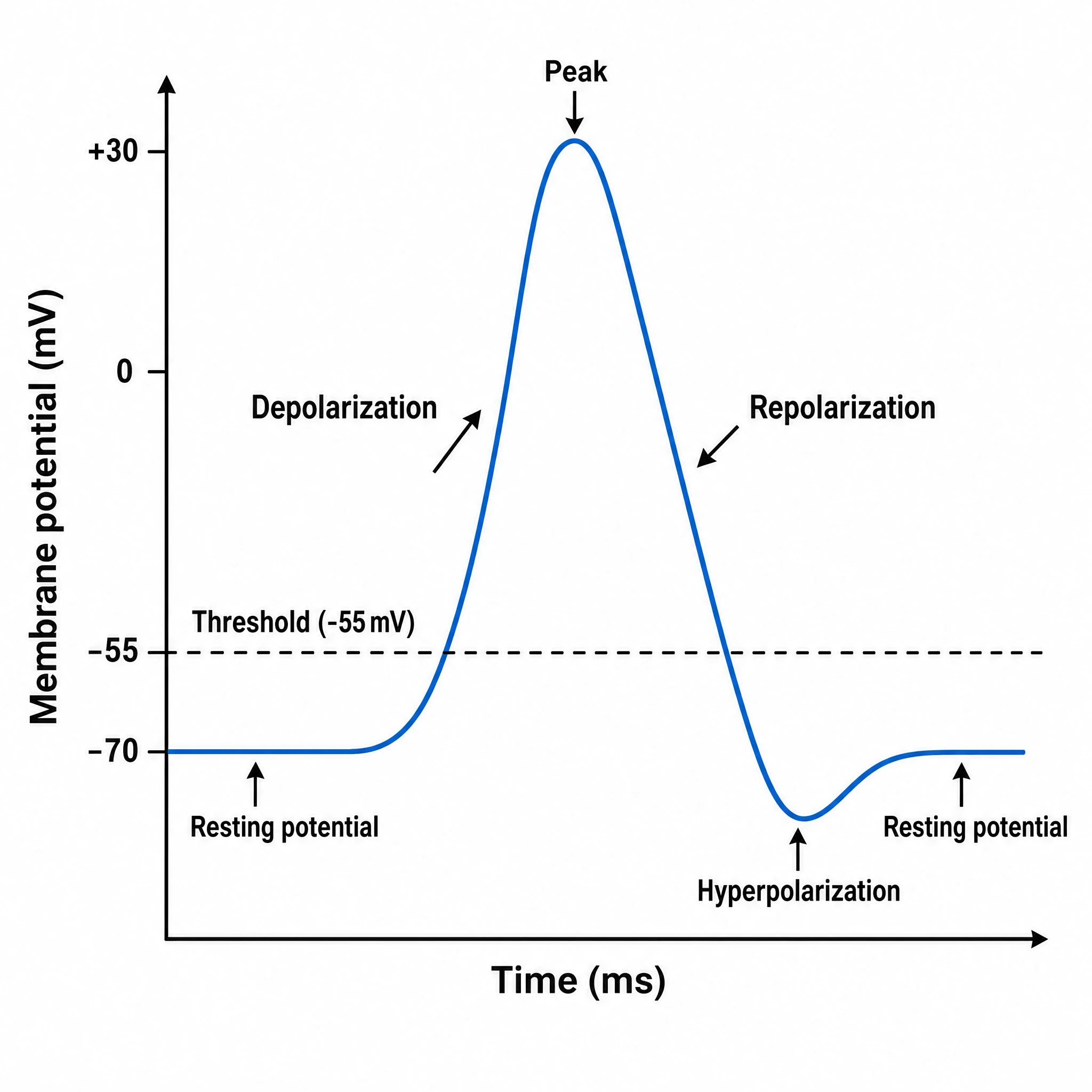
Be able to draw the graph of an action potential and label what is happening at each region.

What are the major (7) anatomical structures of the eye and what are the specific functions of each structure in the process of vision?
there are 7 structures
cornea: responsbile for bending/refracting light
transparent layer of epithelium of stratified squamous epithelium
Iris: colored layer of the eye that has muscles to control the pupil (opening where light passes through)—dilates or constricts depending on light exposure
below cornea
Retina: sensory layer that contains pigments that absorb light and has a neural layer that contain our photoreceptors (rods & cones)
all the way on the back sides of our eye
fovea: looking directly at something—area a greatest visual acuity that contains ONLY cones—high density of color
Lens: concentric layers of cells w/internal lens fibers that contain proteins called crystallines and are covered by dense fibrous capsule
located in the iris
also—crystallins are NOT RENEWABLE and can lead to cloudy vision
optic disc: where the optic nerve leaves the eye; our blind spot of the eye where there are NO PHOTORECEPTORS
optic nerve: brings information from the eye to the brain
What is a cataract?
is when the lens of the eye becomes cloudy instead of clear—which makes it harder for light to passes through and focus properly on the retina, so vision becomes blurry, dim, or even hazy
What is myopia?
light converts before the back of the eye—blurry when looking far
this is when someone is nearsighted—CAN’T see near
What is hyperopia?
the convergence of light behind the eye—blurry near vision
farsighted—CAN’T see near
What is astigmatism?
the surface of the cornea and lens are mismatched in curvature (eyeball curve shape)—causes blurred or distorted vision
vision is not focused in an our normal “even” way
What is the pathway of light through the eye? Include the roles of the cornea and the lens. Why do the lens & cornea lack blood vessels?
the pathway should include cornea → lens → retina
cornea is going to bend/refract that incoming light
lens then changes the shape to focus the light onto the retina
the cornea and lens need to stay clear/transparent so light can pass through without being blocked or scattered
neural pathway: cornea → lens → retina → optic nerve → brain
Which photoreceptor (rod or cone) works best in low light?
rods work best in low light because they are sensitive to light, so they can help you see in dim conditions(outlines figures), but they DO NOT give you color vision
How does vision (light) turn into electrical signals (action potential)?
rods and cones in the retina convert light into electrical signals (AP), the signal then travels to our optic nerve to the brain
How does light change a rod cell? What is different about the membrane potentials in a rod exposed to light and to dark?
in light, rod cells hyeperpolarize and release less glutamate
in darkness, rod cells are more depolarized and releases more glutamate
What causes colorblindness?
when one or more cone photoreceptors systems DO NOT function properly, making certain wavelengths/colors difficult to distinguish
How is visual information transmitted from the eye to the brain?
light is converted into electrical signals in the retina. those signals leave the eye through the optic nerve, travel to the thalamus, and then goes to the occipital cortex where visual information is processed
what are the three types of muscle?
skeletal muscle: voluntary(consciously) muscle, attached to skeleton
ex: bicep and hamstring
smooth muscle: involuntary (no conscious control) movement—think peristalsis, usually covering wall of internal organs
ex: stomach, intestines, and bladder
cardiac muscle: involuntary, only covers wall of the heart
ex: heart muscles: atria and ventricles
What are the anatomical hierarchy of a muscle from largest to smallest?
it is the tendon → facscia → whole muscle → facicle → myocyte (contains muscle fiber) → myofibrils → myofilaments (actin and myosin)
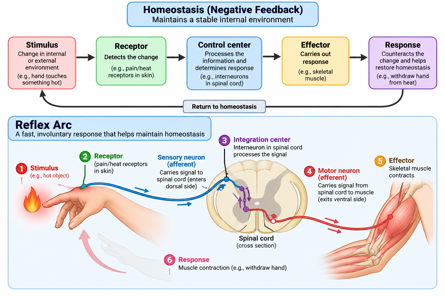
draw a homeostasis diagram with a reflex arc
how does the posterior column pathway bring sensory signals to the brain?
A three neuron chain to get sensory information to the brain
what are the 3 order neurons?
1st order neuron: carries the sensory signal from the receptor in the body into the spinal cord and then up the posterior column to the brainstem
2nd order neuron: starts in the brainstem, crosses to the opposite side of CNS, then travels up to the thalamus
3rd order neuron: starts in the thalamus and carries the signal to the primary somatosensory cortex
what are the areas of primary motor homunculus?
between frontal and parietal lobe
swallowing
tongue
jaw
neck
leg
lips
face
eye
butt
foot
brow
thumb
fingers
hip
toes
wrist
elbow arm knee
what are the homunculus areas for the primary somatosensory area?
pharynx
tongue
teeth and gums
butt
lips
nose
eye
hip
knee
thumb
fingers
forearm
leg
foot
elbow
arm
neck
toes
genitals
What are the motor areas of the cerebral cortex? How do they send motor signals to muscles?
the motor areas are:
premotor cortex (PMA) - organizes motor functions before initiation
supplementary motor cortex (SMA) - learns sequences of movements
posterior parietal cortex (PPC) - movements are targeted accurately towards objects in space
primary motor cortex (M1) - initiates movement
Motor areas send motor signals to muscles in conjunction ( no specific order) of
PPC figuring out where the body is in space and target—uses vision → spatial plan
PMA plans how you’re going to move towards target—choosing muscles and movement pattern based on visual cue
SMA plans and organizes the intention/internal driven or learned movement (habit/sequence)
M1 sends signals to activate specific muscles in a body part
What is the organization or location of the nuclei, myofibrils, sarcoplasmic reticulum, and mitochondria within a myocyte (1 muscle cell)? and draw a diagram of a muscle cell that includes all these organelles?
the nuclei sits at the outer edge under the sarcolemma
myofibrils run the length of the cell
the sarcoplasmic reticulum wraps around the myofibrils to supply ATP

How does a sarcomere shorten during a contraction? What structures of the sarcomere limit sarcomere shortening?
myosin heads pull actin filaments toward the center of the sarcomere, sliding past myosin, which shortens the sarcomere
the limit is when the actin and myosin filaments reach maximum overlap and can’t slide further
How do muscle fibers develop from myoblast stem cells?
many myoblasts fuse together → one muscle fiber forms, the nuclei from those myoblasts remain, making the muscle fiber multinucleated
What role does extracellular Ca2+ play in neurotransmitter
release, and how does nerve signaling trigger a muscular response?
neuron Ca2+ enters the presynaptic terminal and triggers the release of acetylcholine (ACh), which then binds to the ligand-gated ion channels on the muscle cell membrane, causing a muscle action potential
(different Ca2+) Ca2+ is then released from the sarcoplasmic reticulum in the muscle cell to help cause contraction
What neurotransmitter is released at the neuromuscular junction by excited motor neurons to activate skeletal muscle?
ACh is the neurotransmitter released by motor neurons at the neuromuscular junction to activate skeletal muscles
How does nerve signaling trigger a muscular response?
when acetylcholine binds to ligand-gated ion channels on the muscle cell membrane, allowing Na+ to enter and causing a muscle action potential.
that muscle action potential then leads to Ca2+ release from the SR, which helps cause muscle contraction
What is the importance of the sarcoplasmic reticulum?
it stores and releases Ca2+ into the muscle cell when it is stimulated
Ca2+ helps trigger muscle contractions
What are the major steps involved in muscle contraction with regards to actin, myosin, binding sites, troponin, tropomyosin, Ca2+, and energy? (ATP → ADP)? 1-6
entry of Na+ enters (depolarizes) the muscle fiber cell, which initiates a muscle action potential—travels down along the sarcolemma
action potential are in t-tubles which activate voltage-sensitive receptors—triggers Ca2+ release from SR into the cytosol
Ca2+ ions bind to troponin—it changes shape—remving its blocking of tropomyosin, exposing the active myosin binding sites on actin
myosin binds to actin and contraction occurs
myosin heads alternativly attach to exposed binding site on actin and detaches pulling actin filaments towards the center of sarcomere
actin slides past myosin—sarcomere shortens producting contraction
4 (1-2) is the desussation (cross-bridge)
removal of Ca2+ by active transport into the SR after the action potential ends
tropomyosin blocks actin again—contraction ends and muscle fibers relaxes
How does tetanus neurotoxin cause sustained skeletal muscle contractions, and how do sociodemographic factors influence its incidence and risk?
it blocks inhibitory signaling in the nervous system. without inhibitory signals, motor neurons continuously stimulate skeletal muscles—causing sustained involuntary contractions
Socio-demographic index factors also affect lower SDI—lower income/education and fewer health care resources
draw a sarcomere showing thick & thin filaments, & the Z-line?

what is accommodation in eyesight?
the process where lens change shape, so our eyes can focus on objects at different distances—muscles are changing shape of the lens to focus at said distance—decreases with age.
ex: looking at finger/looking past finger
what is the thalamus role in vision?
sensory relay: it receives the visual signal and routes them to the occipital cortex
what is the sequence for seeing in the dark? 1-6
dark current → cGMP is produced which binds to ligand (cGMP) gated channel—keeps the channel open in the dark
Na+ is then going to rush into the rod cell—cell becomes positive
cell resting potential goes from -70 mV to -40 mV (depolarizes)
depolarization triggers rod to release glutamate (neurotransmitter)
production of inhibitory signal (IPSP)
Bipolar cells are then inhibited—bipolar cells DO NOT activate ganglion cells
No synaptic activity here
No signal in optic nerve fiber
what is the sequence for seeing in light? 1-6
dark current ceases—no cGMP production—ligand gated channels are CLOSED
rod cell stays at -70 mV —hyperpolarizes, which STOPS glutamate production
bipolar cells is NOT inhibited, which activates the cell
bipolar cells release a neurotransmitter onto the ganglion cell
ESPS occur (excitation)
excited cell activates ganglion cells to send a signal to the brain (optic nerve fiber → thalamus → occipital cortex)
what is a myocyte?
a muscle cell which contains many myofibrials
how does a reflex arc work? and why is it so fast? how are we sensing movement? 1-5
it
receptor: detects a stimulus; this signal will be sent through an afferent neuron (sensory neuron)
sensory neuron (afferent): carries the signal toward the spinal cord, entering through the dorsal side (back side) of spinal cord
association neuron/interneuron: is sometimes skipped, but it relays the signal within the spinal cord
motor (efferent) neuron: carries the signal out of the spinal cord, usually through the ventral side (front side), towards the effector
effector: a muscle or gland that produces the response, such as a muscle contracting
it is so fast because the spinal cord can process the sensory information and send out a motor response before the brain consciuously processes the stimulus
afferent = arrives at CNS
efferent = exits the CNS
what is the dorsal root ganglion, and what does it do?
it holds sensory neuron cell bodies outside the spinal cord; dorsal root carries afferent signals into the spinal cord
cross-bridge cycle (decussation) - what happens during the cross bridge cycle?
ca2+ binds to tropoin
Tropomyosin moves, exposing binding sites on actin
myosin head attaches to actin → cross-bridge forms
myosin pulls actin towrds the center of sarcomere → power stroke
ATP binds to myosin → myosin detaches from actin
ATP is broken down to ADP + Pi → myosin head rests/energizes
myosin attaches again and the cycle repeats as long as Ca2+ is present
how does a muscle relax?
action potential ends
Ca2+ is actively transported back into the sarcoplasmic reticulum (SR)
this active transport requires energy
Ca2+ moves away from tropoin
tropoin returns to its