Histology exam 2 assignments
 Call Kai
 Learn
 Practice Test
 Spaced Repetition
 Match
 Flashcards
 Knowt Play
1/308

Earn XP
Description and Tags
unit 2 assignments
309 Terms

ID M phase
E

ID G0 phase
B
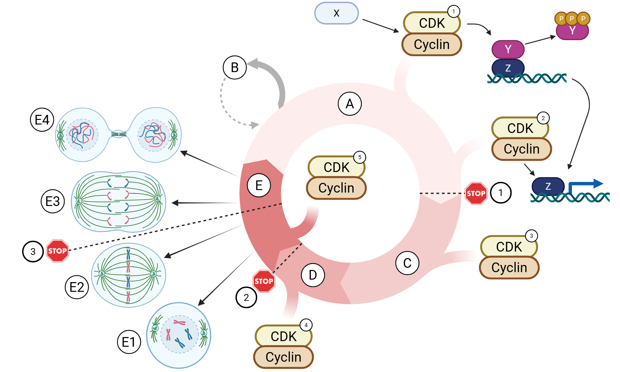
ID G2 phase
D

ID S phase
C

ID G1 phase
A

The cell exits the active cell cycle and enters a quiescent state in which it does not actively divide, although it may re-enter the cycle.
B

The cell grows, synthesizes proteins and organelles, and carries out normal metabolic activities in preparation for DNA replication.
A

The cell undergoes mitosis and cytokinesis, resulting in the formation of two daughter cells.
E

The cell replicates its DNA, producing duplicated chromosomes composed of sister chromatids.
C

The cell continues to grow, synthesizes proteins needed for mitosis, and prepares for entry into the mitotic phase.
D
A researcher collects four cells from a rapidly proliferating tissue and observes the following characteristics:
Cell | Observed Characteristics |
Cell A | DNA replication is actively occurring. |
Cell B | DNA replication is complete, and the cell is preparing for mitosis. |
Cell C | Chromosomes are condensing, and the mitotic spindle is forming. |
Cell D | The cell is growing and synthesizing organelles before DNA replication. |
Which sequence correctly orders these cells according to their progression through the cell cycle?
D → A → B → C
This cyclin-cdk complex maintains a primary role in aiding the G1-to-S phase transition by further promoting the activation of E2F-mediated transcription of machinery involved in DNA replication (e.g., DNA polymerase). In figures provided during lecture, this cyclin-cdk complex is always depicted during the G1-to-S phase transition. This cyclin-cdk complex can also be involved in centrosome duplication
cyclin E-cdk2
This cyclin-cdk complex can be involved in regulation of both the G2 phase and M phase of the cell cycle, although its primary role during early mitotic events involves the phosphorylation of nuclear lamin proteins to allow for nuclear envelope breakdown during prophase of mitosis. In figures provided during lecture, this is depicted as the predominant G2 phase cyclin-cdk complex
cyclin A-cdk1
This cyclin-cdk complex maintains a primary role in phosphorylating Rb, releasing E2F from inhibition to allow transcription of genes needed for DNA replication, thereby contributing to G1-to-S phase progression. In figures provided during lecture, this is depicted as the predominant G1 phase cyclin-cdk complex and is activated by mitogenic factors
cyclin D-cdk4/6
This cyclin-cdk complex maintains a primary role in S phase progression and DNA synthesis through the phosphorylation of proteins involved in DNA replication. In figures provided during lecture, this is depicted as the predominant S phase cyclin-cdk complex. This cyclin-cdk complex can also be involved in centrosome duplication
cyclin A-cdk2
This cyclin-cdk complex forms the mitosis-promoting factor (MPF), which promotes entry into mitosis and is actively involved in mitosis and its completion. In figures provided during lecture, this is depicted as the predominant M phase cyclin-cdk complex
cyclin B-cdk1
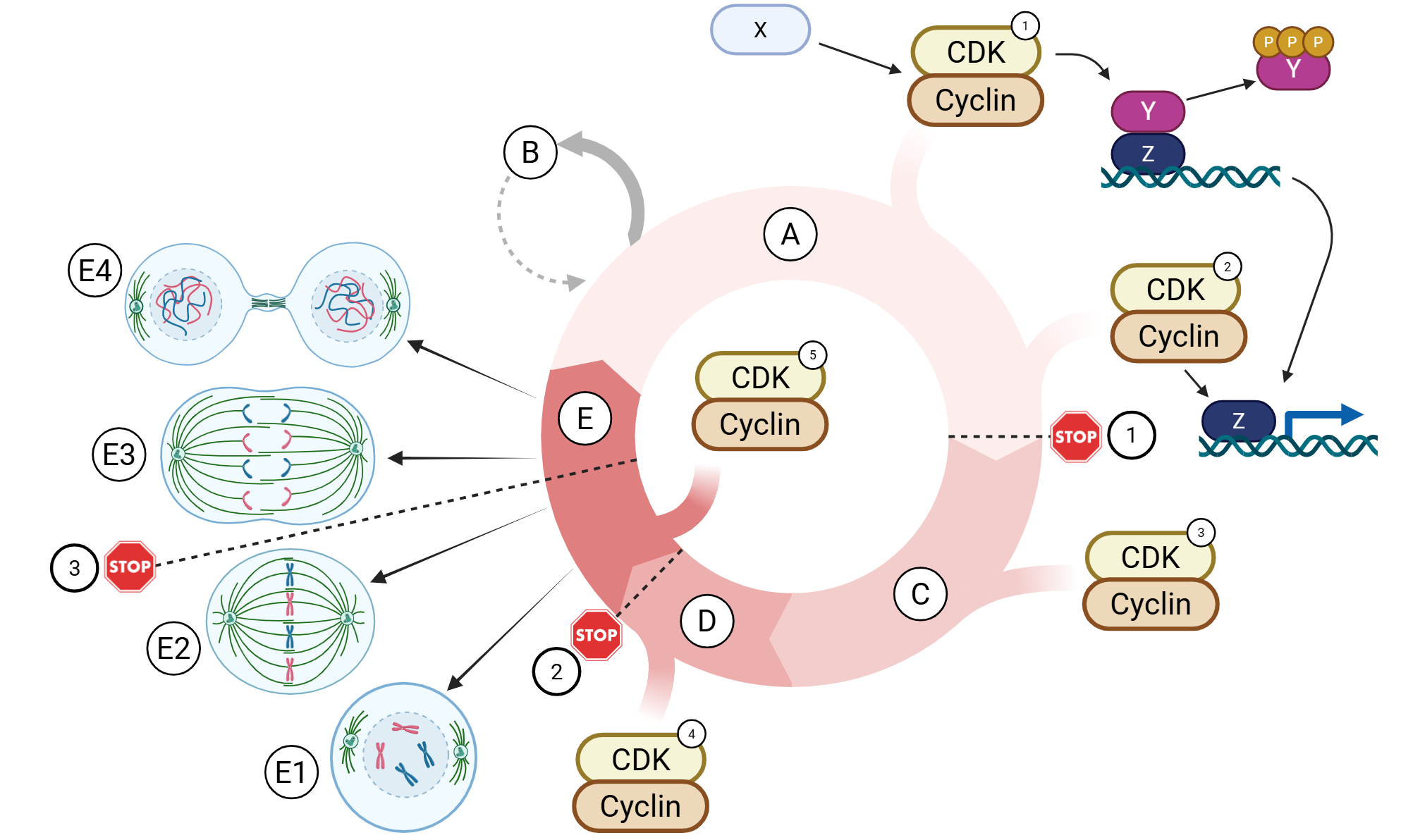
cyclin D-cdk4/6
1

cyclin A-cdk1
4

cyclin B-cdk1
5
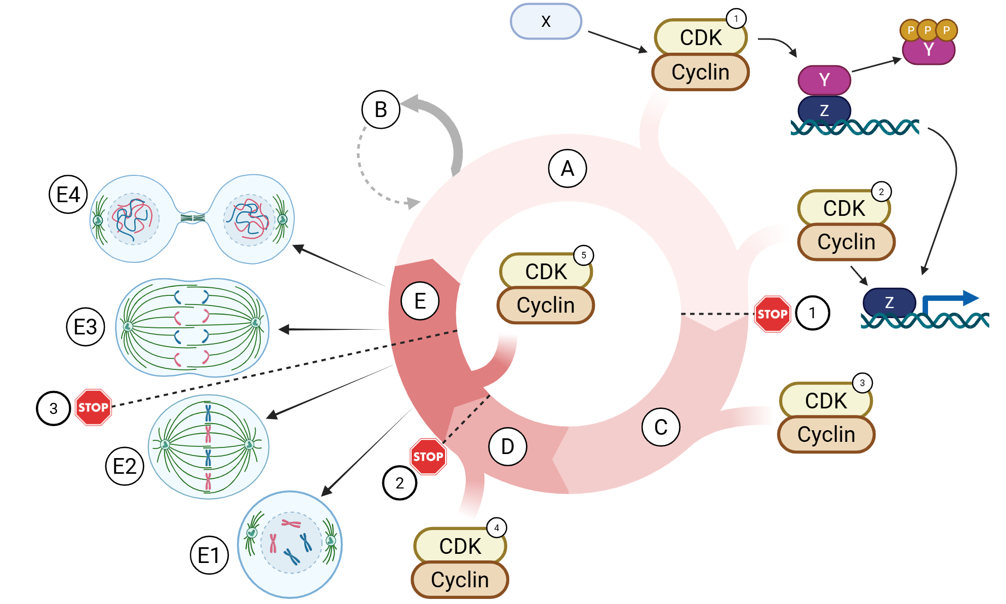
cyclin E-cdk2
2
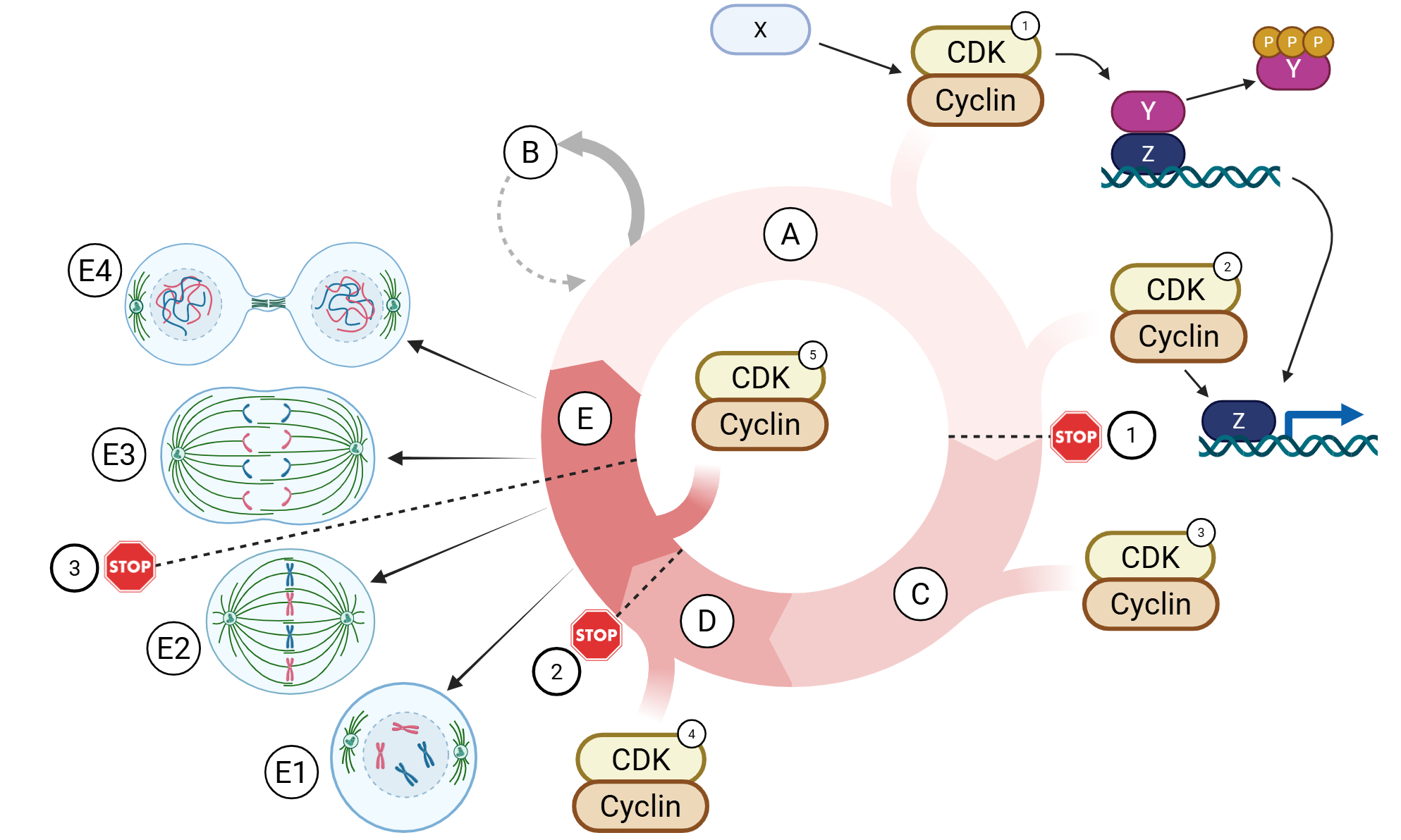
cyclin A-cdk2
3
Two experimental compounds are introduced into separate populations of proliferating cells.
Compound X prevents cyclins from binding to CDKs.
Compound Y increases the production of CDK inhibitors.
Which statement best predicts the effects of these compounds?
Compound X would prevent CDK activation, whereas Compound Y would suppress CDK activity; both could impair cell-cycle progression.

Spindle Checkpoint
STOP 3

G2 Checkpoint
STOP 2

G1 Checkpoint
STOP 1
Located at the G2-to-M transition, this checkpoint regulates whether the cell is ready to enter mitosis.
G2 Checkpoint
Located at the metaphase-to-anaphase transition, this checkpoint regulates whether chromosomes can begin separating.
Spindle Checkpoint
This checkpoint prevents premature anaphase until chromosomes are properly attached to the mitotic spindle.
Spindle Checkpoint
This checkpoint can delay mitotic entry when the cell has not completed the necessary preparations, including successful DNA replication.
G2 Checkpoint
This checkpoint determines whether the cell should commit to division and proceed into S phase, where DNA replication occurs.
G1 Checkpoint
Located at the G1-to-S transition, this checkpoint is referred to as the restriction point or point of no return. It is the primary decision point for whether the cell proceeds with division.
G1 Checkpoint
Arrange events in the correct order to describe how mitogenic stimulation promotes entry into S phase.
A cell enters the G1 phase...
Mitogenic signals stimulate the cell
Cyclin D–CDK4 activity increases.
Rb is phosphorylated and releases E2F.
E2F activates transcription of genes required for DNA replication.
The cell progresses through the G1 restriction point toward S phase.
The cell enters the S phase.
A researcher examines three populations of cells with different regulatory conditions.
Population | Cellular Condition |
A | Rb is dephosphorylated and bound to E2F. |
B | Rb is phosphorylated, and E2F is no longer bound to Rb. |
C | Rb is dephosphorylated, but a mutation prevents it from binding to E2F. |
Which populations would be expected to exhibit increased E2F-dependent transcription of genes required for DNA replication?
Population B and C
Involves DNA replication and chromosome condensation in preparation for chromosome segregation.
Nuclear Cycle
Involves duplication of centrioles and organization of the structures needed for mitotic spindle formation.
Centrosome Cycle
Involves sequential activation of cyclin–CDK complexes that regulate progression through the cell cycle.
Cytoplasmic Cycle
Which statements correctly describe how cyclin–CDK activity coordinates the nuclear and centrosome cycles?
CDK1 contributes to chromosome condensation through phosphorylation-dependent mechanisms.
CDK1 contributes to nuclear envelope breakdown through phosphorylation of nuclear lamins.
CDK2 contributes to initiation of DNA replication through phosphorylation of proteins associated with replication origins.
CDK2 contributes to centriole duplication through phosphorylation of centrosome-associated substrates.

Prophase
E1
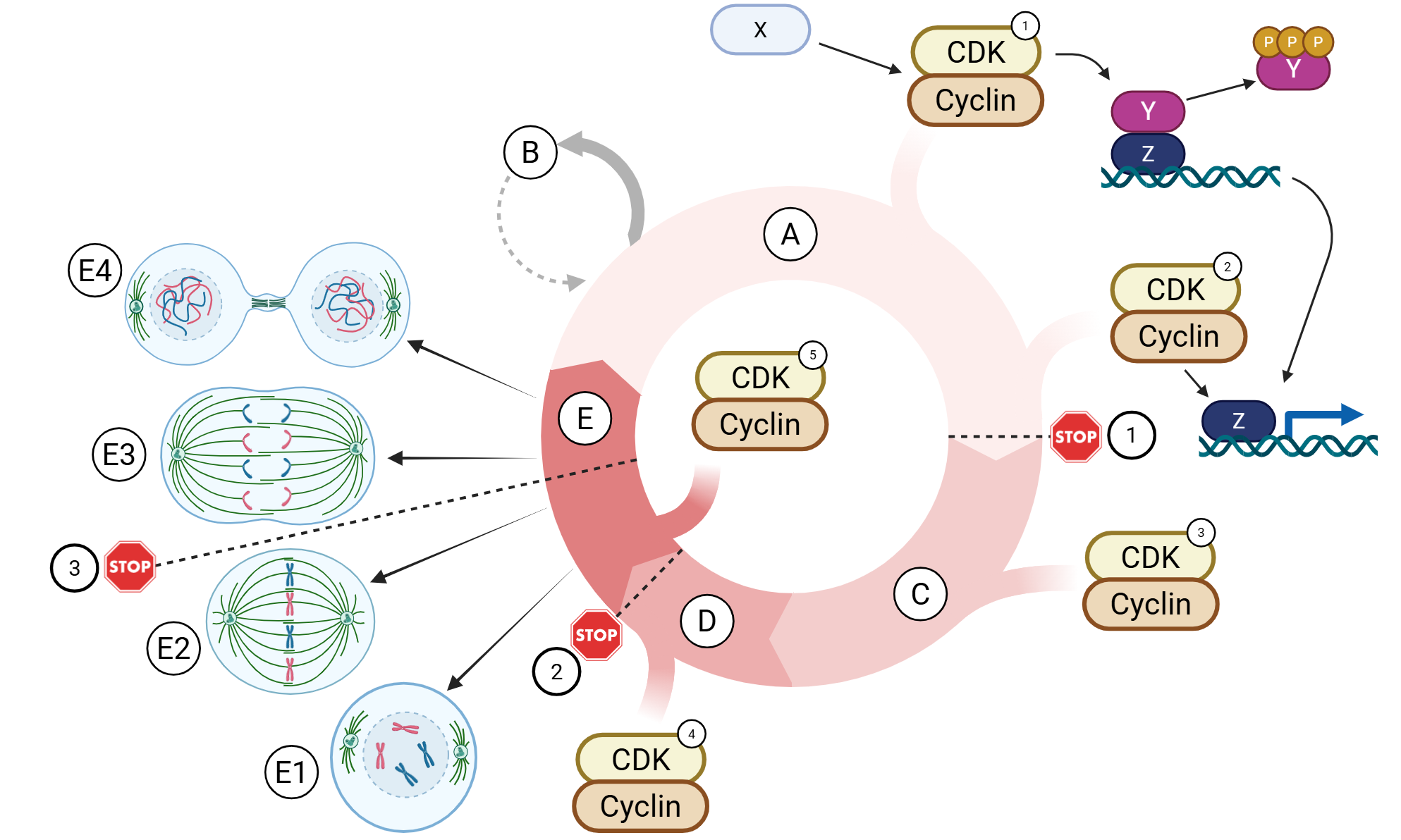
Anaphase
E3

Telophase and Cytokinesis
E4

Metaphase
E2
Separated sister chromatids, now referred to as daughter chromosomes, complete their movement toward opposite poles of the cell.
Anaphase
Nucleoli reappear within the daughter nuclei.
Telophase and Cytokinesis
Chromatin becomes increasingly condensed, resulting in the appearance of individual, discernible chromosomes.
Prophase
Nucleoli disappear as the cell prepares for chromosome segregation.
Prophase
Actin and myosin filaments contract to divide the cytoplasm into two portions surrounding the newly formed nuclei.
Telophase and Cytokinesis
Nuclear envelopes re-form around the separated chromosomes, assisted by dephosphorylation of nuclear lamins.
Telophase and Cytokinesis
Chromosomes aligned at the equatorial plane have established bipolar attachments to spindle microtubules through their kinetochores.
Metaphase
Chromosomes become aligned along the equatorial plane of the cell.
Metaphase
Sister chromatids separate and are drawn toward opposite centrosomes by kinetochore microtubules.
Anaphase
Chromosomes begin to decondense within the newly forming nuclei.
Telophase and Cytokinesis
Kinesin motor proteins facilitate movement of polar microtubules relative to one another, pushing the spindle poles farther apart.
Anaphase
Phosphorylation of nuclear lamins by CDK1–cyclin A contributes to breakdown of the nuclear envelope.
Prophase
A cleavage furrow forms near the equatorial plane as cytokinesis occurs concurrently with this stage.
Telophase and Cytokinesis
Centrosomes move toward opposite poles of the cell and begin organizing microtubules into the mitotic spindle.
Prophase
A checkpoint verifies proper bipolar attachment of chromosomes to the mitotic spindle before chromosome separation begins.
Metaphase
In chromosomes aligned along the equatorial plane, two closely associated sister chromatids can be distinguished, joined at their centromere.
Metaphase
A researcher identifies a mutation that prevents CDK1-mediated phosphorylation of histone H3 during early mitosis. Nuclear lamin phosphorylation and mitotic spindle formation occur normally. Which outcome would MOST likely result from this mutation?
Chromosomes would fail to condense properly, potentially interfering with their organization and segregation during mitosis.
Appears as basophilic ergastoplasm in pancreatic cells
Rough endoplasmic reticulum
Participates in detoxification of harmful lipid-soluble substances
smooth endoplasmic reticulum
Is often found near cytoplasmic deposits of glycogen and lipids
smooth endoplasmic reticulum
Participates in the synthesis of proteins destined for secretion from the cell
smooth endoplasmic reticulum
Appears as basophilic cytoplasmic material called Nissl substance in neurons
rough endoplasmic reticulum
Participates in the synthesis of lipids used in cellular membranes
smooth endoplasmic reticulum
Contains numerous ribosomes attached to the cytosolic surface of its membranes
rough endoplasmic reticulum
Functions in calcium sequestration within skeletal and cardiac muscle cells
smooth endoplasmic reticulum
Is abundant in steroid-producing cells of the gonads and adrenal cortex
smooth endoplasmic reticulum
Is continuous with the outer membrane of the nuclear envelope
rough endoplasmic reticulum
A mutation disrupts the function of the sarcoplasmic reticulum in skeletal muscle cells, reducing its ability to sequester calcium ions. Which statement best explains the relationship between this defect and the smooth ER?
The sarcoplasmic reticulum is a specialized form of smooth ER that participates in intracellular calcium regulation.
Proteins imported into mitochondria
free ribosomes
Proteins incorporated into the Golgi apparatus
membrane-bound ribosomes
Proteins secreted into the extracellular space
membrane-bound ribosomes
Proteins transported into peroxisomes
free ribosomes
Proteins destined for lysosomes
membrane-bound ribosomes
Proteins that remain in the cytosol
free ribosomes
Proteins retained within the rough ER
membrane-bound ribosomes
Proteins incorporated into the plasma membrane
membrane-bound ribosomes
Proteins transported into the nucleus
free ribosomes
Arrange events in the correct order to describe how a newly synthesized secretory protein is directed to the rough ER.
Translation begins on a ribosome in the cytosol, and a hydrophobic signal peptide emerges from the ribosome.
The SRP recognizes and binds the signal peptide, temporarily pausing translation.
The SRP-associated complex binds to an SRP receptor on the cytosolic surface of the rough ER.
Translation resumes, and the growing polypeptide is threaded into the rough ER.
The signal peptide is cleaved from the growing protein by signal peptidase.
Two experimental compounds disrupt different functions of the Golgi apparatus.
Compound X: Inhibits enzymes responsible for modifying carbohydrate groups attached to glycoproteins.
Compound Y: Prevents the TGN from packaging modified cargo into transport vesicles.
Which statement best predicts the consequences of these compounds?
Compound X would impair glycoprotein modification, whereas Compound Y would impair sorting and delivery of processed cargo.
Cargo moves from the Golgi apparatus toward the plasma membrane or lysosomal pathway
anterograde transport
A pancreatic acinar cell stores digestive enzymes in secretory granules until stimulated to release them
regulated secretion
Vesicles return selected proteins and membrane components to the endoplasmic reticulum for reuse
retrograde transport
Modified proteins are packaged into vesicles at the trans-Golgi network and transported toward their downstream destinations
anterograde transport
Secretory products are stored in vesicles until an extracellular signal stimulates their release
regulated secretion
Newly synthesized proteins and lipids are continuously delivered to the plasma membrane without requiring a specific stimulus
constitutive secretion
Cargo is transported from the Golgi apparatus back toward the endoplasmic reticulum
retrograde transport
A cell continuously secretes extracellular matrix proteins into the surrounding tissue
constitutive secretion
A researcher identifies a mutation that prevents vesicle-associated SNARE proteins from interacting with complementary SNARE proteins on target membranes. Vesicle formation, cargo packaging, and transport remain functional. Which outcome would most likely result from this mutation?
Vesicles would successfully reach their target membranes but exhibit impaired fusion because complementary membrane recognition proteins cannot interact.
Forms the protein coat surrounding transport vesicles carrying lysosomal enzymes
Clathrin
Serves as the Golgi region where lysosomal enzymes are sorted and packaged for transport
trans Golgi network
Is recycled after releasing its associated lysosomal enzyme cargo
mannose-6-phosphate receptor
Recognizes and binds appropriately tagged lysosomal enzymes
mannose-6-phosphate receptor
A carbohydrate modification that identifies enzymes destined for lysosomes
mannose-6-phosphate
Must be present on lysosomal enzymes for recognition by their specific sorting receptors
mannose-6-phosphate
A researcher examines the uptake of low-density lipoprotein (LDL) by hepatocytes through receptor-mediated endocytosis. Which of the following statements accurately describe this process? Select all that apply.
Adaptins contribute to cargo selection by interacting with cargo receptors.
Dynamin facilitates separation of clathrin-coated vesicles from the plasma membrane.
Internalized LDL receptors can be recycled to the plasma membrane for subsequent use.
LDL binds to specific receptors located on the plasma membrane.
A lysosomal hydrolase is packaged into a vesicle at the trans-Golgi network
Clathrin
A Golgi-associated protein is transported back to the endoplasmic reticulum
COPI
A resident Golgi enzyme is transported between Golgi cisternae
COPI
An LDL–receptor complex is internalized from the plasma membrane
Clathrin