Patho Exam 3
 Call Kai
 Learn
 Practice Test
 Spaced Repetition
 Match
 Flashcards
 Knowt Play
1/76

There's no tags or description
Looks like no tags are added yet.
77 Terms
Heart Diagram
Aorta
Brachiocephalic artery, Right subclavian artery, right common carotid artery
Left common carotid artery
Left subclavian artery
Tricuspid → Mitral Valve

Wiggers Diagram
1 - All 4 relaxed
2 - Atria contract; ventricles fill
3 - Ventricular Systole begins
3a - Isovolumetric contraction, up the hill
3b- Ventricular ejection
4- Blood against semilunar valves closure, blood flows into relaxed atria; dicrotic notch
5 - Late ventricular diastole - Isovolumetric relaxation

AV Closure (Tricuspid and Mitral Valves)
S1, Lub
Semilunar (Pulmonary and Aortic) closer
S2,/Dub, Ventricular diastole begins 2/3
Preload
Amount of blood in ventricle is full
LV = 5-12
Optimal action (most effective)
Excessive stretch ↓ venous return
Less optimal stretch → ↓ preload, SV, and CO
Afterload SVR
Resistant blood flow out of the ventricle
Systemic Vascular Resistance 800- 1,400
Inotropy/Contractility
Ventricular
Negative - hypoxic cells, lower ATP
Positive - increased calcium
SV
Stroke Volume: blood amount ventricular ejection
EF
Ejection Fraction (55-75%)
CO
Blood pumped/minute
HR x SV = 4- 6 L/min at rest, 8 if active/healthy
Cardiac Index (CI)
Adjusted, body surface area: 2.5–4.0
HR
Frequency of left ventricular contractions = 60–100
Tachycardia shortens diastole, reducing ventricular filling
Increased HR =
Lower Ventricle fill, Lower SV, Lower CO
BP = CO x SVR
CO 4-6, up to 8
SVR 800-1,400
Circulatory System
Pulmonary: R heart → Pulmonary artery → Lungs → Pulmonary veins (large, thin, elastic) → L heart | low pressure 25/10 mean is 15
Systemic - L heart → Aorta → Organs/tissues (nutrients/wastes in blood/cells→ Vena Cava → R heart
Blood vessels layers
Tunica intima: Endothelial cells
Tunica media: Smooth muscle, elastic tissue, diameter
Tunica externa (adventitia): Connective tissue, collagen support & strength
Arteries/Capillary Network
Away from the heart
Arterioles: Small branches of arteries; smooth muscle regulates blood flow
Metarterioles: Connect arterioles to capillaries; regulate blood flow
Capillaries: Thin endothelial layer (tunica intima); exchange of fluids, nutrients, gases, and wastes between the blood and interstitial space (ISS).

Veins
Return blood to the heart; thin, less muscle/elasticity
Venous valves: Prevent backflow in from the limbs (none in head & neck)
Venules: Small vessels, capillary beds, carry it into larger veins, very thin
Blood Pressure
Systolic <120
Diastolic <80
Pulse Pressure
Systolic - diastolic = 40
Mean arterial Pressure (MAP)
Tissue blood flow
(S+ (D2) / 3 = 70-110
Dyslipidemia
Primary (Familial) Hypercholesterolemia: >LDL cholesterol → tissues, tendons, cornea.
Heterozygous: One gene; LDL = 190–400 mg/dL.
Homozygous: Two genes; LDL = 400–1,000+ mg/dL.
Optimal: <100 mg/dL
Very high: >190 mg/dL
Secondary Hyperlipidemia - Lifestyle, diet, obesity, T2DM, families with similar lifestyles
Blood Lipids
Cholesterol - Waxy, fat-like substance; cell membranes + phospholipid
75% - body, 25%- food
Desirable: <200 mg/dL,
High cholesterol: >240 mg/dL
Triglycerides - Stored body fat, energy reserve
Normal: <150 mg/dL
High: 200–499 mg/dL
Very high: >500 mg/dL
Chylomicrons - Large lipoprotein carrier for these two

Lipoprotein’s (carry fat through the blood)
LDL Cholesterol → body tissues.
Optimal: <100 mg/dL
Very high: >190 mg/dL
HDL Cholesterol → liver/removal.
Low: <40 men, <50 women, Protective: >60
VLDL - Triglycerides from the liver → body tissues
Acute Arterial Occlusion & Peripheral artery disease
Acute Arterial Occlusion - An embolus breaks off, travels through the bloodstream, and blocks another vessel. Sudden, severe pain, pallor, pulselessness, paresthesia, and paralysis. Emergency
Peripheral Artery Disease (PAD) - Reduced arterial blood flow to the extremities; intermittent claudication: Calf pain, cool hairless skin, decreased pulse, poor wound healing, rubor

Raynaud disease
Vasospasm, arteries, cold, females, pale/white, number fingers, avoid triggers
Atheroscleoris
Chronic Inflammatory disease; buildup of lipid-rich plaques in arterial walls/intima; Stable or unstable, elevated LDL and VLDL cholesterol
Damage to the endothelium occurs (initial step); vascular inflammation; lipids and inflammatory cells diffuse into the endothelium, releasing growth factors; smooth muscle proliferation; artery becomes changed or distorted; released cytokines cause the accumulation of monocytes and other WBCs
Elevated levels of serum cholesterol LDL-C overwhelm healthy endothelium, abnormal endothelial metabolism
Causes CAD
Prevent/treat: heart-healthy diet, exercise, and statins
Fatty streaks: Flat, yellow lipid deposits (child)
Fibrous plaque: Raised, grayish-white plaque that thickens/narrow the arterial wall
Thrombosis: A blood clot forms when a plaque ruptures or erodes, potentially blocking blood flow to the heart or brain → MI or ischemic stroke.
Stable plaque: Thicker fibrous/calcified cap and smaller lipid core; less likely to rupture
Vulnerable/unstable plaque: Thin cap, larger lipid core, inflammation. Rupture, thrombus formation, UA or MI.

High-Sensitivity C-Reactive Protein (hs-CRP)
Chronic systemic inflammation contributes to CAD + MI
<1 low risk >3 high risk >10 test again infection
Homocysteine
Amino acids damage the endothelial lining
Eat B6, B12, and B2 (riboflavin)
Ankle Branchial Index (ABI)
Highest (Systolic) ankle BP
Highest arm BP
= 1.0-1.4
Severe PAD <0.5
HTN
Hypertensive Crisis/Emergency: Severe increase, acute organ damage, or stroke
White Coat Hypertension: BP is elevated in a clinical setting but normal at home
Primary Hypertension: High BP, no cause
Secondary Hypertension: another condition → medications, kidney disease, or RAAS dysfunction. Increases systemic vascular resistance (SVR) and afterload.
May be asymptomatic until organ damage occurs. Patients may have difficulty adhering to medications because the effects are not immediately noticeable.
Normal <120/<80
Elevated: 120-129/<80
Stage 1: 130-139/80-89
Stage 2: >140/>90
Goal
18-59 <140/90
80+ <150/90
Aneurysms
Aortic Aneurysm (Dilation/Pouch): Weak & bulging of the aortic wall (Atherosclerosis risk)
Abdominal Aortic Aneurysm: asymptomatic; abdominal pain. Rupture risk: >4 cm
Ascending Aortic Aneurysm: May cause chest/back pain, hoarseness, swollen face
Aortic Dissection: A tear in the inner aortic wall (enlargement or rupture) - False Aneurysms
Pathway of Conduction
1) SA node
2) Atrial depolarization (P wave)
3) Atrioventricular (AV) node
4) Bundle of His
5) Right and left bundle branches
6) Purkinje fibers
Ventricular depolarization (QRS complex) >0.12 seconds
PR interval: From the beginning of the P wave to the beginning of the QRS complex. Normal: 0.12–0.20 seconds (3–5 small boxes).

Dysrhythmias
Atrial flutter: Sawtooth-shaped flutter (F) waves.
Atrial fibrillation: No distinct, organized P waves
Ventricular tachycardia (VT): Up-and-down QRS complexes
Asystole: No activity; confirm with another lead & check the monitor connections

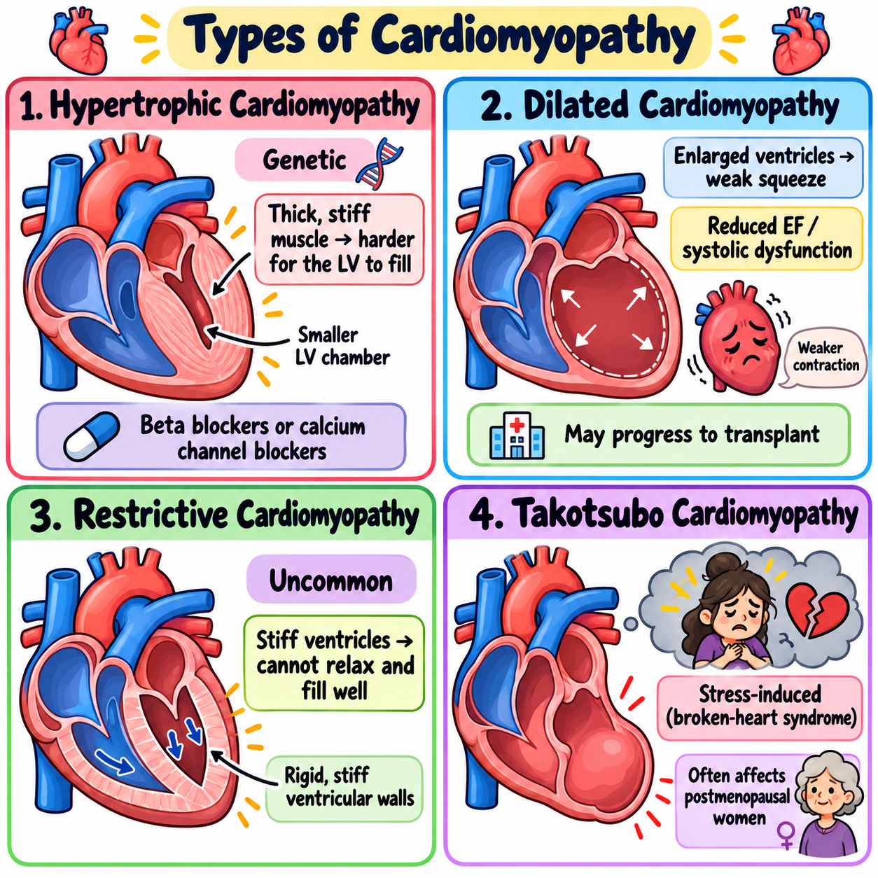
Cardiomyopathies (Heart muscle disease)
Hypertrophic cardiomyopathy: Genetic; thickens and stiffens, left ventricular filling. treat: beta/calcium channel blockers
Dilated cardiomyopathy: Common + transplant. Ventricles enlarge = reduced EF, systolic dysfunction
Restrictive cardiomyopathy: Uncommon, ventricles become stiff, can’t relax or fill
Takotsubo (stress-induced) cardiomyopathy: broken-heart syndrome, women post-menopause. Stress; only acute complications
Infective Endocarditis (IE)
Heart valve regurgitation (Staphylococcus aureus)
Risk factors: Prosthetic heart valves, previous endocarditis, congenital heart disease, implanted cardiac devices, and injection drugs
B — Blood cultures
E — Echocardiographic
T — Temperature >38/100.4
O — Osler nodes tender
M — Murmur/micro evidence
E — Embolic phenomena + emergency
Rheumatic Heart Disease
Disease post-group A streptococcal throat infection
Heart effects: Inflammation can cause mitral valve regurgitation/stenosis
Features: Fever, polyarthritis, and carditis. Aschoff bodies
Treatment note: Aspirin relieve symptoms, but not the underlining
Valve Disorders
Aortic stenosis: Left ventricular outflow; murmur radiating to carotids.
Aortic regurgitation: Blood flows back into LV during diastole; high-pitched, diastolic decrescendo murmur.
Mitral stenosis: Narrow mitral opening impairs LV filling during diastole; atrial fibrillation may occur; diastolic rumble & snap.
Mitral regurgitation: Blood leaks back into the left atrium during systole; holosystolic murmur at the apex, radiating to the axilla.

Heart Failure (HF)
The heart can’t meet the body’s needs, leading to dyspnea, fatigue, and fluid retention.
HFrEF (reduced), systolic HF: LVEF <40%; end-systolic volume rises, the ventricle dilates, and SV falls.
Left-sided backup: pulmonary congestion
Right-sided backup: peripheral edema (diuretic therapy)
HFpEF (preserved), diastolic HF: LVEF >50%; elevated filling pressures; the ventricle is stiff
HFmrEF (Mildly reduced) LVEF 41–49%;
Pericardium
Two-layer sac around the heart; protects, stable, barrier to infection, reduces friction, lubricates
15–50 mL of fluid
Pericarditis
Inflammation of the thin pericardial sac; fibrin-rich inflammation, viral or idiopathic: pericardium may thicken (men) Acute <4-6 weeks, Chronic >3 months
Radiating chest pain (tripod position), ECG, friction rub
WideST elevation, PR depression
Inflammation increases Erythrocyte sedimentation rate (ESR)

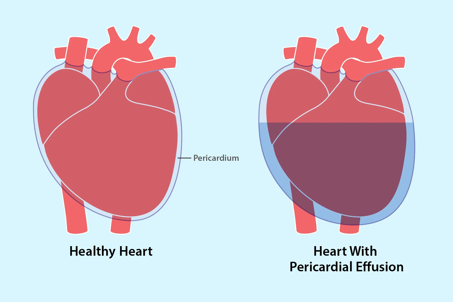
Pericardial Effusion
Fluid in the pericardial space → inflammation, infection, or cancer
Slow accumulation = no symptoms
Rapid accumulation = tamponade (Squeeze, water-bottle sign, emergency)
Echocardiography identifies
Chronic effusion: water-bottle-shaped
Cardiac tamponade
Emergency
Fluid in the pericardial sac limits diastolic filling → collapse or arrest.
Tachycardia, swinging heart, water bottle sign, large effusion
Urgent drainage: image-guided pericardiocentesis or a subxiphoid approach.
Signs: Beck’s triad: JVD, Narrowed pulse pressure, Muffled heart sounds

Constrictive Pericarditis
Fibrous fluid, calcified scar tissue, and a thickened pericardium restrict ventricular expansion & diastolic filling, reducing SV and CO
Findings include fatigue, peripheral/pedal edema, ascites, and venous congestion as blood backs up into the systemic veins

Angina
Stable: Predictable pressure, squeezing, or tightness with exertion or stress; relieved by rest or nitroglycerin
Unstable (UA): New or worsening chest pain; ischemia without a rise in cardiac troponin, indicating no acute myocardial injury. ECG normal or ST depression; emergency.
Vasospastic (Prinzmetal) Angina
Transient coronary spasms, @rest or at night; ST elevation; dysrhythmias; not MI
Acute coronary syndrome
Reduction in myocardial blood flow
Stable, unstable, variant angina, NSTEMI, and STEMI
Chest discomfort + SOB

Coronary Artery Disease
Reduced blood flow through the coronary arteries (atherosclerosis, angina, MI, etc.)
Left coronary circulation flow is about 225 mL/min.

Myocardial Infarction
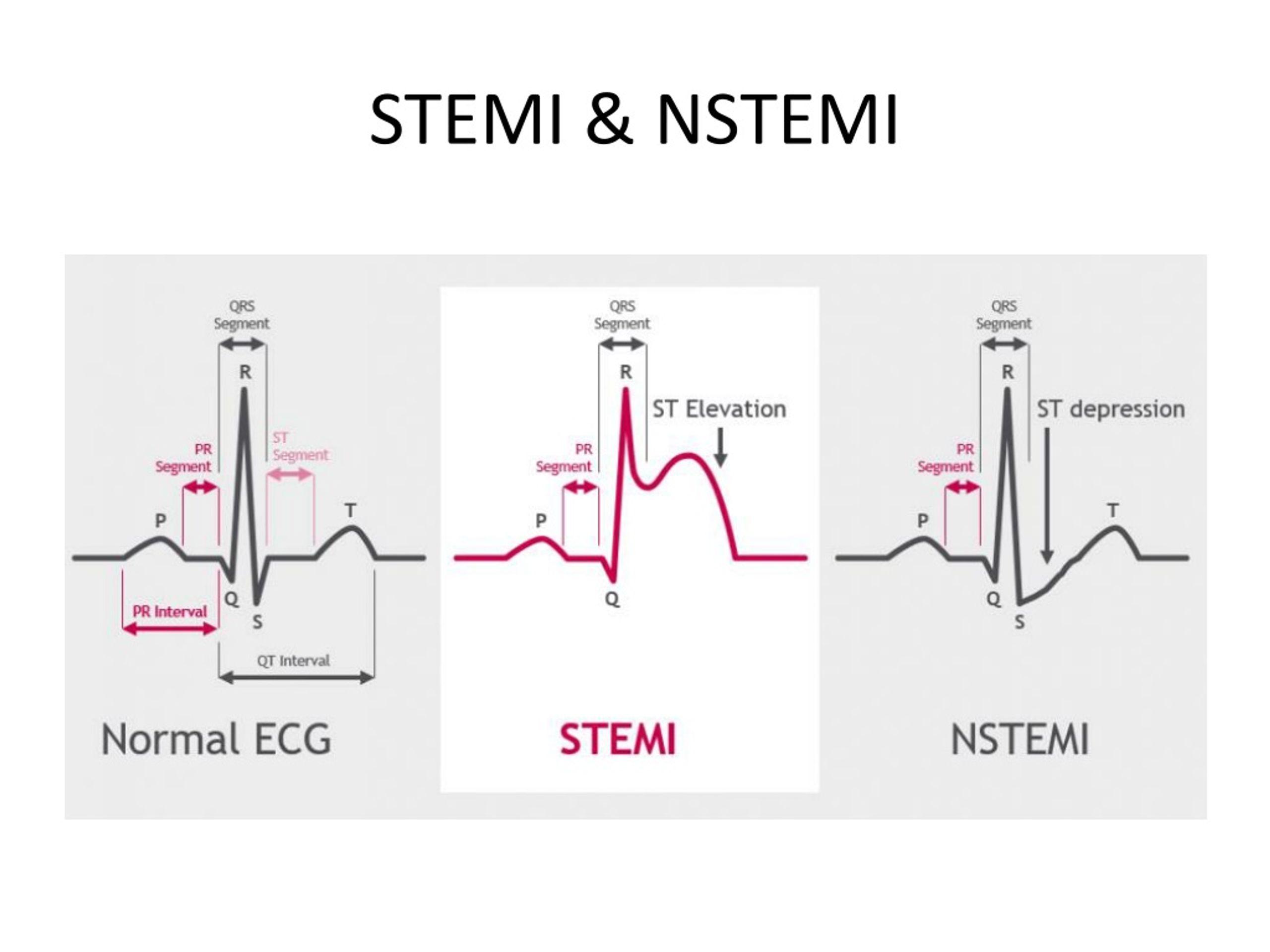
Unstable angina: ischemia without detectable myocardial necrosis
Troponin: protein released when heart muscle cells are injured NSTEMI + STEMI
NSTEMI: myocardial infarction without qualifying ST elevation. subendocardial
STEMI: myocardial infarction with qualifying ST elevation; emergency, transmural
PCI hospital: Blood flow within 90 minutes; 120+ minutes → Fibrinolysis for eligible STEMI patients
Necrotic myocardium → scar. Ventricular septal or free-wall rupture may occur 3–7 days later
Sudden cardiac death: rapid symptoms within 1 hour.
Endocrine Glands
Exocrine: Ducts, surface, secretory; not part of the endocrine system.
Endocrine: Ductless; secrete specific hormones (Chemical messengers) into the bloodstream. Hormones bind to receptors to be delivered to body tissues and organs.
Pancreas and Liver have both

Endocrine & Thyroid glands
Hypothalamus: “Master switchboard” controls the endocrine system. It secretes hormones to anterior pituitary. TRH stimulates the anterior pituitary to secrete TSH, which stimulates the thyroid gland to produce and release thyroid hormones (T3 and T4)
Thalamus → Hypothalamus (pea-sized) → pituitary gland
Pituitary gland: “Master gland” controls the functions of many target glands and cells.
Anterior pituitary: FLAT PG — FSH, LH, ACTH, TSH, prolactin, and GH.
Posterior pituitary: ADH and oxytocin
Thyroid Gland: Two lobes + TH storage
TRH → TSH → T3 and T4 → bone growth, heart function, and metabolism. (Negative feedback)

Serum Osmolality at 265, the body stops vasopressin, what will happen?
More urine is excreted; serum will increase
Parathyroid Glands
Blood calcium/phosphate homeostasis
Stops osteoblast activity (forms new bone) and osteoclast activity (breaks down old bone).
Low serum Ca²⁺ → Increases PTH release, raising calcium by promoting bone breakdown (Osteoclast)
High serum Ca²⁺ → Decreases PTH release, reducing calcium by building bone (Osteoblast)

Thymus
Behind the sternum, Immune & T cells before birth/childhood
After puberty, the thymus decreases in size and is gradually replaced with fatty tissue
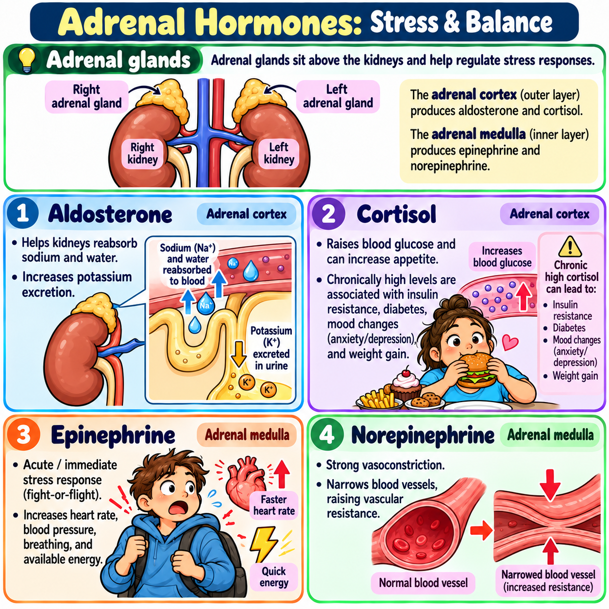
Adrenal glands
Above renal/kidney, regulates stress
Aldosterone - sodium and water reabsorption and potassium excretion
Cortisol - glucocorticoid; increases BG & appetite; high levels → insulin resistance, diabetes, depression, anxiety, and weight gain
Epinephrine - acute stress
Norepinephrine - potent/strong vasoconstriction
Pancreas: Exocrine vs. Endocrine Function
Exocrine/Ducts: 99%; digestive enzymes
Endocrine/Bloodstream: 1%; hormones & insulin 18–40 units/day or 0.5–1 unit/hour
Gonads: Ovaries and testes.
Endocrine tissues: heart releases atrial natriuretic peptide (ANP) → diuresis in the kidneys → release renin.
Release of Hormones
Steady-state release: (TH) Steady rate; blood hormone levels fluctuate very little
Intermittent release: (GH) Large amounts only as needed
Cyclic release: Fluctuates in a recurring cycle (Estrogen)
Diurnal release: Time of day → Cortisol higher (7–8 AM) & lower (2–4 AM).
Growth Hormone
Endocrine disorders can change personal appearance
GHs: GH, TH, insulin, and androgens/sex hormones
Linear bone growth in kids GH 2nd half of night
Short stature is defined as a height below 3% of population
Familial/constitutional short stature are variations (not disease)
TH bound to
globulin = inactive
Free T3 or T4 hormones, not protein-bound, enter target cells/active.
Hyperthyriodism
Excessive TH: women, smoking, iodine deficiency or excess
Graves Disease – common, autoimmune; TSH increases thyroid hormone (TH); hypertension (HTN), goiter, ophthalmopathy/exophthalmos (bulging eyes), hypermetabolic. Treatment: Radioactive iodine, beta blockers for symptoms.
Thyroid Storm – Rare, life-threatening, triggered by stress or trauma, high fever, tachycardia, agitation, psychosis, emergency. Treatment: Stabilize hemodynamic/circulatory status, then aggressively treat everything
Hypothyriodism
Shortage of dietary iodine
Goiter - 1st visible sign
Hashimoto’s Thyroiditis – Autoimmune, iodine normal, most common cause in the US. Everything slow, more menstrual bleeding, anemia.
Postpartum Thyroiditis – Inflammation after giving birth, often undiagnosed. First - hyperthyroidism. Second - 4–8 months later, the thyroid has lost most of its hormones; lasts 6–12 months, hypothyroidism. More likely permanent hypothyroidism within 5–10 years.
Manifestations of Hypothyroidism – Weakness, fatigue, weight gain, cold intolerance. Babies/Cretinism: jaundice, umbilical hernia, and sleepiness. Maternal iodine deficiency: impaired growth and intellectual disability.
Myxedema – Rare, non-pitting, mucus-type edema of the shins, feet, and eyes.
Myxedema Coma – Life-threatening; hypothyroidism in every cell → IV TH

Adrenal Cortical Insuffiency
Addison Disease – Adrenal insufficiency, hypocortisolism (everything low) low aldosterone (helps retain sodium and water), sodium and water loss, Lifelong hormone replacement therapy, medical alert bracelet
Cushing Syndrome – Excessive amounts of glucocorticoids (hypercortisolism): obesity, moon face, delayed healing, high blood sugar, infections. Treatment: Stop excessive steroid use, remove tumor surgically, radiation.
DM test
Hyperglycemia → inability to transport glucose into cells, breakdown of fats and proteins.
Blood Tests
Fasting Plasma Glucose (FPG): Morning, 8 hours of fasting; 80–100 normal, >126 DM.
Random Plasma Glucose: >200 DM.
Oral Glucose Tolerance Test (OGTT): 75 g of concentrated glucose, 2 hours later; <40 mg/dL normal, >200 DM
Capillary Whole Blood Glucose Monitoring
Hemoglobin A1C: Glucose molecules become irreversibly bound to the hemoglobin. A1C: 120-day RBC lifespan. No fasting required. <5.7% is good; >6.5% DM
Goal: DM should keep A1C <7%
Urine Test: Renal tubular maximum is approximately 180, the kidneys can no longer reabsorb all the glucose
Prediabetes
Blood glucose is elevated but does not meet the criteria for DM. Early treatment → diet, exercise, and weight loss of 7% of body weight. Exercise 30 mins/day x 5 days
T1DM
Autoimmune: insulin-producing pancreatic beta cells within the islets of Langerhans.
Ketosis and ketoacidosis
Dependent on exogenous insulin to live; rarely diagnosed <9, peaks around 12
High BG greater than 180 mg/dL and breakdown of body fats and proteins
Glucagon (opposite of insulin) a high BG level fails to suppress the production of glucagon, further increasing hyperglycemia.
Polyuria, polydipsia, polyphagia, weight loss.
Latent diabetes
Subtype of T1DM, adulthood, genetic, T1.5DM still produces some insulin for the first 6 months, age 35+, autoantibodies present against islet beta cells
T2DM
Chronic, persistent hyperglycemia; insulin supply is insufficient to meet needs; 45+ abdominal obesity & PCOS; insulin levels are high, low, or normal.
Treatment: Diet and exercise; aerobic activity 90–150 minutes per week, metformin first-line, goal A1C <7%.
Insulin Resistance (Metabolic Syndrome)
Excess body fat (T2DM), insulin resistance impair glucose regulation, compensatory increase in beta-cell → hyperinsulinemia
Maturity-Onset Diabetes of the Young (MODY)
Autosomal dominant inheritance (single-gene mutation), family history of DM (T2DM)
Gestational Diabetes
Glucose intolerance during pregnancy
Oral glucose drink, BG 2 hours later, larger babies, higher risk of fetal abnormalities.
Chronic Complications of Diabetes
Neuropathy: loss of sensation; high blood sugar damages nerves over time
Vascular: microangiopathy, thickening/hardening of capillaries/small arteries, strokes, heart attacks
Infections: bacteria thrive in high-glucose states.
Claudication, gangrene, amputation
T1DM Acute Life-Threatening Complication
Diabetic Ketoacidosis – Hyperglycemia, increased ketone concentration, blood glucose (BG) of 400–800 mg/dL, Kussmaul respirations and lethargy
Give Insulin, low Na+ & Cl-, high K+, dehydration NS 2 L over 1-1.5hrs
Hyperosmolar Hyperglycemic State (HHS)
Elderly T2DM w/ infection, MI, or stroke, CHOs, neurological deficits (not common)
Hypoglycemia
Low blood glucose <70: anxiety/irritability/hangry
Subcutaneous (SubQ) insulin can cause
Apple, grape, or cranberry juice; no orange juice if kidney disease 15/15 rule
Numbers to Memorize
Left Ventricle Preload: 5-12
Systemic Vascular Resistance: 800-1,400
Ejection Fraction: 55-75%
CO: 4-6 8 active/healthy
Cardiac Index: 2.5-4
Pulmonary low pressure: 25/10 mean 15
Blood Pressure: <120/<80
Pulse Pressure: 40
Mean arterial Pressure S + (2D) / 3 = 70-110
Heterozygous Primary: 190-400
Homozygous Primary: 400-1,000+
LDL: 100-190
Cholesterol: 200-240
Triglycerides 150-500
HDL: 40 Men, 50 Women, 60 Protective
High-sensitivity C-reactive Protein: <1 low risk >3 high risk >10 test again infection
Ankle Branchial Index: 1.0-1.4 normal, <0.5 Severe PAD
Heart Failure: r <40, p >50, mr 41-49
Pericardium: 15-50
Left coronary circulation flow is about 225 mL/min
Anterior pituitary: FLAT PG: FSH, LH, ACTH, TSH, Prolactin, and GH
Short Stature - below 3%
Fasting BG >126
Infective Endocarditis: BE TOME Blood cultures, echocardiographic, temp >38/100.4, oslet nodes tender, Murmur/micro evidence, embolic phenomena, emergency
Coarctation of the aorta
Physical Defects (Coarctation of the Aorta): A congenital condition where the aortic arch is narrowed, increasing systemic vascular resistance (SVR) and afterload. This creates a major resistance to forward blood flow.
